
14: The Endocrine System
- Page ID
- 12534
Structures of the Endocrine System
The endocrine system consists of cells, tissues, and organs that secrete hormones as a primary or secondary function. The endocrine gland is the major player in this system. The primary function of these ductless glands is to secrete their hormones directly into the surrounding fluid. The interstitial fluid and the blood vessels then transport the hormones throughout the body. The endocrine system includes the pituitary, thyroid, parathyroid, adrenal, and pineal glands (Figure 14.1). Some of these glands have both endocrine and non-endocrine functions. For example, the pancreas contains cells that function in digestion as well as cells that secrete the hormones insulin and glucagon, which regulate blood glucose levels. The hypothalamus, thymus, heart, kidneys, stomach, small intestine, liver, skin, female ovaries, and male testes are other organs that contain cells with endocrine function. Moreover, adipose tissue has long been known to produce hormones, and recent research has revealed that even bone tissue has endocrine functions.

Figure 14.1 Endocrine System Endocrine glands and cells are located throughout the body and play an important role in homeostasis.
The ductless endocrine glands are not to be confused with the body’s exocrine system, whose glands release their secretions through ducts. Examples of exocrine glands include the sebaceous and sweat glands of the skin. As just noted, the pancreas also has an exocrine function: most of its cells secrete pancreatic juice through the pancreatic and accessory ducts to the lumen of the small intestine
Download for free at http://cnx.org/contents/14fb4ad7-39a1-4eee-ab6e-3ef2482e3e22@11.1
LAB 14 EXERCISE 14-1

On the person, draw and label the following endocrine organs:
- Thyroid
- Adrenals
- Pancreas
- Ovaries
- Hypothalamus
- Pituitary
LAB 14 EXERCISE 14-2
ENDOCRINE SYSTEM – CNS AND HISTOLOGY

Identify this organ and related structures Label the following: Thyroid, parathydroid
3
5
1
2
Organ:
4
6
5
LAB 14 EXERCISE 14-3
IDENTIFY THE ORGANS AND THEIR RELATED
STRUCTURES
4
1
6
7
2
8

Organ:
3
Organ:
Adrenal/suprarenal
LAB 14 EXERCISE 14-4
- Obtain a slide of each of the tissues listed below from the slide box at your table.
- Follow the checklist above to set up your slide for viewing.
- View the slide on the objective which provides the best view. Find the representative object.
- In the circle below the name, draw a representative sample of the tissue, taking care to correctly and clearly draw their true shape in the slide. If it is a stratified epithelium draw all the layers. Draw your structures proportionately to their size in your microscope’s field of view.
- Fill in the blanks next to your drawing and identify the structures listed on the last page under, “Histology”
Repeat this for each of the tissue types seen below.

Pituitary
Pancreas

Thyroid
Parathyroid
Adrenal/suprarenal


LAB 14 EXERCISE 14-5
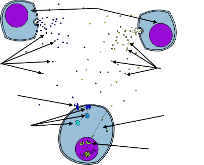
HORMONE ACTION:
LABEL THE FOLLOWING: TARGET CELL * ENDOCRINE CELLS (X2) * WATER-SOLUBLE HORMONE * STEROID HORMONE * STEROID HORMONE RECEPTOR * CELL-SURFACE HORMONE RECEPTOR * SECOND MESSENGER SYSTEM
4
1
5
2
3
6
7
MODELS: Torso, Mid-Sagittal Head
Found on models:
Hypothalamus
Pituitary gland
- Infundibulum
Pancreas
Pineal gland
Adrenal glands
Testis
Ovary
Thyroid gland
- Ithsmus
Histology:
Pituitary
- Anterior pituitary
- Posterior pituitary
- Infundibulum
Pancreas
- Endocrine pancreas
Pancreatic islet of Langerhans
Capillary in pancreatic islet
- Exocrine pancreas
acini
duct
Thyroid gland
- Follicular cell
- Parafollicular cell
- Colloid
- Thyroid follicle
Parathyroid gland
Adrenal glands
- Capsule of suprarenal gland
- Suprarenal cortex
Zona fasciculata
Zona glomerulosa
Zona reticularis
- Suprarenal medulla