
17: Blood Vessels and Circulation
- Page ID
- 12537
Skills to Develop
- Compare and contrast the three tunics that make up the walls of most blood vessels
- Distinguish between elastic arteries, muscular arteries, and arterioles on the basis of structure and location
- Describe the basic structure of a capillary bed, from the supplying metarteriole to the venule into which it drains
- Explain the structure of venous valves in the large veins of the extremities
Shared Structures
Different types of blood vessels vary slightly in their structures, but they share the same general features. Arteries and arterioles have thicker walls than veins and venules because they are closer to the heart and receive blood that is surging at a far greater pressure (Figure 17.1). Each type of vessel has a lumen—a hollow passageway through which blood flows. Arteries have smaller lumens than veins, a characteristic that helps to maintain the pressure of blood moving through the system. Together, their thicker walls and smaller diameters give arterial lumens a more rounded appearance in cross section than the lumens of veins.

Figure 17.1 Structure of Blood Vessels (a) Arteries and (b) veins share the same general features, but the walls of arteries are much thicker because of the higher pressure of the blood that flows through them. (c) A micrograph shows the relative differences in thickness. LM × 160. (Micrograph provided by the Regents of the University of Michigan Medical School © 2012)
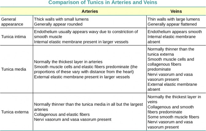
Table 17.1 compares and contrasts the tunics of the arteries and veins.
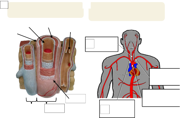
LAB 17 EXERCISES 17-1
B
A Label the following: Tunica interna * Tunica media * Tunica externa * Valve * Vasa vasorum
1
6
* Artery * Vein.
Label the following (Include Left/Right on all pages): Aorta * L common carotid a. * Brachiocephalic trunk * L subclavian a.
2
4
1
3
4
3
5
2
7
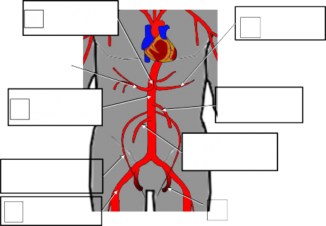
LAB 17 EXERCISES 17-2
ARTERIES OF THE ABDOMEN
5
9
4
Label the arteries (Include Left/Right where appropriate): Celiac trunk * Common hepatic a. * Gonadal a. * Superior mesenteric a. * Inferior mesenteric a. * Internal iliac a. * External iliac a. * Common Hepatic a. * Splenic a. * Abdominal aorta.
1
6
2
3
7
8
LAB 17 EXERCISES 17-3
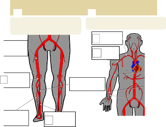
ARTERIES OF THE LIMBS
A
B
Label the following arteries (Include Left/Right where appropriate): Popliteal a.
* Posterior tibial a. * Anterior tibial a. * Fibular a. * Femoral a.
Label the following (Include Left/Right where appropriate): Ulnar a. * Radial a. * Brachial a. * Subclavian a. * Axillary a.
1
1
2
4
|
3 |
|
|
4 |
|
|
5 |
|
2
3
5
ARTERIES OF THE HEAD
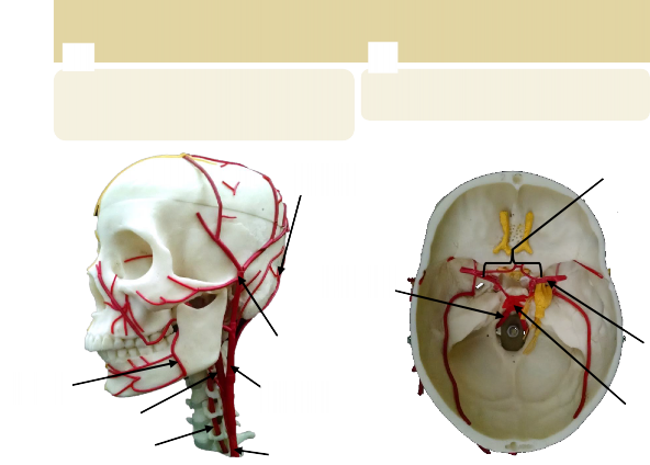
LAB 17 EXERCISES 17-4
B
A
Identify the following: Superficial temporal a.
3
7
* Facial a. * Common carotid a. * Vertebral a. * Internal carotid a. * External carotid a. * Occipital a.
Identify the following: Circle of Willis * Basilar a. * Vertebral a. * Internal carotid a.
4
2
1
5
3
1
2
6
4
LAB 17 EXERCISES 17-5
VEINS (AND ONE ARTERY)
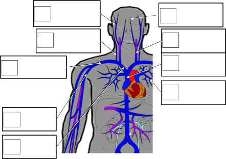
Label the following veins (Include Left/Right where appropriate): Superficial temporal v. * External jugular v. * Internal jugular v. * Axillary v. * Subclavian v. * Brachiocephalic v * Facial v * Superior vena cava * Pulmonary a.
1
6
2
7
3
8
9
4
5
LAB 17 EXERCISES 17-6
VEINS
A
B
Identify the veins (Include Left/Right where appropriate): Basilic v. * Cephalic v. * Brachial v. * Axillary v. * Median cubital v. * Radial v. * Ulnar v.
Label the veins (Include Left/Right where appropriate): Femoral v. * Common iliac v. * External iliac v. * Great saphenous v. * Inferior vena cava.
1
1
5
4
3
6
5
3
2
2
4
7

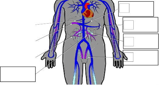
LAB 17 EXERCISES 17-7
VEINS

7
6
Label the veins: R. Hepatic v. * Hepatic Portal v. * Gastric v. * Splenic v. * Inferior mesenteric v. * Superior mesenteric v. * L Gonadal v. * L Renal v. *
5
|
1 |
|
|
2 |
|
|
3 |
|
4
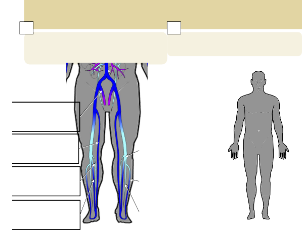
LAB 17 EXERCISES 17-8
8
VEINS & PRESSURE POINTS
A
B
4
3
2
Label the following (Include Left/Right where appropriate): Internal iliac v. * Great saphenous v. * Small saphenous v. * Popliteal v. * Anterior tibial v. * Posterior tibial v. * Fibular v.
Label 4 common pulse pressure points with names and arrows (places where you could find a pulse on a patient)
1
|
5 |
|
|
6 |
|
|
7 |
|
MODELS: Flat Body, Skull with Arteries & Nerves, Torso, Leg, Arm, and Sagittal Head
Blood vessel histology:
- Tunica externa
- Tunica media
- Tunica interna
- Vasa vasorum
- Valves
- Varicose vein
Arteries
Aorta
- Ascending * arch * descending * thoracic * abdominal
Brachiocephalic trunk
Common carotid (skull)
- External carotid (skull)
Superior thyroid (torso)
Facial (skull)
Superficial temporal (skull)
Occipital (skull)
- Internal carotid
- Circle of Willis (skull)
Subclavian
- Vertebral artery
Basilar artery (skull)
- Axillary artery
- Brachial artery
Radial artery
Ulnar artery
Celiac trunk
Left & right gastric artery
Splenic
Common hepatic a.
- Hepatic
- Superior mesenteric
Suprarenal
Renal
Gonadal
Inferior mesenteric
Common iliac
Internal iliac
External iliac
Femoral
Popliteal
Anterior tibial Posterior tibial
Veins –
Superior vena cava
Brachiocephalic
Internal jugular
- Superior sagittal sinus
- Straight sinus
External jugular
Subclavian
- Axillary
- Cephalic
- Brachial
- Radial
- Ulnar
- Basilic
- Median cubital
Inferior vena cava
- Median cubital
- Hepatic
- Suprarenal
- Renal
- Gonadal
- Common iliac
Internal iliac
External iliac
Great saphenous
Femoral
- Popliteal
- Anterior tibial
- Posterior tibial
Fibular
Small saphenous
Inferior mesenteric
Splenic
Superior mesenteric
Hepatic portal vein