
10.8: Liver Tumour
- Page ID
- 14851

ACR – Gastrointestinal – Liver Lesion – Initial Characterization
Case 1
Hepatocellular Carcinoma
Clinical:
History– This patient was a chronic alcoholic. An ultrasound performed for vague right upper quadrant pain suggested there was a solitary lesion in segment 8 of the liver.
Symptoms – Pain had resolved.
Physical – Non-contributory.
DDx:
Hepatocellular Carcinoma of the Liver (HCC)
Imaging Recommendation
ACR – Gastrointestinal – Liver Lesion – Initial Characterization, Variant 7
CT Abdomen = MR Abdomen
ODIN Link for Liver Lesion imagins (CT and MRI), Figure 10.12A and B: mistr.usask.ca/odin/?caseID=20170123135553121

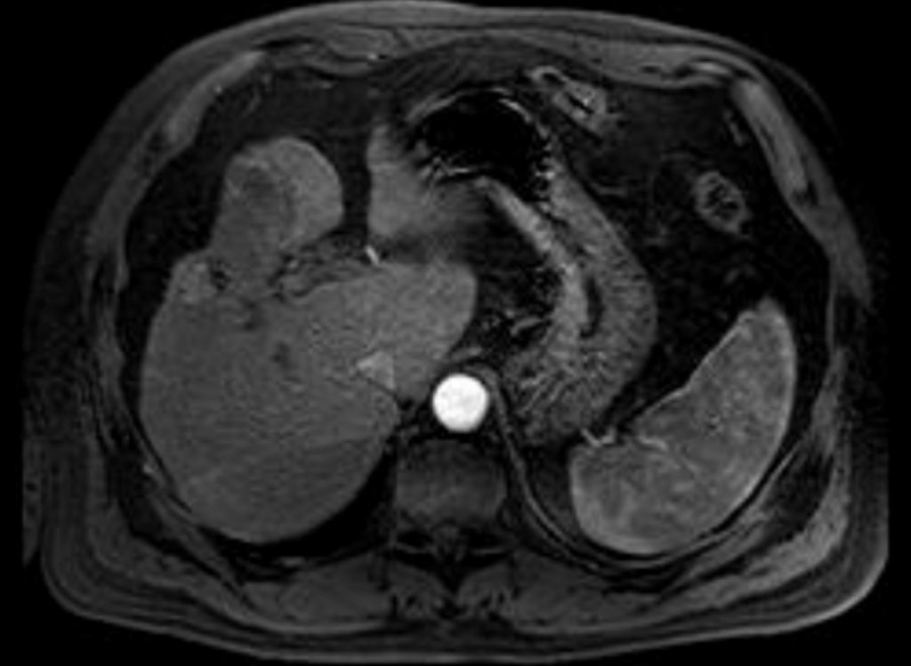
Image Assessment
Findings:
Both CT and MRI demonstrate a poorly defined, low-grade, contrast enhancing, lesion in segment 8. The liver was small and irregular.
Interpretation:
HCC
Diagnosis:
Hepatocellular Carcinoma – HCC – Biopsy Proven
Discussion:
Due to the high prevalence of benign focal hepatic lesions in adults, liver lesion characterization is an important objective of diagnostic imaging. Incidental liver masses are often discovered in healthy adults during routine imaging procedures, often abdominal ultrasounds performed for other reasons, as well as, during staging of a known malignancy, and they need to be characterized.
Common benign liver masses include cysts, biliary hamartomas, and hemangiomas; common malignant tumors include metastases and hepatocellular carcinomas (HCCs). Less common liver tumors include focal nodular hyperplasia (FNH), hepatocellular adenoma, fibrolamellar HCC, intrahepatic cholangiocarcinoma, biliary cystadenoma and cystadenocarcinoma, lymphoma, stromal tumors, a variety of sarcomas, hemangioendothelioma, and hepatoblastoma, the latter occurring in children.
On occasion, non-tumorous masses may mimic liver tumors. These mimics include focal fat deposition or sparing, abscess, hematoma, vascular shunts such as the ones to treat portal venous-hepatic venous malformations, peliosis hepatitis and transient hepatic attenuation differences on computed tomography (CT), or transient hepatic intensity differences on magnetic resonance imaging (MRI).
Patients with cirrhosis are a special group in whom certain benign (regenerating nodules), premalignant (dysplastic nodules), malignant (HCC), and nontumorous (confluent hepatic fibrosis) masses are more prevalent.
MRI is rapidly becoming the imaging modality of choice for liver lesions characterization after Ultrasound or CT have been completed.
Imaging findings may include:
- Most often normal
- A distended abdomen may be a sign of ascites.
Attributions
Figure 10.12A Axial CT of Liver by Dr. Brent Burbridge MD, FRCPC, University Medical Imaging Consultants, College of Medicine, University of Saskatchewan is used under a CC-BY-NC-SA 4.0 license.
Figure 10.12B Axial MRI of Liver by Dr. Brent Burbridge MD, FRCPC, University Medical Imaging Consultants, College of Medicine, University of Saskatchewan is used under a CC-BY-NC-SA 4.0 license.