
11.2: Normal Pregnancy
- Page ID
- 14855

ACR Women’s Imaging – Assessment of Fetal Well-Being
Case
Normal Obstetrical Ultrasound – 20 weeks
Clinical:
History – This was a 20 week gestation, routine, obstetrical ultrasound.
Symptoms – Normal pregnancy.
Physical – Active, live fetus, uterine fundus at the level of the umbilicus.
DDx:
Normal Pregnancy
Imaging Recommendation
ACR Women’s Imaging – Assessment of Fetal Well-Being, Variant 1
Ultrasound
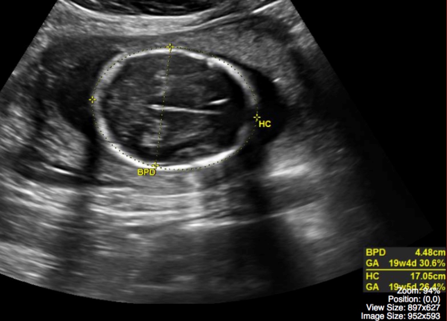
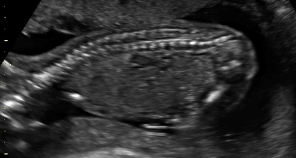
ODIN Link for Normal Pregnancy images (Ultrasound), Figure 11.4A and B: mistr.usask.ca/odin/?caseID=20170126222052150
Imaging Assessment
Findings:
Single, live fetus in breech presentation. The placenta was not previa. No fetal abnormalities. The estimated gestational age by ultrasound was 19 weeks, 6 days.
Interpretation:
Normal pregnancy.
Diagnosis:
Normal Obstetrical Ultrasound – 20 weeks
Discussion:
Despite the fact that many fetal deaths occur in women without identifiable risk factors, there is no convincing evidence that routine antenatal testing, in low-risk pregnancies, improves perinatal outcome. This is true for all imaging modalities, including the Biophysical Profile (BPP), Modified Biophysical Profile (mBPP), Doppler assessment of umbilical blood flow, and functional fetal echocardiography. Although there is a paucity of data on the use of the BPP, mBPP, and cardiovascular function testing in low-risk patients, a review of 5 trials involving 14,185 women concluded the use of umbilical artery Doppler US in low-risk or unselected populations had no maternal or perinatal benefits.
Consequently, routine antenatal fetal surveillance by any imaging modality is not recommended in pregnancies at low risk for intrauterine fetal demise.
CAR Imaging Referral Guidelines:
The scan at 18 – 20 weeks is recommended for fetal anatomy. However, screening at this time has not been shown to alter perinatal mortality except where selective termination of pregnancy is applied in the presence of gross fetal abnormality and in cases where fetal therapy or direction of delivery to a high-risk center has proven useful. Ultrasound has a proven value in assessing placenta previa, intrauterine growth restriction, incompetent cervix, and fetal demise at any stage of pregnancy.
For More Information:
CAR – Referral Guidelines: Obstetrics and Gynaecology –
www.car.ca/uploads/standards%20guidelines/car-referralguidelines-i-en_20120918.pdf
Attributions
Figure 11.4A Fetal ultrasound demonstrating head circumference measurements by Dr. Brent Burbridge MD, FRCPC, University Medical Imaging Consultants, College of Medicine, University of Saskatchewan is used under a CC-BY-NC-SA 4.0 license.
Figure 11.4B Fetal ultrasound demonstrating the spine by Dr. Brent Burbridge MD, FRCPC, University Medical Imaging Consultants, College of Medicine, University of Saskatchewan is used under a CC-BY-NC-SA 4.0 license.