
11.4: Placenta Previa
- Page ID
- 14857

ACR Women’s Imaging – Second and Third Trimester Vaginal Bleeding
Case
Placenta Previa – Posterior
Clinical:
History – This pregnant patient reported sporadic, small amounts of blood per vagina for 2 days.
Symptoms – Nil
Physical – Normal.
DDx:
Incompetent Cervix
Vaginitis
Placenta Previa
Ectopic Pregnancy
Imaging Recommendation
ACR Women’s Imaging – Second and Third Trimester Vaginal Bleeding, Variant 1
Obstetrical Ultrasound
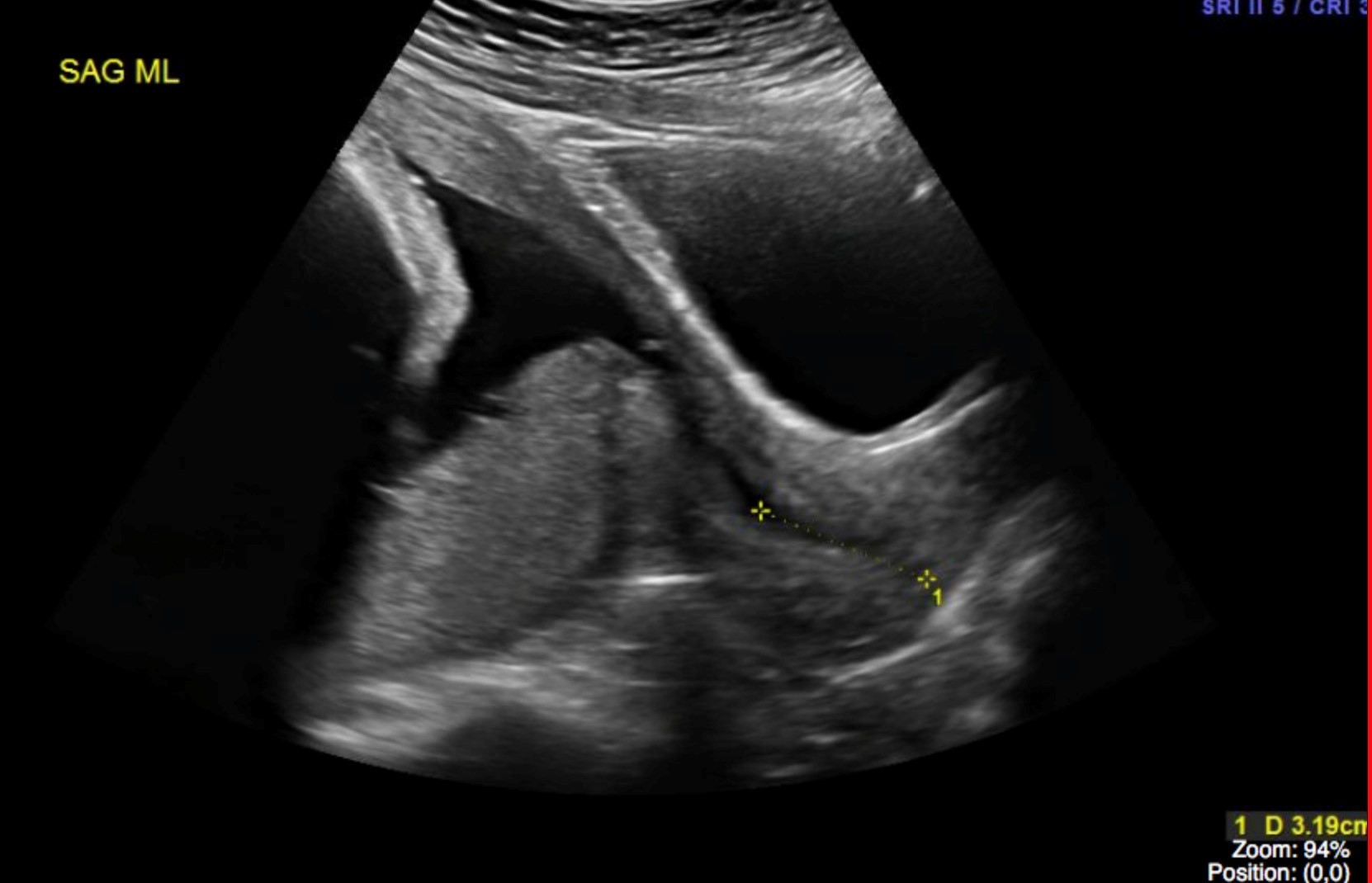
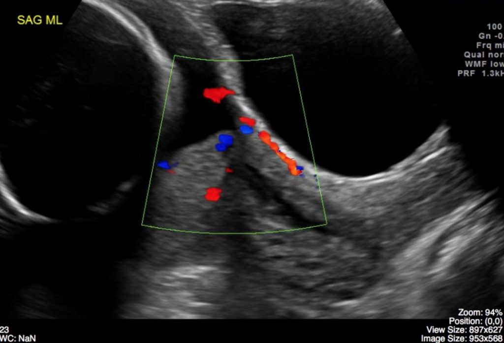
ODIN Link for Placenta Previa images (Ultrasound), Figure 11.6A and B: mistr.usask.ca/odin/?caseID=20170126224802586
Imaging Assessment
Findings:
A single liver fetus was seen in cephalic presentation. The placenta was posterior and previa. Normal otherwise. The cervix measured 3.2 cm in length. Estimated Gestational Age by US – 30 weeks.
Interpretation:
Posterior Placenta Previa
Diagnosis:
Placenta Previa – Posterior
Discussion:
The underlying cause of placenta previa is unknown. There is a clear association between placental implantation in the lower uterine segment and prior endometrial damage and uterine scarring from curettage, surgery (cesarean delivery), prior placenta previa, or multiple prior pregnancies.
At least 90% of placentas identified as being “low lying” in early pregnancy ultimately resolve by the third trimester. The term “placental migration” is widely used to describe this phenomenon. The placenta clearly does not move, rather, it is likely that the placenta grows toward the better blood supply at the fundus (a process known as trophotropism), leaving the distal portions of the placenta, closer to the relatively poor blood supply of the lower segment, to regress and atrophy. As the uterus grows and expands to accommodate the developing fetus, there is differential growth of the lower segment, and this may further increase the distance between the lower edge of the placenta and the cervix.
Bleeding from placenta previa may occur before labor as a result of development of the lower uterine segment and effacement of the cervix with advancing gestation. Pre-labor uterine contractions may also produce bleeding, as may intercourse or injudicious vaginal examination. Once labor begins, significant bleeding will occur as the cervix dilates and the placenta is forced to separate from the underlying decidua.
Attributions
Figure 11.6A Sagittal midline ultrasound of the cervix and placenta by Dr. Brent Burbridge MD, FRCPC, University Medical Imaging Consultants, College of Medicine, University of Saskatchewan is used under a CC-BY-NC-SA 4.0 license.
Figure 11.6B Sagittal midline doppler ultrasound of the cervix and placenta in a pregnant patient by Dr. Brent Burbridge MD, FRCPC, University Medical Imaging Consultants, College of Medicine, University of Saskatchewan is used under a CC-BY-NC-SA 4.0 license.