
14.4: Glenohumeral Dislocation – Anterior and Posterior
- Page ID
- 14883

ACR – MSK – Acute Shoulder Pain
Case 1
Anterior Dislocation
Clinical:
History – This patient collided with another player on the soccer field. He immediately experienced moderate right shoulder pain. He has never had shoulder problems before.
Symptoms – He reports having a painful right shoulder with limited range of motion.
Physical – The shoulder was deformed and there is a vacant space where the humeral head should be. There was very limited, painful, range of motion.
DDx:
Humeral Dislocation
Humeral Fracture
Imaging Recommendation
ACR – MSK – Acute Shoulder Pain, Variant 1
X-rays
ODIN Link for Anterior Humeral Disloation images, Figure 14.5A and B: mistr.usask.ca/odin/?caseID=20150209141141444
Imaging Assessment
Findings:
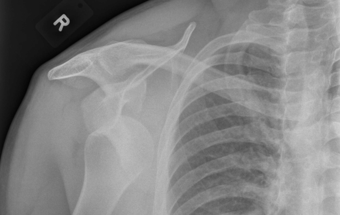
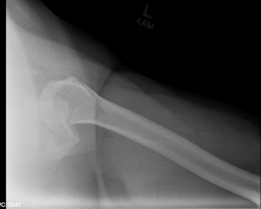
The humerus was dislocated anteriorly and resides in a subcoracoid location. No fractures identified.
Interpretation:
Anterior humeral dislocation
Diagnosis:
Anterior Dislocation of the Humerus
Discussion:
Glenohumeral joint dislocation accounts for >50% of all dislocations in the body. Anterior/subcoracoid shoulder dislocation is most common form of shoulder dislocation (96%).
Mechanism: Direct blow to a externally rotated, abducted, and extended arm.
Age – Younger individuals
Anterior humeral dislocation may be associated with:
- Hill-Sachs defect (50%), is a depression fracture of the posterolateral surface of the humeral head from impaction of the head against the glenoid rim. Best demonstrated on the anteroposterior projection with the arm internally rotated.
- Bankart lesion is a fracture of anterior aspect of inferior glenoid rim. Only the cartilaginous portion of the glenoid labrum may be injured, which may only be visible on MRI.
- Fracture of greater tuberosity (15%).
Case 2
Posterior Dislocation
Clinical:
History – This patient was accidentally electrocuted while installing an oven. He has had recurrent shoulder dislocations secondary to a seizure disorder.
Symptoms – Burns on his hands from the cross-current. Confusion. Painful, immobile right shoulder.
Physical – The shoulder was deformed and almost immobile. Mobility attempts cause severe pain.
Laboratory – None.
DDx:
Posterior humeral dislocation
Humeral fracture
Imaging Recommendation
ACR – MSK – Acute Shoulder Pain, Variant 1
X-rays
ODIN Link for Posterior Humeral Dislocation images, Figure 14.6A and B:mistr.usask.ca/odin/?caseID=20150209145146474
Imaging Assessment
Findings:
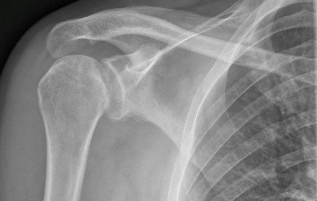
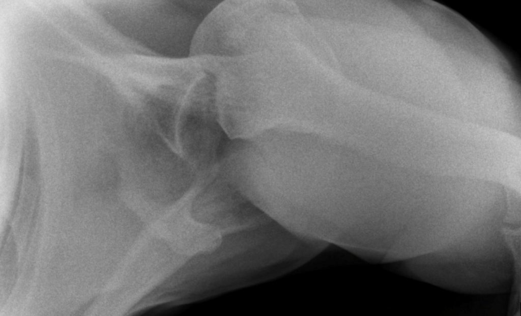
The humerus was vertically oriented and had the appearance of a light bulb on the AP view. There was a posterior dislocation of the humerus. The head/neck junction was sclerotic and had a deep groove where it had chronically impacted the glenoid rim at the time of previous dislocations. The acromioclavicular joint was fused due to arthritic change.
Interpretation:
Posterior Dislocation of the Humerus
Diagnosis:
Posterior Dislocation with Reverse Hills-Sachs Deformity
Discussion:
Posterior humeral dislocations account for 14% of all glenohumeral dislocations. The patient most often has a seizure disorder.
The humeral head is fixed in internal rotation and looks like a light bulb on all views of the shoulder. Look at the axillary or Y-view to see if the head still lies within the glenoid fossa. On the Y-view (an oblique view of the shoulder), the head will lie lateral to the glenoid in a posterior dislocation.
May be associated with:
A Reverse Hills-Sachs deformity where an anterior-medial humeral head depression is formed from chronic impaction of the humeral head upon the glenoid.
Attributions
Figure 14.5B X-ray of the shoulder displaying dislocation of the humeral head by Dr. Brent Burbridge MD, FRCPC, University Medical Imaging Consultants, College of Medicine, University of Saskatchewan is used under a CC-BY-NC-SA 4.0 license.
Figure 14.5C X-ray of the humerus and glenohumeral joint displaying anterior dislocation of the humeral head by Dr. Brent Burbridge MD, FRCPC, University Medical Imaging Consultants, College of Medicine, University of Saskatchewan is used under a CC-BY-NC-SA 4.0 license.
Figure 14.6A X-ray of the shoulder displaying dislocation of the humeral head by Dr. Brent Burbridge MD, FRCPC, University Medical Imaging Consultants, College of Medicine, University of Saskatchewan is used under a CC-BY-NC-SA 4.0 license.
Figure 14.6B X-ray of the glenohumeral joint displaying posterior dislocation by Dr. Brent Burbridge MD, FRCPC, University Medical Imaging Consultants, College of Medicine, University of Saskatchewan is used under a CC-BY-NC-SA 4.0 license.