
14.10: Ankle Trauma, Fractures
- Page ID
- 14876

ACR – MSK – Acute Ankle Trauma
Case
Tri-malleolar fracture of the ankle
Clinical:
History – This 27 year old male caught the toe of his shoe under third base when sliding. He was in severe pain.
Symptoms – The patient was in severe pain. He reported that he could not bear weight on his ankle.
Physical – He had a deformed ankle with fracture blisters present. Severe tenderness was detected on palpation. Pulses were normal.
DDx:
Ankle dislocation
Ankle fracture
Imaging Recommendation
ACR – MSK – Acute Trauma to the Ankle, Variant 1
X-rays
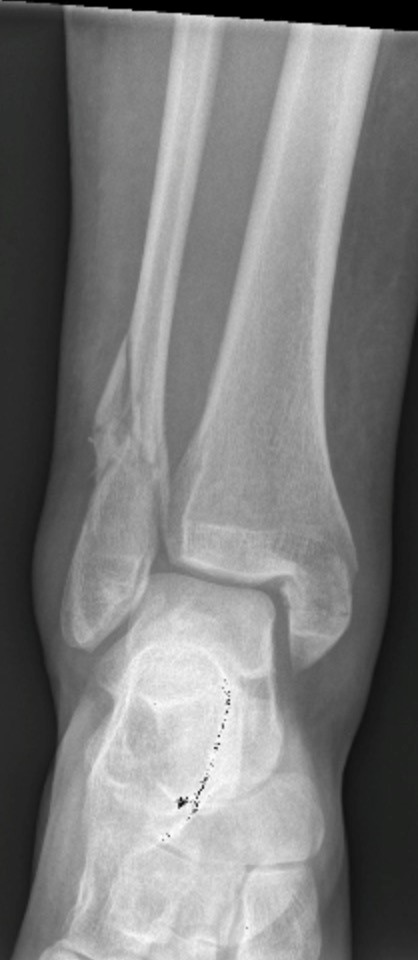
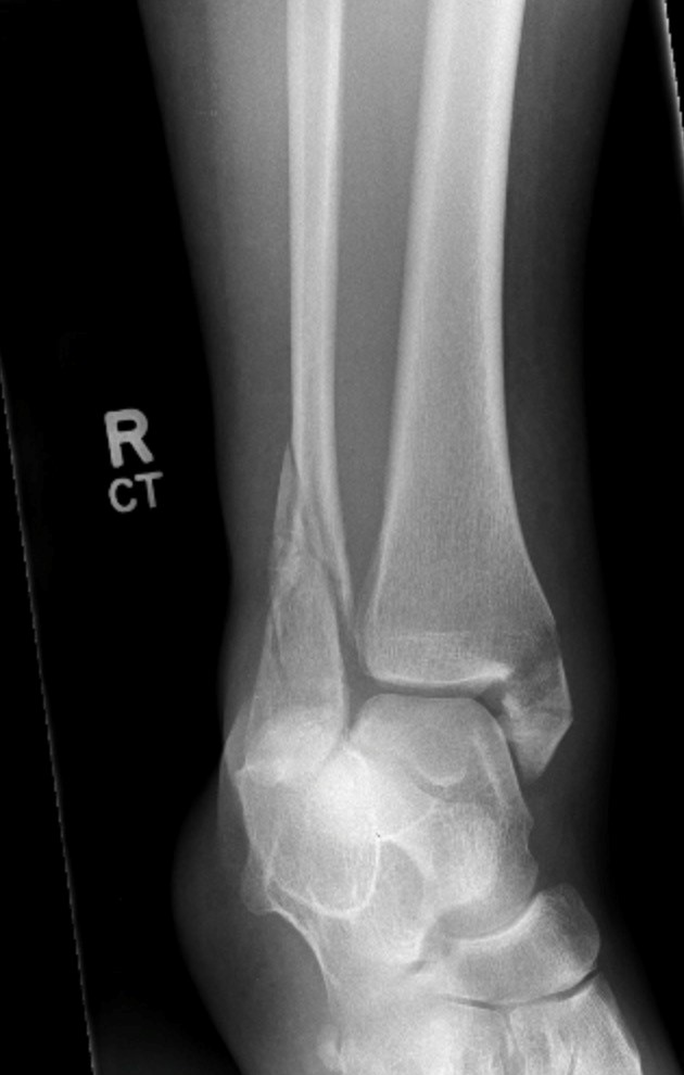
ODIN Link for Ankle Fracture images, Figure 14.14A and B: mistr.usask.ca/odin/?caseID=20150317215510072
Imaging Assessment
Findings:
There was a transverse fracture of the medial malleolus associated with an oblique fracture of the fibula above the level of the ankle joint. A small, minimally displaced, posterior malleolar fracture was also seen on the lateral view. The fibular fracture was mildly laterally displaced.
Interpretation:
Tri-malleolar fracture of the ankle
Diagnosis:
Tri-malleolar fracture of the ankle
Discussion:
Given the relatively low incidence of fracture in patients experiencing ankle trauma, the Ottawa Ankle Rule (OAR) criteria were established to identify those patients with sufficiently low probability of fracture that they can safely be treated without radiographic evaluation. Subsequently, the validation and cost-effectiveness of these guidelines have been confirmed in numerous studies and have been extended to a variety of outpatient practice settings.
The OAR guidelines recommend ankle radiographs in patients with the following clinical findings:
- inability to bear weight immediately after the injury and take 4 steps in the ED or;
- bone tenderness at the posterior edge and tip of either malleolus.
An evaluation of the traumatized ankle should consist of anteroposterior (AP), lateral, and mortise views of the ankle. Additional views can be added to the minimal series in questionable cases. In the setting of suspected deltoid ligament disruption following supination-external rotation injuries of the ankle, a gravity-stress view has been shown to be as reliable and is perceived to be more comfortable than x-rays obtained with manual stress. The fifth metatarsal base distal to the tuberosity should be seen on at least one projection. The use of a pertinent clinical history for the site of point tenderness will decrease the miss rate for subtle fractures by approximately 50%.
X-ray findings may include:
- Generalized or localized soft tissue swelling.
- Fractures may be seen in the medial malleolus, the lateral malleolus, the posterior tibia and in the fibula.
- Joint space widening, the ankle mortice, may be present.
- A joint effusion may be seen.
- The relationship of the fibula to the distal tibia may be disrupted.
Attributions
Figure 14.14A X-ray of the ankle, displaying fractures by Dr. Brent Burbridge MD, FRCPC, University Medical Imaging Consultants, College of Medicine, University of Saskatchewan is used under a CC-BY-NC-SA 4.0 license.
Figure 14.14B X-ray of the ankle, displaying fractures by Dr. Brent Burbridge MD, FRCPC, University Medical Imaging Consultants, College of Medicine, University of Saskatchewan is used under a CC-BY-NC-SA 4.0 license.