
15.3: Urinary Tract Infection and Suspected Vesico-Ureteral Reflux
- Page ID
- 14893

ACR – Pediatric – Urinary Tract Infection
Case
Vesico-ureteric reflux
Clinical:
History – This 3 year old female has had 2 culture proven urinary tract infections.
Symptoms – None at present.
Physical – No abnormalities.
DDx:
Normal
Vesico-ureteric reflux
Urinary tract calculus
Bladder diverticulum
Imaging Recommendation
ACR – Pediatric – Urinary Tract Infection
Renal ultrasound was normal.
Cysto-urethrography.
ODIN for Cysto-Urethrogram images, Figure 15.6A and B: mistr.usask.ca/odin/?caseID=20160503232955033


Imaging Assessment
Findings:
There was ureteric reflux as the bladder progressively filled. The collecting system on the left was more dilated and the fornices of the calyces were blunted. No other bladder abnormalities. The bladder emptied completely on voiding.
Interpretation:
Vesico-ureteric reflux. Grade 3 on the left and Grade 2 on the right.
Diagnosis:
Vesico-ureteric reflux.
Discussion:
Urinary tract infection (UTI) is a common disease of childhood. The investigation of UTI in children has been the subject of debate and controversy for many years. Most agree that the first imaging modality to be used should be an ultrasound examination to exclude obstruction, structural abnormalities, or renal calculi.
The role of 99m Tc dimercaptosuccinic acid scintigraphy (DMSA) in the diagnosis of acute pyelonephritis is becoming increasingly important. Many argue that if the DMSA study is normal at the time of acute UTI, no further investigation is required because the kidneys have not been involved and thus there will be no late sequelae. Others use the acute DMSA study to determine the intensity of antibiotic therapy.
The importance of the role of vesico-ureteric reflux (VUR) is being debated. Some physicians will only proceed to cystography to detect VUR if the DMSA study is abnormal, whereas others advocate a more aggressive approach and employ cystography for recurrent infection. VUR can be identified using radiological and scintigraphic techniques. Although the radiological cystogram is the gold standard and in a male patient it is the optimal imaging study to exclude posterior urethral valves, radionuclide cystograms are advantageous in other situations.
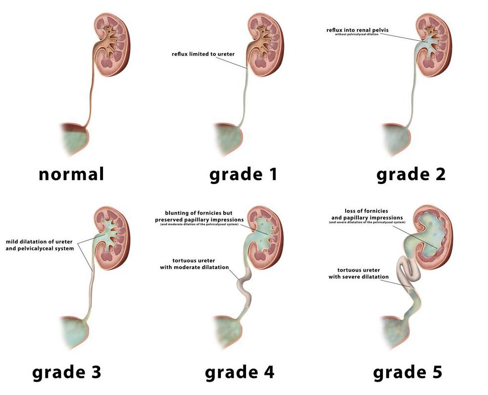
Grading of the vesico-ureteric reflux helps to determine treatment strategies i.e. medical vs. surgical.
Vesico-ureteric Reflux Grading
Attributions
Figure 15.6A Pelvis x-ray of the bladder filled with contrast by Dr. Brent Burbridge MD, FRCPC, University Medical Imaging Consultants, College of Medicine, University of Saskatchewan is used under a CC-BY-NC-SA 4.0 license.
Figure 15.6B Pelvis x-ray of the bladder with contrast and bilateral vesico-ureteric reflux by Dr. Brent Burbridge MD, FRCPC, University Medical Imaging Consultants, College of Medicine, University of Saskatchewan is used under a CC-BY-NC-SA 4.0 license.
Figure 15.7 Vesico-ureteric reflux grading. Originally published at https://radiopaedia.org/cases/illustration-vesicoureteric-reflux-grading under a Creative Commons Attribution-Non-commercial-Share Alike 3.0 Unported License.