
16.4: Testicular Tumour
- Page ID
- 14901

Case
Mixed Germ Cell Tumour, Testicle
Clinical:
History – This 45 year old male discovered a testicular mass.
Symptoms – Scrotal mass
Physical – The left testicle was enlarged and lobulated.
DDx:
Testicular mass
Epididymal mass
Testicular appendix
Imaging Recommendation
Scrotal Ultrasound
Imaging Assessment
Findings:
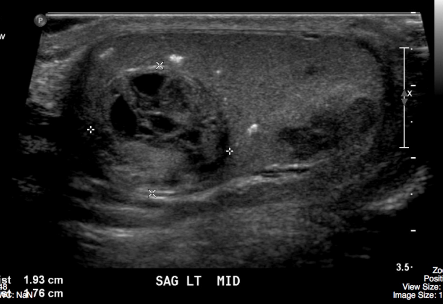
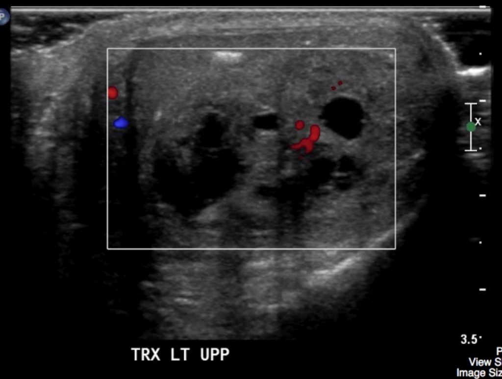
The images revealed a mixed cystic and solid mass of the left testicle with increased Doppler signal associated with the mass.
Interpretation:
Testicular mass
Diagnosis:
Mixed Germ Cell Tumour – Pathology proven
Discussion:
Although carcinoma of the testicle is relatively uncommon, representing only 1% of all malignancies occurring in men, it is the most frequent malignancy in men between the ages of 20 and 34, accounting for 10% to 14% of cancer incidence in that age group. The National Cancer Institute estimates that there will be about 8,430 new cases of testicular cancer in the U.S. and about 380 deaths from the disease in 2015.
Over 90% of testicular tumors are of germ cell origin and are malignant. Of these, 40% are seminomas. The nonseminomatous tumours are clinically more aggressive and include embryonal cell carcinoma (15% to 20%), teratoma (5% to 10%), and choriocarcinoma (<1%). Testicular cancer has an excellent prognosis, with 10-year survival rates exceeding 96%. Non–germ-cell tumours are typically benign and have their origin from the Leydig and Sertoli cells or from connective tissue stroma.
Scrotal US is frequently used, and should always be the initial imaging modality in assessing patients with scrotal masses. This study can often differentiate fluid-filled spermatoceles and hydroceles from solid intratesticular tumours. Often times, the diagnosis of a testicular mass is apparent by clinical evaluation, and US can be used for confirmation.
US findings may include:
- US is used to distinguish between intra-testicular masses, which are more commonly malignant, and extra-testicular masses, which are more commonly benign.
- US can also be used to accurately differentiate intra-testicular solid masses, which are often malignant, from cystic lesions, which are usually benign and may include tubular ectasia of the rete testes, simple cysts, and tunica albuginea cysts.
- Solid masses usually appear hypoechoic relative to the adjacent testicular parenchyma, and internal vascularity is usually detectable with color Doppler imaging.
- In comparison, cysts appear anechoic, with no internal vascularity, and usually demonstrate posterior acoustic enhancement.
- At imaging, a solid intra-testicular mass with internal vascularity is suggestive of a testicular tumor in the appropriate clinical setting.
- US has been shown to have a 92%–98% sensitivity and a 95%–99.8% specificity for testicular malignancy.
Attributions
Figure 16.4A Sagittal ultrasound of the left testicle revealed a cystic and solid mass by Dr. Brent Burbridge MD, FRCPC, University Medical Imaging Consultants, College of Medicine, University of Saskatchewan is used under a CC-BY-NC-SA 4.0 license.
Figure 16.4B Transverse doppler ultrasound of the left testicular tumor by Dr. Brent Burbridge MD, FRCPC, University Medical Imaging Consultants, College of Medicine, University of Saskatchewan is used under a CC-BY-NC-SA 4.0 license.