
17.1: Head and Neck
- Page ID
- 14905

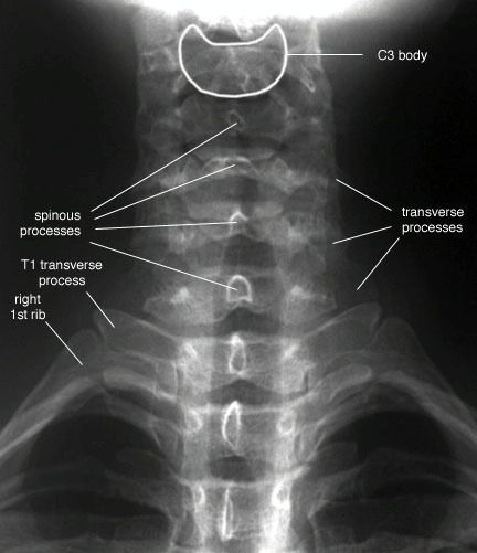
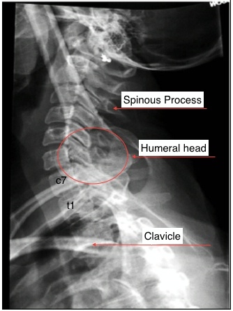
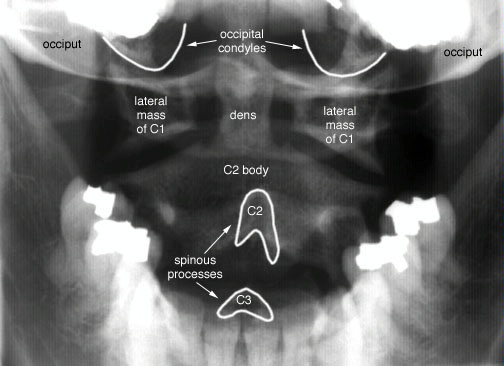
The following are normal x-rays of the cervical spine (C-spine):
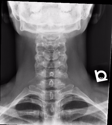
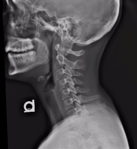
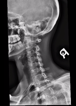
ODIN Link to C-Spine X-Rays – mistr.usask.ca/odin/?caseID=20160503232057810
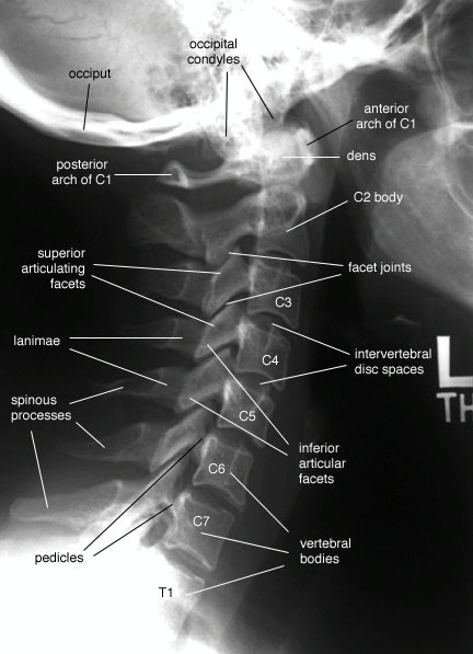
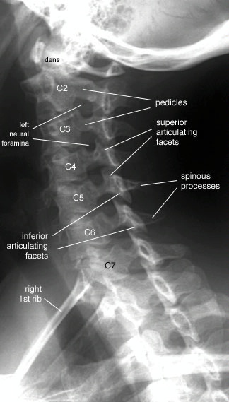
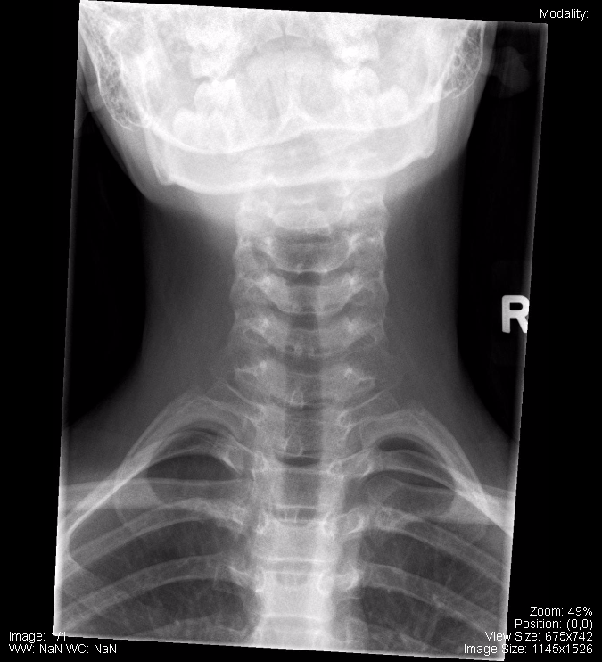
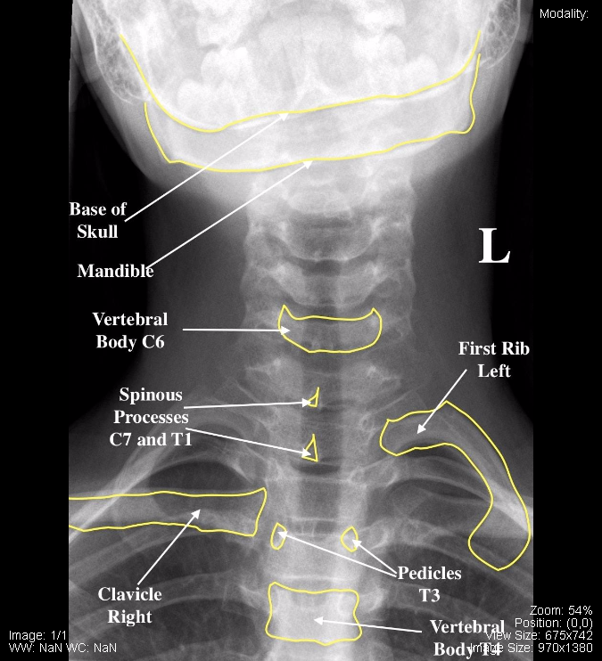
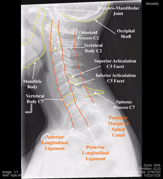
Normal Adult C-Spine, Labelled
ODIN Link to Normal, Adult Cspine Images, Labelled: mistr.usask.ca/odin/?caseID=20170406001750724
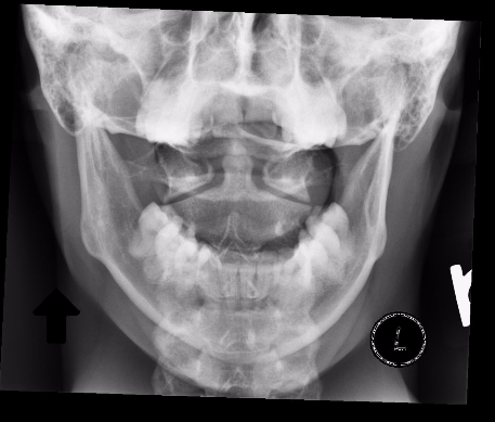
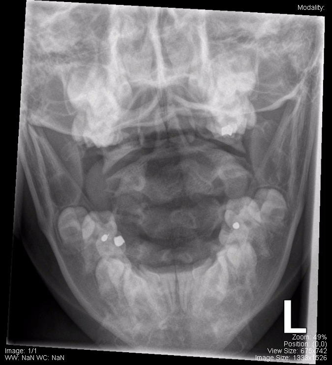
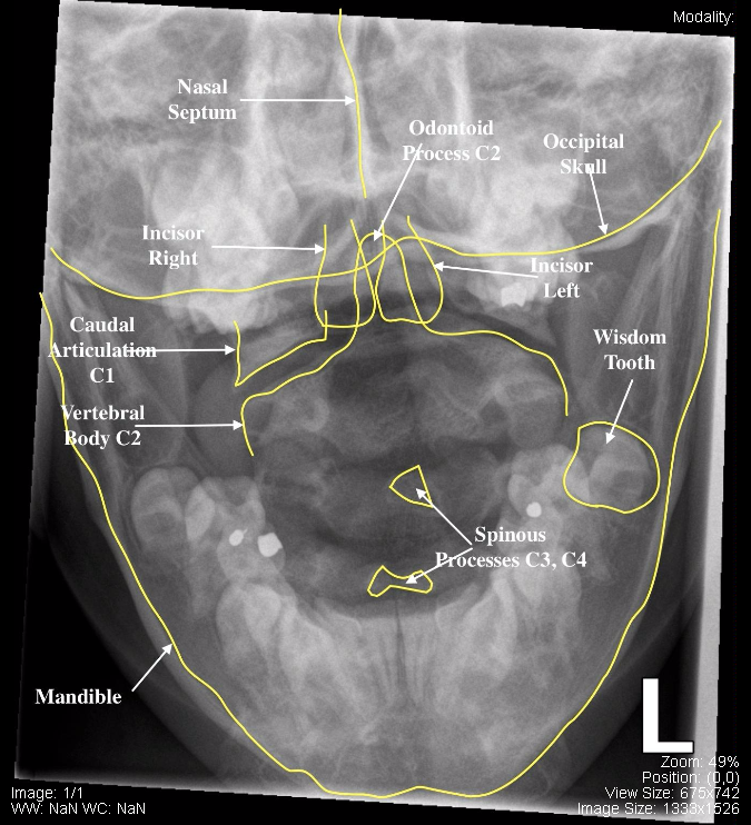
The following are normal images of the Pediatric C-Spine, unlabelled and labelled:

ODIN Link to Pediatric C-Spine – mistr.usask.ca/odin/?caseID=20170825145159975
The following are normal CT images of the brain:
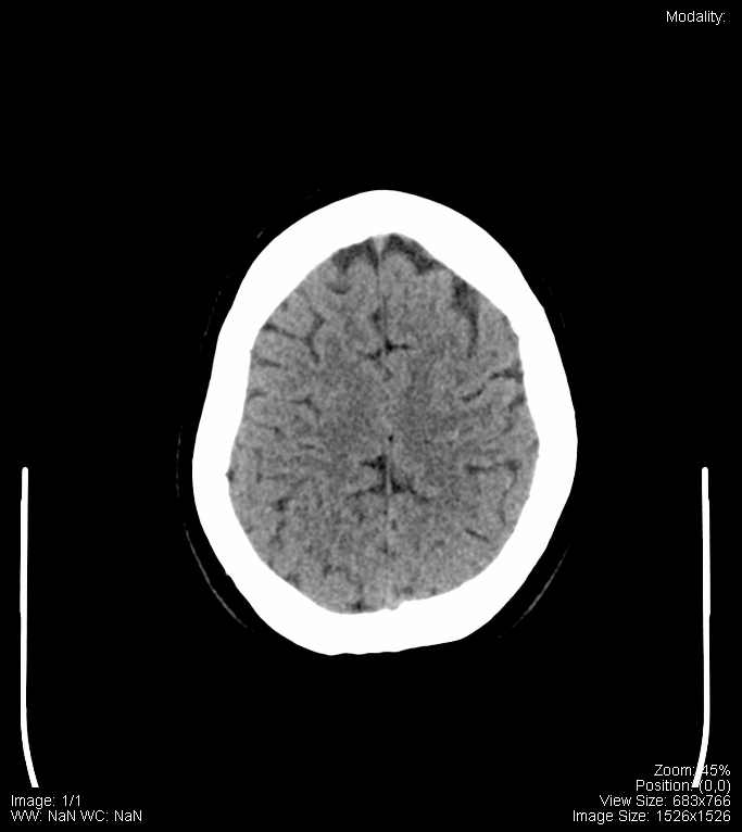
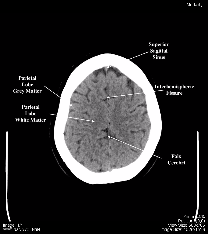
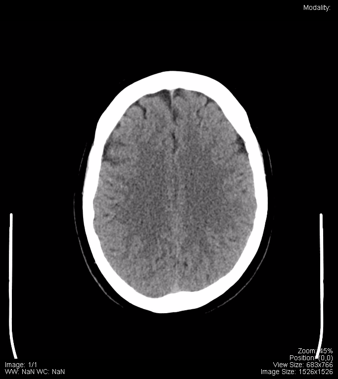
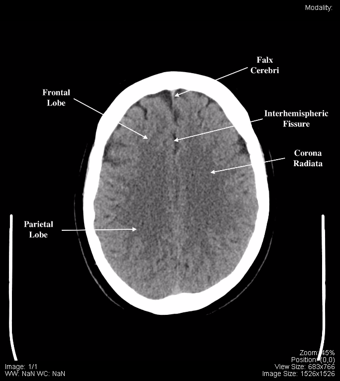
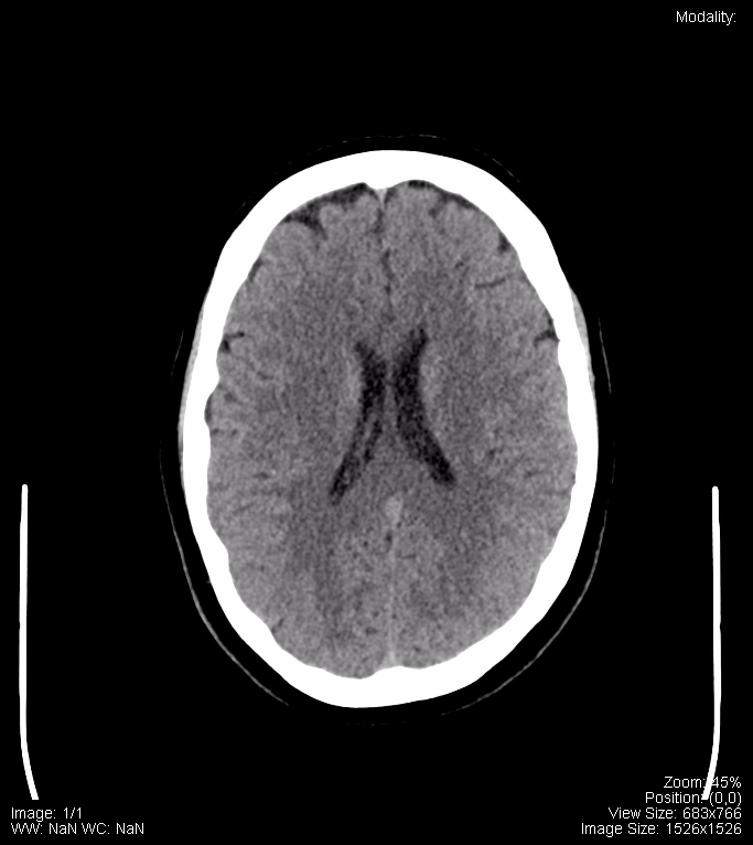
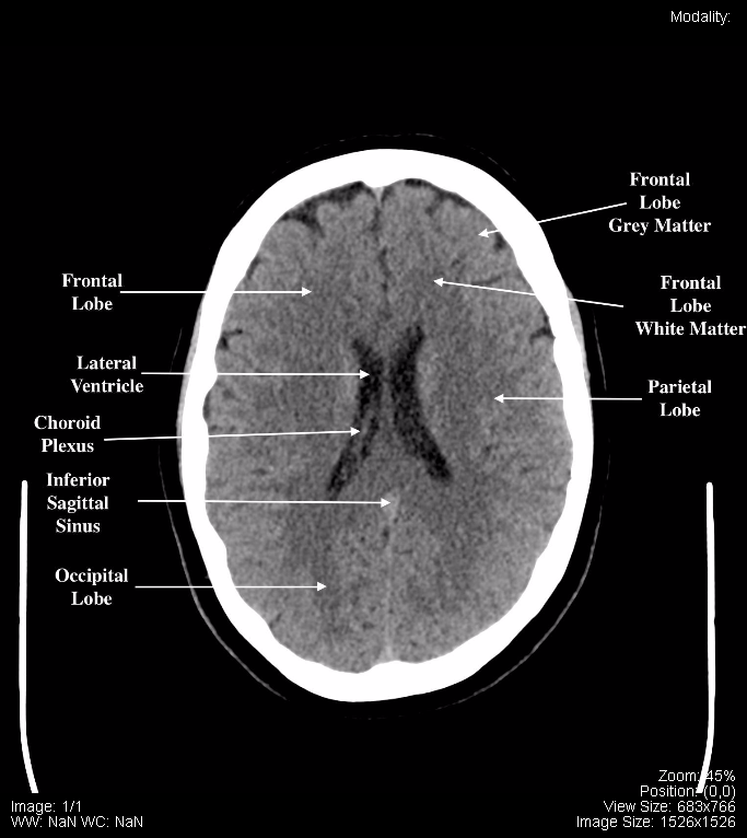
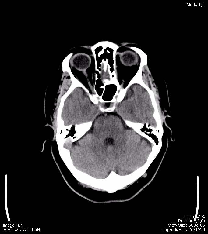
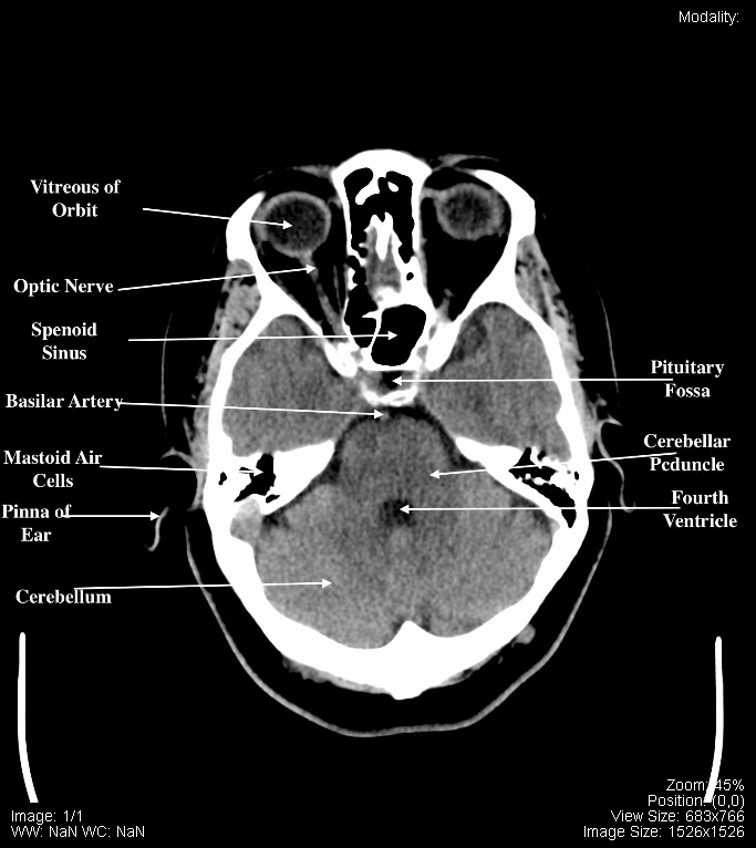
The ODIN link provides the full set of images and they can be viewed with different level/window settings. The brain level and window was utilized for the labelled brain CT images (including labelled and unlabelled). The images above are sample thumbnails.
ODIN Link to Brain CT – mistr.usask.ca/odin/?caseID=20170825124325726
The following are normal CT images of the C-Spine (axial, coronal, and sagittal):
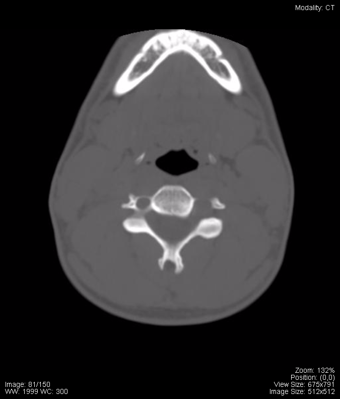
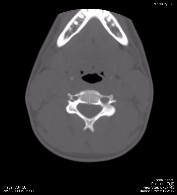
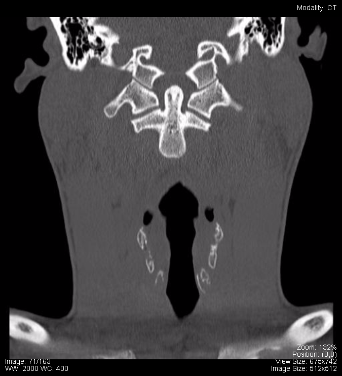
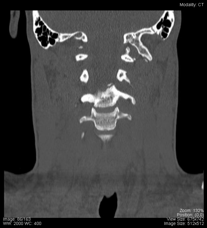
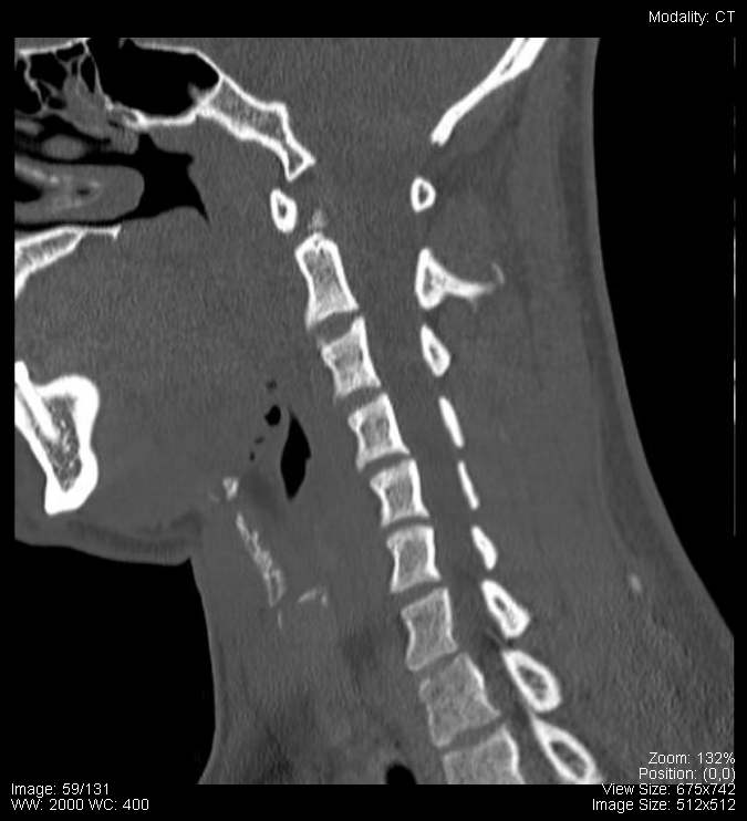
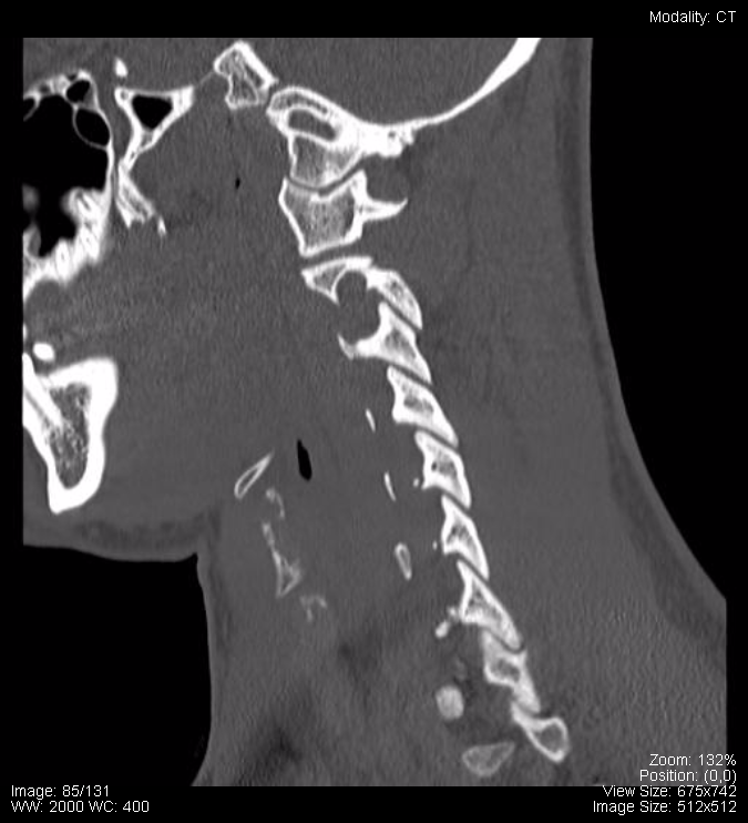
The ODIN link provides the full set of images and they can be viewed with different level/window settings. The brain level and window was utilized for the labelled brain CT images (including labelled and unlabelled). The images above are sample thumbnails.
ODIN Link to C-Spine CT – mistr.usask.ca/odin/?caseID=20170724143602846
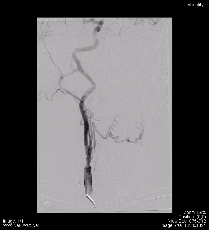
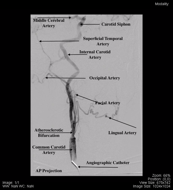
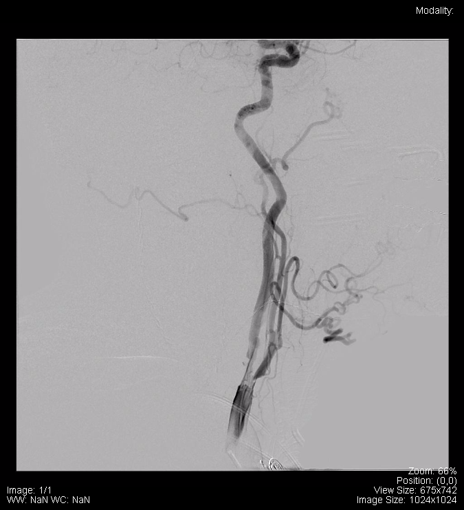
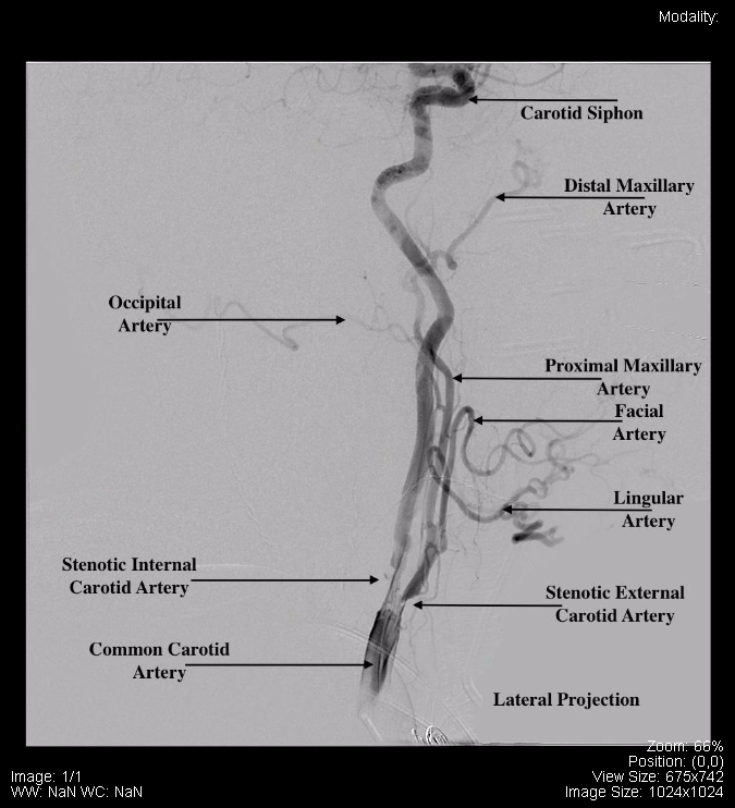
The following Digital Subtraction Angiography (DSA) is not normal because of the Stenotic Carotid Artery. Otherwise, the following DSA displays the vasculature in the head and neck:
ODIN Link to Images – mistr.usask.ca/odin/?caseID=20170825125327526
Attributions
All figures in “Chapter 17: Head and Neck” by Dr. Brent Burbridge MD, FRCPC, University Medical Imaging Consultants, College of Medicine, University of Saskatchewan is used under a CC-BY-NC-SA 4.0 license.