
17.4: Pelvis
- Page ID
- 14908

\( \newcommand{\vecs}[1]{\overset { \scriptstyle \rightharpoonup} {\mathbf{#1}} } \) \( \newcommand{\vecd}[1]{\overset{-\!-\!\rightharpoonup}{\vphantom{a}\smash {#1}}} \)\(\newcommand{\id}{\mathrm{id}}\) \( \newcommand{\Span}{\mathrm{span}}\) \( \newcommand{\kernel}{\mathrm{null}\,}\) \( \newcommand{\range}{\mathrm{range}\,}\) \( \newcommand{\RealPart}{\mathrm{Re}}\) \( \newcommand{\ImaginaryPart}{\mathrm{Im}}\) \( \newcommand{\Argument}{\mathrm{Arg}}\) \( \newcommand{\norm}[1]{\| #1 \|}\) \( \newcommand{\inner}[2]{\langle #1, #2 \rangle}\) \( \newcommand{\Span}{\mathrm{span}}\) \(\newcommand{\id}{\mathrm{id}}\) \( \newcommand{\Span}{\mathrm{span}}\) \( \newcommand{\kernel}{\mathrm{null}\,}\) \( \newcommand{\range}{\mathrm{range}\,}\) \( \newcommand{\RealPart}{\mathrm{Re}}\) \( \newcommand{\ImaginaryPart}{\mathrm{Im}}\) \( \newcommand{\Argument}{\mathrm{Arg}}\) \( \newcommand{\norm}[1]{\| #1 \|}\) \( \newcommand{\inner}[2]{\langle #1, #2 \rangle}\) \( \newcommand{\Span}{\mathrm{span}}\)\(\newcommand{\AA}{\unicode[.8,0]{x212B}}\)
The following is a normal ultrasound of the testicles:
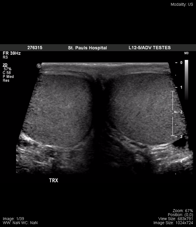
Transverse – both testicles
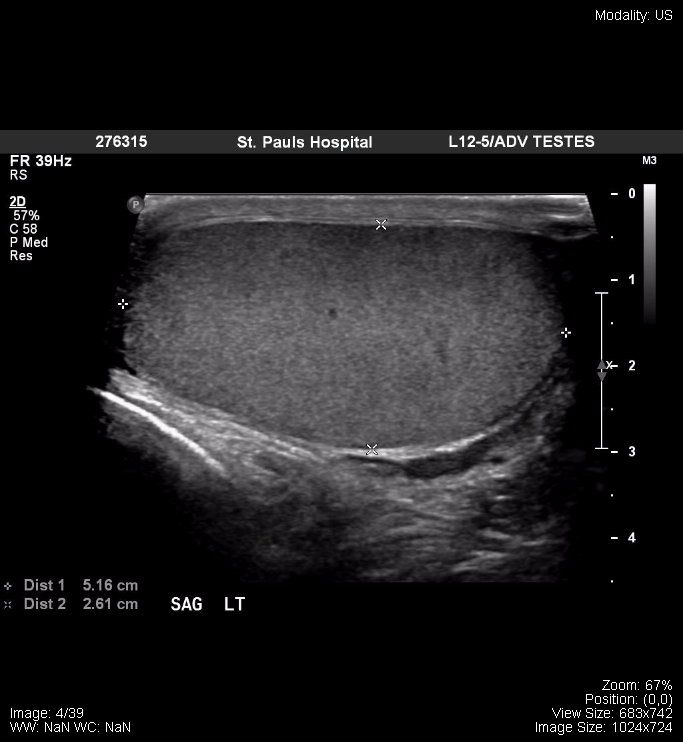
Sagittal view, left testicle, with measurements
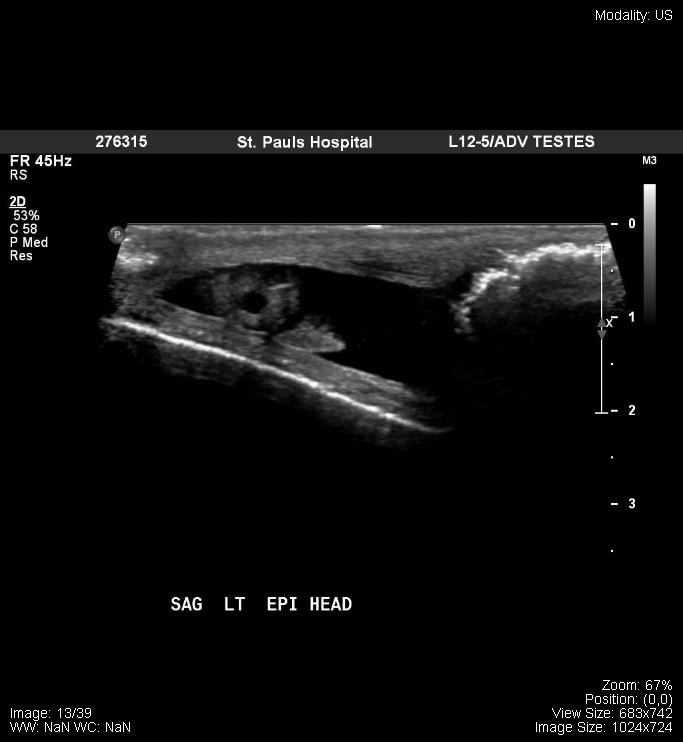
Sagittal view, left epididymis, head
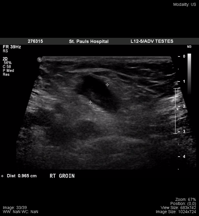
Right Groin
ODIN Link to Images: mistr.usask.ca/odin/?caseID=20170803094403042
The following are from a normal female pelvic ultrasound:
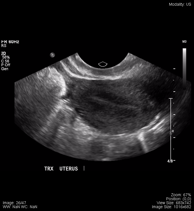
Transverse Uterus |
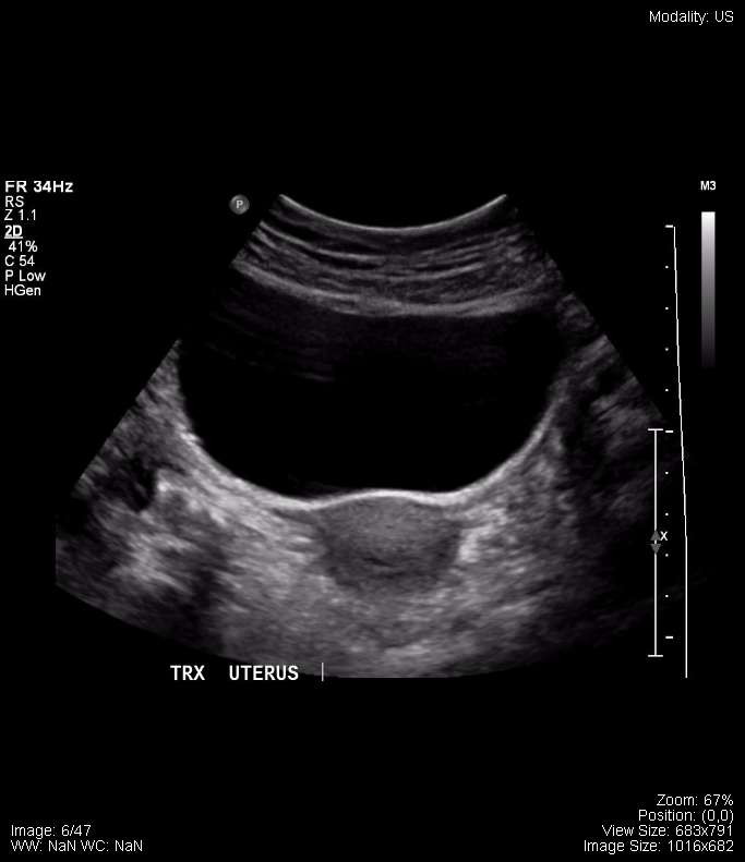
Transverse Uterus 2 |
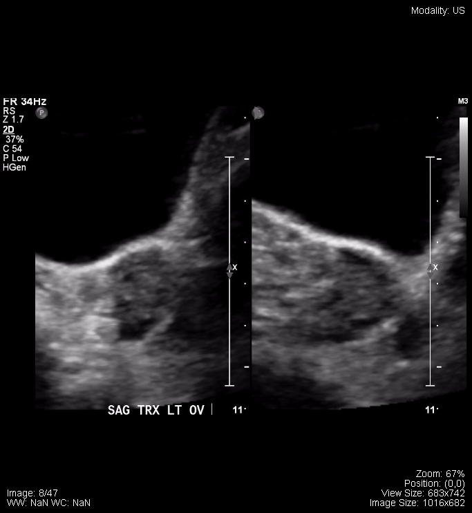
Sagittal Ovary |
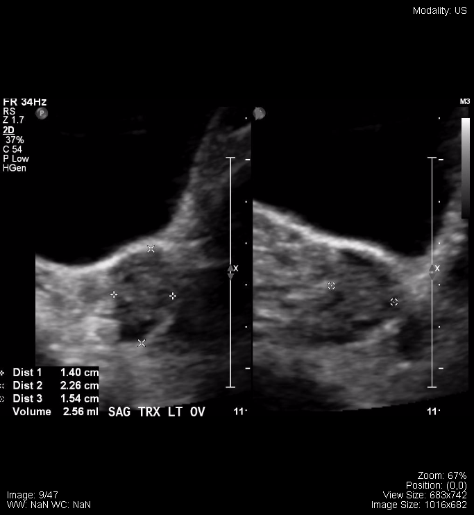
Sagittal Ovary, with measurements |
ODIN Link to Images: mistr.usask.ca/odin/?caseID=20170731231759071
The following are samples from a normal pregnancy ultrasound:
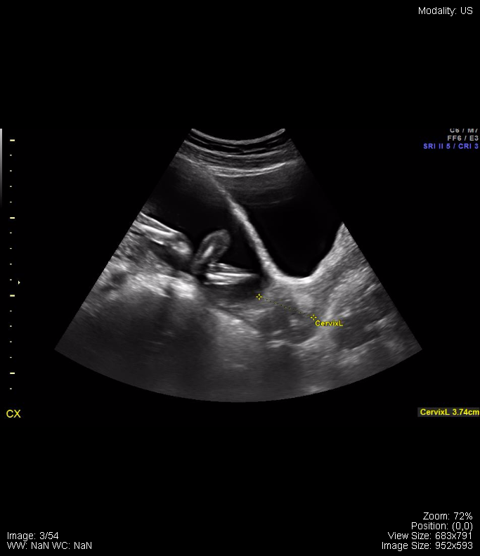
Pregnancy Ultrasound
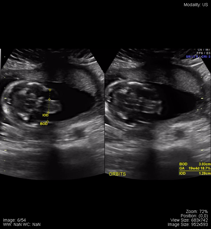
Fetal Orbits with measurements
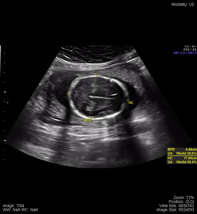
Fetal Head with measurements
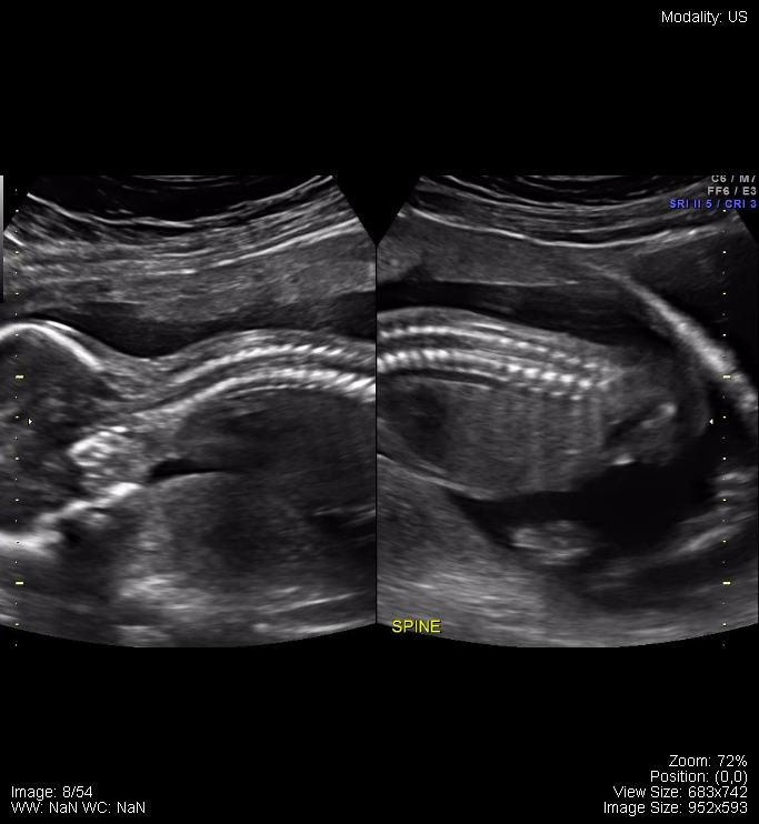
Fetal spine with measurements
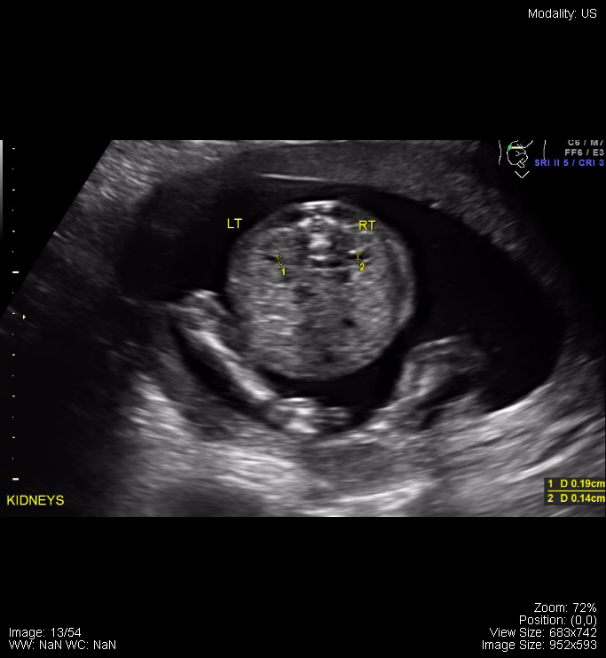
Fetal Kidneys with measurements
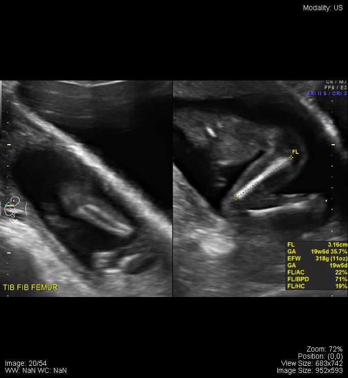
Fetal Legs with femur measurements
ODIN Link to Images: mistr.usask.ca/odin/?caseID=20170126222052150
Attributions
All figures in “Chapter 17: Pelvis” by Dr. Brent Burbridge MD, FRCPC, University Medical Imaging Consultants, College of Medicine, University of Saskatchewan is used under a CC-BY-NC-SA 4.0 license.