
12.7A: Cervical Plexus
- Page ID
- 7687
The cervical plexus is the plexus of the ventral rami of the first four cervical spinal nerves.
- Describe the cervical plexus and its function
Key Points
- The cervical plexus describes the plexus of the ventral rami of the first four cervical spinal nerves that arise from the cervical spinal column in the neck.
- The cervical spinal nerves that form the cervical plexus are located laterally (farther from the median line) to the transverse processes of the prevertebral skeletal muscles of the neck from the medial side, and vertebral (closer to the vertebral column) to these muscles from the lateral side.
- The cervical plexus forms an anastomosis, a connection, with the accessory nerve, the hypoglossal nerve, and the sympathetic trunk.
- The cervical plexus is located in the neck, internal to the sternocleidomastoid, an anterior neck muscle.
Key Terms
- sympathetic trunk: Also called the sympathetic chain or gangliated cord, these are a paired bundle of nerve fibers that run from the base of the skull to the coccyx.
- plexus: A network or interwoven mass, especially of nerves, blood vessels, or lymphatic vessels.
- platysma: A superficial muscle that overlaps the sternocleidomastoid.
- cervical plexus: A plexus of the ventral rami of the first four cervical spinal nerves that are located from the C1 to C4 cervical segment in the neck. They are located laterally to the transverse processes of the prevertebral muscles from the medial side and vertebral (scalenus, levator scapulae, splenius cervicis muscles) from the lateral side.
Structure and Distribution
The cervical plexus is a plexus of the ventral rami of the first four cervical spinal nerves located from the C1 to C4 cervical segment in the neck. They are located laterally to the transverse processes between prevertebral muscles from the medial side and anteriolateral to the m. scalenus and m. levator scapulae.
There is anastomosis with the accessory nerve, hypoglossal nerve, and sympathetic trunk. It is located deep in the neck, near the sternocleidomastoid muscle.
Nerves formed from the cervical plexus innervate the back of the head, as well as some neck muscles. The branches of the cervical plexus emerge from the posterior triangle at the nerve point, a point that lies midway on the posterior border of the sternocleidomastoid.
Branches and Their Functions
The cervical plexus has two types of branches: cutaneous and muscular.
Cutaneous branches include:
- The lesser occipital nerve, or small occipital nerve, is a cutaneous spinal nerve that arises between the second and third cervical vertebrae, along with the greater occipital nerve. It innervates the scalp in the lateral area of the head posterior to the ear.
- The great auricular nerve originates from the cervical plexus and is composed of branches from spinal nerves C2 and C3. It provides sensory innervation for the skin over the parotid gland and mastoid process, and both surfaces of the outer ear.
- The transverse cervical nerve (superficial cervical or cutaneous cervical) arises from the second and third cervical nerves, turns around the posterior border of the sternocleidomastoideus about its middle, then passes obliquely forward beneath the external jugular vein to the anterior border of the muscle, where it perforates the deep cervical fascia and divides beneath the platysma into ascending and descending branches that are distributed to the antero-lateral parts of the neck.
- The supraclavicular nerves (descending branches) arise from the third and fourth cervical nerves. They emerge beneath the posterior border of the sternocleidomastoideus, and descend in the posterior triangle of the neck beneath the platysma and deep cervical fascia.
Muscular branches include:
- Ansa cervicalis (loop formed from C1–C3), geniohyoid (C1 only), thyrohyoid (C1 only), sternothyroid, sternohyoid, omohyoid: The ansa cervicalis is a loop of nerves that are part of the cervical plexus.
- The phrenic nerve (C3–C5, but primarily C4) is a nerve that originates in the neck and passes down between the lung and heart to reach the diaphragm.
- Segmental branches (C1–C4) innervate the anterior and middle scalenes.
There are two additional branches that are formed by the posterior roots of the spinal nerves:
- Preauricular nerve (from the posterior roots of C2–C3).
- Postauricular nerve (from the posterior roots of C3–C4).
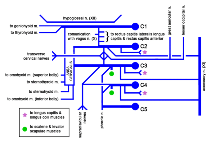
Cervical plexus: Diagram of the cervical plexus.