
17.1G: Fibrous Skeleton of the Heart
- Page ID
- 7817
The cardiac skeleton, also known the heart’s fibrous skeleton, consists of dense connective tissue in the heart that separates the atria from the ventricles.
- Describe composition and function of the heart’s fibrous skeleton
Key Points
- The cardiac skeleton consists of four bands of dense connective tissue, called fibrous rings, that surround the base of the pulmonary trunk, aorta, and mitral and tricuspid valves.
- The heart ‘s fibrous skeleton stops the flow of electrical currents between the chambers of the heart so that it only flows through the atrioventricular (AV) bundle. This causes a delay in depolarization so that the ventricles contract after they fill with blood.
- The AV bundle is a bundle of electrically-connected cardiomyocytes that transmit impulses from the AV node to the apex of the heart. It is located in the center of the cardiac skeleton.
- The cardiac skeleton consists mainly of the protein collagen, a glycoprotein found in all connective tissues.
Key Terms
- collagen: A glycoprotein that forms elongated fibers, usually found in the extracellular matrix of connective tissue.
- fibrous rings: Four dense bands of tough elastic tissue that encircle the bases of the valves of the heart.
The cardiac skeleton, or fibrous skeleton of the heart, is the structure of dense connective tissue that separates the atria from the ventricles. The fibrous skeleton provides critical support for the heart and separates the flow of electrical impulses through the heart.
Fibrous Ring Structure
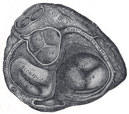
Fibrous Rings of the Heart: Transverse section of the heart showing the fibrous rings surrounding the valves.
The primary structure of cardiac skeleton consists of four dense bands of tough elastic tissue called fibrous rings that encircle the bases of the heart valves. The fibrous skeleton is composed primarily of collagen, a protein found in every type of connective tissue in the human body. There are four fibrous rings:
- The aortic ring encircles the aortic valve. It provides support for the aortic valve so that it is open, yet does not have backflow.
- The pulmonary ring encircles the pulmonary valve. Similar to the aortic ring, it provides structural support for the pulmonary valve.
- The left fibrous ring encircles the bicuspid valve. This ring is the thickest and strongest of all the fibrous rings due to the thickness of the left ventricle, which requires more structural support than the other chambers of the heart. It also surrounds the coronary arteries and AV node.
- The right fibrous ring encircles the tricuspid valve. It also surrounds the coronary arteries and AV node.
The fibrous skeleton provides a great amount of structural and functional support for the valves of the heart by enabling them to stay open and provides a point of attachment for the valves to the wall of the heart.
Electrical Functions
The fibrous skeleton of the heart acts as an insulator for the flow of electrical current across the heart. It stops the flow of electricity between the different chambers of the heart so that electrical impulses do not flow directly between the atria and ventricles. The sinoatrial (SA) node lies on the top of the heart, while the AV node is located at the center of the fibrous skeleton, the only path by which electricity can flow through the heart.
This electrical separation is essential for cardiac function, because electrical impulses flow from the top of the heart to the bottom of the heart. The separation allows the AV node and AV bundle to delay the wave of depolarization so that the atria can contract and assist in ventricular filling before the ventricles themselves depolarize and contract. Without the fibrous skeleton of the heart, the heart’s ability to pump blood would be considerably less efficient since the ventricles would contract before filled to capacity. The fibrous skeleton of the heart also protects against cardiac arrhythmias.
LICENSES AND ATTRIBUTIONS
CC LICENSED CONTENT, SHARED PREVIOUSLY
- Curation and Revision. Authored by: Boundless.com. Provided by: Boundless.com. License: CC BY-SA: Attribution-ShareAlike
CC LICENSED CONTENT, SPECIFIC ATTRIBUTION
- Human heart. Provided by: Wikipedia. Located at: en.Wikipedia.org/wiki/Human_heart%23Structure. License: CC BY-SA: Attribution-ShareAlike
- Human Physiology/The cardiovascular system. Provided by: Wikibooks. Located at: en.wikibooks.org/wiki/Human_P...em%23The_Heart. License: CC BY-SA: Attribution-ShareAlike
- heart. Provided by: Wiktionary. Located at: en.wiktionary.org/wiki/heart. License: CC BY-SA: Attribution-ShareAlike
- myocardium. Provided by: Wiktionary. Located at: en.wiktionary.org/wiki/myocardium. License: CC BY-SA: Attribution-ShareAlike
- ventricle. Provided by: Wiktionary. Located at: en.wiktionary.org/wiki/ventricle. License: CC BY-SA: Attribution-ShareAlike
- Diagram of the human heart (cropped). Provided by: Wikipedia. Located at: en.Wikipedia.org/wiki/File:Di...%23globalusage. License: CC BY-SA: Attribution-ShareAlike
- Fibrous pericardium. Provided by: Wikipedia. Located at: en.Wikipedia.org/wiki/Fibrous_pericardium. License: CC BY-SA: Attribution-ShareAlike
- Serous pericardium. Provided by: Wikipedia. Located at: en.Wikipedia.org/wiki/Serous_pericardium. License: CC BY-SA: Attribution-ShareAlike
- Pericardium. Provided by: Wikipedia. Located at: en.Wikipedia.org/wiki/Pericardium. License: CC BY-SA: Attribution-ShareAlike
- Human Physiology/The cardiovascular system. Provided by: Wikibooks. Located at: en.wikibooks.org/wiki/Human_P...%23Pericardium. License: CC BY-SA: Attribution-ShareAlike
- pericardium. Provided by: Wiktionary. Located at: en.wiktionary.org/wiki/pericardium. License: CC BY-SA: Attribution-ShareAlike
- fibrous pericardium. Provided by: Wikipedia. Located at: en.Wikipedia.org/wiki/fibrous%20pericardium. License: CC BY-SA: Attribution-ShareAlike
- serous pericardium. Provided by: Wikipedia. Located at: en.Wikipedia.org/wiki/serous%20pericardium. License: CC BY-SA: Attribution-ShareAlike
- Diagram of the human heart (cropped). Provided by: Wikipedia. Located at: en.Wikipedia.org/wiki/File:Di...%23globalusage. License: CC BY-SA: Attribution-ShareAlike
- Transverse Thorax. Provided by: Wikimedia. Located at: upload.wikimedia.org/wikipedi...01/Gray968.png. License: Public Domain: No Known Copyright
- Epicardium. Provided by: Wikipedia. Located at: en.Wikipedia.org/wiki/Epicardium. License: CC BY-SA: Attribution-ShareAlike
- Myocardium. Provided by: Wikipedia. Located at: en.Wikipedia.org/wiki/Myocardium. License: CC BY-SA: Attribution-ShareAlike
- Human Physiology/The cardiovascular system. Provided by: Wikibooks. Located at: en.wikibooks.org/wiki/Human_P...em%23The_Heart. License: CC BY-SA: Attribution-ShareAlike
- Endocardium. Provided by: Wikipedia. Located at: en.Wikipedia.org/wiki/Endocardium. License: CC BY-SA: Attribution-ShareAlike
- epicardium. Provided by: Wiktionary. Located at: en.wiktionary.org/wiki/epicardium. License: CC BY-SA: Attribution-ShareAlike
- endocardium. Provided by: Wiktionary. Located at: en.wiktionary.org/wiki/endocardium. License: CC BY-SA: Attribution-ShareAlike
- cardiomyocyte. Provided by: Wiktionary. Located at: en.wiktionary.org/wiki/cardiomyocyte. License: CC BY-SA: Attribution-ShareAlike
- endothelial cell. Provided by: Wikipedia. Located at: en.Wikipedia.org/wiki/endothelial%20cell. License: CC BY-SA: Attribution-ShareAlike
- Diagram of the human heart (cropped). Provided by: Wikipedia. Located at: en.Wikipedia.org/wiki/File:Di...%23globalusage. License: CC BY-SA: Attribution-ShareAlike
- Transverse Thorax. Provided by: Wikimedia. Located at: upload.wikimedia.org/wikipedi...01/Gray968.png. License: Public Domain: No Known Copyright
- Heart ant wall infarction. Provided by: Wikimedia. Located at: commons.wikimedia.org/wiki/Fi...infarction.jpg. License: CC BY: Attribution
- Atrium (heart). Provided by: Wikipedia. Located at: en.Wikipedia.org/wiki/Atrium_(heart). License: CC BY-SA: Attribution-ShareAlike
- Ventricle (heart). Provided by: Wikipedia. Located at: en.Wikipedia.org/wiki/Ventricle_(heart). License: CC BY-SA: Attribution-ShareAlike
- Human Physiology/The cardiovascular system. Provided by: Wikibooks. Located at: en.wikibooks.org/wiki/Human_P...Heart_Chambers. License: CC BY-SA: Attribution-ShareAlike
- systole. Provided by: Wiktionary. Located at: en.wiktionary.org/wiki/systole. License: CC BY-SA: Attribution-ShareAlike
- pulsatile. Provided by: Wikipedia. Located at: en.Wikipedia.org/wiki/pulsatile. License: CC BY-SA: Attribution-ShareAlike
- diastole. Provided by: Wiktionary. Located at: en.wiktionary.org/wiki/diastole. License: CC BY-SA: Attribution-ShareAlike
- Diagram of the human heart (cropped). Provided by: Wikipedia. Located at: en.Wikipedia.org/wiki/File:Di...%23globalusage. License: CC BY-SA: Attribution-ShareAlike
- Transverse Thorax. Provided by: Wikimedia. Located at: upload.wikimedia.org/Wikipedia/commons/0/01/Gray968.png. License: Public Domain: No Known Copyright
- Heart ant wall infarction. Provided by: Wikimedia. Located at: commons.wikimedia.org/wiki/File:Heart_ant_wall_infarction.jpg. License: CC BY: Attribution
- Heart diagram-en. Provided by: Wikipedia. Located at: en.Wikipedia.org/wiki/File:Heart_diagram-en.svg. License: CC BY-SA: Attribution-ShareAlike
- Human Physiology/The cardiovascular system. Provided by: Wikibooks. Located at: en.wikibooks.org/wiki/Human_Physiology/The_cardiovascular_system%23The_Cardiovascular_Pathways. License: CC BY-SA: Attribution-ShareAlike
- Aorta. Provided by: Wikipedia. Located at: en.Wikipedia.org/wiki/Aorta. License: CC BY-SA: Attribution-ShareAlike
- Inferior vena cava. Provided by: Wikipedia. Located at: en.Wikipedia.org/wiki/Inferior_vena_cava. License: CC BY-SA: Attribution-ShareAlike
- Vena cavae. Provided by: Wikipedia. Located at: en.Wikipedia.org/wiki/Vena_cavae. License: CC BY-SA: Attribution-ShareAlike
- Pulmonary artery. Provided by: Wikipedia. Located at: en.Wikipedia.org/wiki/Pulmonary_artery. License: CC BY-SA: Attribution-ShareAlike
- Superior vena cava. Provided by: Wikipedia. Located at: en.Wikipedia.org/wiki/Superior_vena_cava. License: CC BY-SA: Attribution-ShareAlike
- Pulmonary vein. Provided by: Wikipedia. Located at: en.Wikipedia.org/wiki/Pulmonary_vein. License: CC BY-SA: Attribution-ShareAlike
- venae cavae. Provided by: Wikipedia. Located at: en.Wikipedia.org/wiki/venae%20cavae. License: CC BY-SA: Attribution-ShareAlike
- pulmonary vessels. Provided by: Wikipedia. Located at: en.Wikipedia.org/wiki/pulmonary%20vessels. License: CC BY-SA: Attribution-ShareAlike
- aorta. Provided by: Wiktionary. Located at: en.wiktionary.org/wiki/aorta. License: CC BY-SA: Attribution-ShareAlike
- Diagram of the human heart (cropped). Provided by: Wikipedia. Located at: en.Wikipedia.org/wiki/File:Diagram_of_the_human_heart_(cropped).svg%23globalusage. License: CC BY-SA: Attribution-ShareAlike
- Transverse Thorax. Provided by: Wikimedia. Located at: upload.wikimedia.org/Wikipedia/commons/0/01/Gray968.png. License: Public Domain: No Known Copyright
- Heart ant wall infarction. Provided by: Wikimedia. Located at: commons.wikimedia.org/wiki/File:Heart_ant_wall_infarction.jpg. License: CC BY: Attribution
- Heart diagram-en. Provided by: Wikipedia. Located at: en.Wikipedia.org/wiki/File:Heart_diagram-en.svg. License: CC BY-SA: Attribution-ShareAlike
- Illu pulmonary circuit. Provided by: Wikipedia. Located at: en.Wikipedia.org/wiki/File:Illu_pulmonary_circuit.jpg. License: Public Domain: No Known Copyright
- Illu systemic circuit. Provided by: Wikimedia. Located at: commons.wikimedia.org/wiki/Fi...ic_circuit.svg. License: CC BY-SA: Attribution-ShareAlike
- Syncytium. Provided by: Wikipedia. Located at: en.Wikipedia.org/wiki/Syncytium. License: CC BY-SA: Attribution-ShareAlike
- T-tubules. Provided by: Wikipedia. Located at: en.Wikipedia.org/wiki/T-tubules. License: CC BY-SA: Attribution-ShareAlike
- Myocardium. Provided by: Wikipedia. Located at: en.Wikipedia.org/wiki/Myocardium. License: CC BY-SA: Attribution-ShareAlike
- Sarcomere. Provided by: Wikipedia. Located at: en.Wikipedia.org/wiki/Sarcomere. License: CC BY-SA: Attribution-ShareAlike
- Excitation-contraction coupling. Provided by: Wikipedia. Located at: en.Wikipedia.org/wiki/Excitat...ction_coupling. License: CC BY-SA: Attribution-ShareAlike
- sarcomere. Provided by: Wiktionary. Located at: en.wiktionary.org/wiki/sarcomere. License: CC BY-SA: Attribution-ShareAlike
- cardiomyocyte. Provided by: Wiktionary. Located at: en.wiktionary.org/wiki/cardiomyocyte. License: CC BY-SA: Attribution-ShareAlike
- myoglobin. Provided by: Wiktionary. Located at: en.wiktionary.org/wiki/myoglobin. License: CC BY-SA: Attribution-ShareAlike
- Diagram of the human heart (cropped). Provided by: Wikipedia. Located at: en.Wikipedia.org/wiki/File:Diagram_of_the_human_heart_(cropped).svg%23globalusage. License: CC BY-SA: Attribution-ShareAlike
- Transverse Thorax. Provided by: Wikimedia. Located at: upload.wikimedia.org/Wikipedia/commons/0/01/Gray968.png. License: Public Domain: No Known Copyright
- Heart ant wall infarction. Provided by: Wikimedia. Located at: commons.wikimedia.org/wiki/File:Heart_ant_wall_infarction.jpg. License: CC BY: Attribution
- Heart diagram-en. Provided by: Wikipedia. Located at: en.Wikipedia.org/wiki/File:Heart_diagram-en.svg. License: CC BY-SA: Attribution-ShareAlike
- Illu pulmonary circuit. Provided by: Wikipedia. Located at: en.Wikipedia.org/wiki/File:Illu_pulmonary_circuit.jpg. License: Public Domain: No Known Copyright
- Illu systemic circuit. Provided by: Wikimedia. Located at: commons.wikimedia.org/wiki/File:Illu_systemic_circuit.svg. License: CC BY-SA: Attribution-ShareAlike
- Right_Ventricular_hypertrophy.svg. Provided by: Wikipedia. Located at: en.Wikipedia.org/wiki/Ventricular_hypertrophy#/media/File:Right_Ventricular_hypertrophy.svg. License: CC BY-SA: Attribution-ShareAlike
- Cardiac_Muscle.png. Provided by: Wikipedia. Located at: en.Wikipedia.org/wiki/Cardiac_muscle#/media/File:Cardiac_Muscle.png. License: CC BY-SA: Attribution-ShareAlike
- Cardiac skeleton. Provided by: Wikipedia. Located at: en.Wikipedia.org/wiki/Cardiac_skeleton. License: CC BY-SA: Attribution-ShareAlike
- collagen. Provided by: Wiktionary. Located at: en.wiktionary.org/wiki/collagen. License: CC BY-SA: Attribution-ShareAlike
- fibrous rings. Provided by: Wikipedia. Located at: en.Wikipedia.org/wiki/fibrous%20rings. License: CC BY-SA: Attribution-ShareAlike
- cardiac skeleton. Provided by: Wikipedia. Located at: en.Wikipedia.org/wiki/cardiac%20skeleton. License: CC BY-SA: Attribution-ShareAlike
- Diagram of the human heart (cropped). Provided by: Wikipedia. Located at: en.Wikipedia.org/wiki/File:Diagram_of_the_human_heart_(cropped).svg%23globalusage. License: CC BY-SA: Attribution-ShareAlike
- Transverse Thorax. Provided by: Wikimedia. Located at: upload.wikimedia.org/Wikipedia/commons/0/01/Gray968.png. License: Public Domain: No Known Copyright
- Heart ant wall infarction. Provided by: Wikimedia. Located at: commons.wikimedia.org/wiki/File:Heart_ant_wall_infarction.jpg. License: CC BY: Attribution
- Heart diagram-en. Provided by: Wikipedia. Located at: en.Wikipedia.org/wiki/File:Heart_diagram-en.svg. License: CC BY-SA: Attribution-ShareAlike
- Illu pulmonary circuit. Provided by: Wikipedia. Located at: en.Wikipedia.org/wiki/File:Illu_pulmonary_circuit.jpg. License: Public Domain: No Known Copyright
- Illu systemic circuit. Provided by: Wikimedia. Located at: commons.wikimedia.org/wiki/File:Illu_systemic_circuit.svg. License: CC BY-SA: Attribution-ShareAlike
- Right_Ventricular_hypertrophy.svg. Provided by: Wikipedia. Located at: en.Wikipedia.org/wiki/Ventricular_hypertrophy#/media/File:Right_Ventricular_hypertrophy.svg. License: CC BY-SA: Attribution-ShareAlike
- Cardiac_Muscle.png. Provided by: Wikipedia. Located at: en.Wikipedia.org/wiki/Cardiac_muscle#/media/File:Cardiac_Muscle.png. License: CC BY-SA: Attribution-ShareAlike
- Gray495. Provided by: Wikipedia. Located at: en.Wikipedia.org/wiki/File:Gray495.png. License: Public Domain: No Known Copyright