
17.4A: Electrical Events
- Page ID
- 7829
Cardiac contraction is initiated in the excitable cells of the sinoatrial (SA) node by both spontaneous depolarization and sympathetic activity.
- Describe the electrical events of the heart
Key Points
- The sinoatrial (SA) and atrioventricular (AV) nodes make up the intrinsic conduction system of the heart by setting the rate at which the heart beats.
- The SA node generates action potentials spontaneously.
- The SA node fires at a normal rate of 60–100 beats per minute (bpm), and causes depolarization in atrial muscle tissue and subsequent atrial contraction.
- The AV node slows the impulses from the SA node, firing at a normal rate of 40-60 bpm, and causes depolarization of the ventricular muscle tissue and ventricular contraction.
- Sympathetic nervous stimulation increases the heart rate, while parasympathetic nervous stimulation decreases the heart rate.
Key Terms
- pacemaker: A structure that sets the rate at which the heart beats. Under normal conditions, the SA node serves this function for the heart.
- atrioventricular (AV) node: The bundle of conducting tissue that receives impulses from the SA node and delays them before stimulating depolarization in the muscles of the ventricles.
The heart’s activity is dependent on the electrical impulses from the sinoatrial (SA) node and atrioventricular (AV) node, which form the intrinsic conduction system of the heart. The SA and AV nodes act as a pacemaker for the heart, determining the rate at which it beats, even without signals from the larger nervous system of the human body. The SA and AV nodes initiate the electrical impulses that cause contraction within the atria and ventricles of the heart.
Sinoatrial Node
The SA node is a bundle of nerve cells located on the outer layer of the right atria. These cells are specialized to undergo spontaneous depolarization and generation of action potentials without stimulation from the rest of the nervous system. The SA node nerve impulses travel through the atria and cause direct muscle cell depolarization and contraction of the atria. The SA node stimulates the right atria directly and stimulates the left atria through the Bachmann’s bundle. The SA node impulses also travel to the AV node, which stimulates ventricular contraction.
The SA node generates its own action potentials, but may be influenced by the autonomic nervous system. Without autonomic nervous stimulation, the SA node will set the heart rate itself, acting as the primary pacemaker for the heart. The SA node fires to set a heart rate in a range of 60–100 beats per minute (bpm), a normal range that varies from person to person.
Atrioventricular Node
The AV node is a bundle of conducting tissue (not formally classified as nerve tissue) located at the junction between the atria and ventricles of the heart. The AV node receives action potentials from the SA node, and transmits them through the bundle of His, left and right bundle branches, and Purkinje fibers, which cause depolarization of ventricular muscle cells leading to ventricular contraction. The AV node slightly slows the neural impulse from the SA node, which causes a delay between depolarization of the atria and the ventricles.
The normal firing rate in the AV node is lower than that of the SA node because it slows the rate of neural impulses. Without autonomic nervous stimulation, it sets the rate of ventricular contraction at 40–60 bpm. Certain types of autonomic nervous stimulation alter the rate of firing in the AV node. Sympathetic nervous stimulation still increases heart rate, while parasympathetic nervous stimulation decreases heart rate by acting on the AV node.
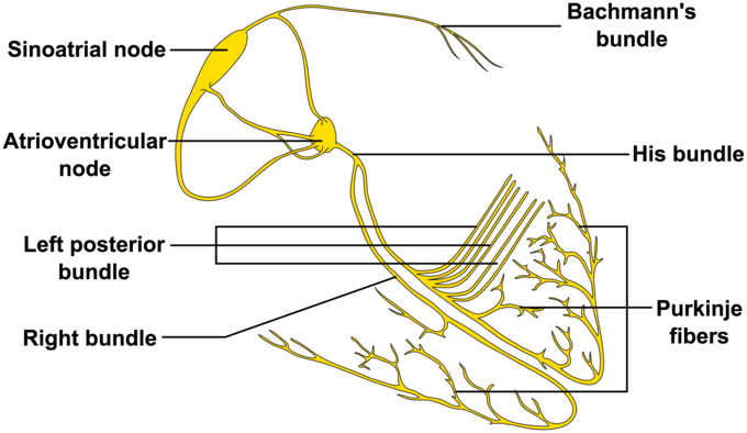
The Cardiac Conduction System: The system of nerves that work together to set the heart rate and stimulate muscle cell depolarization within the heart.