
22.5C: Muscularis
- Page ID
- 8039
The muscularis is responsible for the segmental contractions and peristaltic movements in the gastrointestinal (GI) tract.
- Identify the function of muscularis in the GI tract
Key Points
- The muscularis, or muscularis externa, consists of an inner circular muscular layer and a longitudinal outer muscular layer. The coordinated contractions of these layers is called peristalsis, which propels the food through the GI tract.
- Between the two muscle layers is the myenteric or Auerbach’s plexus, which controls peristalsis.
- In the colon, the muscularis externa is much thicker because the feces are large and heavy, requiring more force to push along.
- The stomach has a third layer of muscularis externa: the inner oblique layer. This helps churn the chyme in the stomach.
- Peristaltic activity in the muscularis externa is regulated by the enteric nervous system and the autonomic nervous system.
Key Terms
- muscularis externa: A region of muscle in many organs in the vertebrate body, adjacent to the submucosa membrane. It is responsible for gut movement such as peristalsis.
- oblique layer: This layer is responsible for creating the motion that churns and physically breaks down the food.
- tiniae coli: These are three, separate longitudinal ribbons of smooth muscle on the outside of the ascending, transverse, descending, and sigmoid colons.
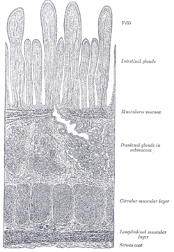
The gastrointestinal (GI) tract is composed of four layers of tissue, known as tunics. Each layer has different structures and functions. From the inside out they are called the mucosa, submucosa, muscularis externa, and serosa.
Structure of the Muscularis Externa
Muscularis mucosa of the submucosa: The muscularis mucosa is adjacent to the submucosa, and should not be confused with the muscularis externa.
The muscularis externa is responsible for segmental contractions and peristaltic movement in the GI tract. These muscles cause food to move and churn together with digestive enzymes down the GI tract. The muscularis externa consists of an inner circular layer and a longitudinal outer muscular layer. It should not be confused with a thin layer of muscle known as the muscularis mucosa, which lies within the submucosa, a layer of tissue adjacent to the muscularis externa. The muscularis mucosa is made up of smooth muscle, and is most prominent in the stomach.
Within the muscularis externa, the circular muscle layer prevents food from traveling backward, while the longitudinal layer shortens the tract. The layers are not truly longitudinal or circular, rather the layers of muscle are helical with different pitches. The inner circular is helical with a steep pitch and the outer longitudinal is helical with a much shallower pitch.
The coordinated contractions of these layers is called peristalsis. Between the two muscle layers is the myenteric or Auerbach’s plexus, which controls peristalsis. Peristaltic activity is regulated by these nerve cells, and the rate of peristalsis can be modulated by the rest of the autonomic nervous system.
The thickness of muscularis externa varies in each part of the tract. In the colon, for example, the muscularis externa is much thicker because the feces are large and heavy, and require more force to push along. The outer longitudinal layer of the colon thins out into three discontinuous longitudinal bands known as tiniae coli (bands of the colon). This is one of the three features helping to distinguish between the large and small intestine.
General Structure of the gut wall: General structure of the gut wall—the muscularis externa is labeled circular muscle and longitudinal muscle here.
Occasionally in the large intestine (two to three times a day), there will be mass contraction of certain segments, moving a lot of feces along. This is generally when one gets the urge to defecate.
The pylorus of the stomach has a thickened portion of the inner circular layer: the pyloric sphincter. Alone among the GI tract, the stomach has a third layer of muscularis externa. This is the inner oblique layer, and helps churn the chyme in the stomach.