
27.3B: Bilaminar Embryonic Disc Development
- Page ID
- 8272
The floor of the amniotic cavity is formed by the embryonic disc.
- Identify the bilaminar embryonic disc
Key Points
- In humans, the formation of the embryonic disc occurs after implantation and prior to embryonic folding (between about day 14 to day 21 post-fertilization).
- The embryonic disc is derived from the epiblast layer, which lies between the hypoblast layer and the amnion. The epiblast layer is derived from the inner cell mass.
- The formation of the bilaminar embryonic disc precedes gastrulation.
Key Terms
- optic disc: The location where ganglion cell axons exit the eye to form the optic nerve.
- bilaminar: Formed of, or having, two laminae, or thin plates.
The floor of the amniotic cavity is formed by the embryonic disc, which is composed of a layer of prismatic cells and the embryonic ectoderm. It is derived from the inner cell mass and lies adjacent to the endoderm.
In humans, the formation of the embryonic disc occurs after implantation and prior to embryonic folding (between about day 14 to day 21 post-fertilization). The embryonic disc is derived from the epiblast layer, which lies between the hypoblast layer and the amnion. The epiblast layer is derived from the inner cell mass.
Embryonic Disc Development
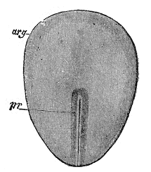
Surface view of a rabbit embryo: A drawing of a rabbit embryo that identifies the embryonic disc and the primitive streak (arg—embryonic disc; pr—primitive streak).
The embryonic disc forms during early development. By the blastocyst stage, the embryo is a hollow ball of cells with the inner cell mass (embryoblast) off to one side, and the blastocystic cavity fills the remainder of the sphere.
As the embryo progresses in implantation, a small space appears in the embryoblast and forms the amniotic cavity. Simultaneously, morphological changes occur in the embryoblast that result in the formation of a flat, almost circular bilaminar plate of cells that include the epiblast and the hypoblast—the embryonic disk.
The epiblast forms the floor of the amniotic cavity and is continuous with the amnion. The hypoblast forms the roof of the exocoelomic cavity and is continuous with the thin exocoelomic membrane.
The formation of the bilaminar embryonic disc precedes gastrulation. As gastrulation progresses, the embryonic disc becomes trilaminar and the notochord is formed. Through the process of neurulation, the notochord induces the formation of the central nervous system in the embryonic disc.