
11.2: Organization and Functions of the Nervous System
- Page ID
- 22336

By the end of this section, you will be able to:
- Understand the basic structure of a neuron
- Distinguish between gray and white matter and relate their differences to the structure of neurons
- Identify the structural and functional divisions of the nervous system
- Identify the organs of the central and peripheral divisions of the nervous system
- Understand the basic functions and control of the nervous system
The picture you have in your mind of the nervous system probably includes the brain, the nervous tissue contained within the cranium in the cranial cavity, and the spinal cord, the extension of nervous tissue within the vertebral column. That suggests it is made of two organs—and you may not even think of the spinal cord as an organ—but the nervous system is a very complex structure. Within the brain, many different and separate regions are responsible for many different and separate functions. It is as if the nervous system is composed of many organs that all look similar and can only be differentiated using tools such as the microscope or an electrophysiology apparatus. In comparison, it is easy to see that the stomach is different than the esophagus or the liver, so you can imagine the digestive system as a collection of specific organs. For this reason, you will look at the organization of the nervous tissue first and the structures within it. Then you will look at the structural divisions of the nervous system by regions where the organs are located, and then the functional divisions by the specific functions achieved by the organs within it.
Organization of the Nervous Tissue
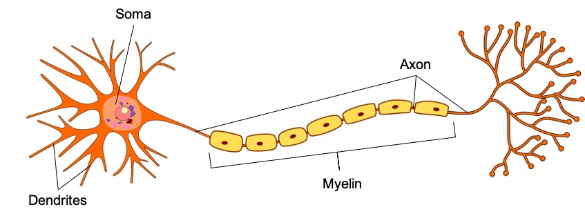
Nervous tissue contains two basic types of cells: neurons and glial cells. A glial cell is one of a variety of cells that provide a framework of tissue that supports the neurons and their activities. The neuron is the more functionally important of the two, in terms of the communicative function of the nervous system. To describe the functional divisions of the nervous system, it is important to understand the basic structure of a neuron (Figure \(\PageIndex{1}\)). Neurons are cells and therefore have a soma, or cell body, but they also have extensions of the cell; each extension is generally referred to as a process. There is one important process that every neuron has called an axon, which is the fiber that connects a neuron with its target. Axons are partially insulated by a lipid-rich substance called myelin. Another type of process that branches off from the soma is the dendrite. Dendrites are responsible for receiving most of the input from other neurons.
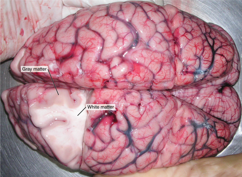
Looking at nervous tissue, there are regions that predominantly contain cell bodies and regions that are largely composed of just axons. These two regions within nervous system structures are often referred to as gray matter (the regions with many cell bodies and dendrites) or white matter (the regions with many axons). Figure \(\PageIndex{2}\) demonstrates the appearance of these regions in the brain and spinal cord. The colors ascribed to these regions are what would be seen in “fresh,” or unstained, nervous tissue. Gray matter is not necessarily gray. It can be pinkish because of blood content, or even slightly tan, depending on how long the tissue has been preserved. But white matter is white because axons are insulated by the lipids of the myelin. Lipids can appear as white (“fatty”) material, much like the fat on a raw piece of chicken or beef. Actually, gray matter may have that color ascribed to it because next to the white matter, it is just darker—hence, gray. Cell bodies of neurons cluster in regions called nuclei or ganglia, depending on their location. Axons of neurons also tend to bundle up in structures called tracts (columns) or nerves (fibers), depending on their location.
Structural Divisions of the Nervous System
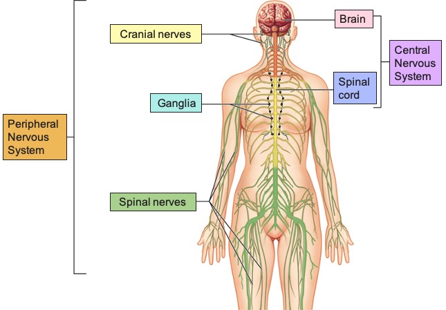
The nervous system can be divided into two major regions: the central and peripheral nervous systems. The central nervous system (CNS) is composed of the brain and spinal cord, and the peripheral nervous system (PNS) is composed of ganglia (structures containing neurons outside of the CNS) and nerves (bundles of axons from CNS and PNS neurons) (Figure \(\PageIndex{3}\)). The brain is contained within the cranial cavity of the skull, and the spinal cord is contained within the vertebral cavity of the vertebral column. It is a bit of an oversimplification to say that the CNS is what is inside these two cavities and the peripheral nervous system is outside of them, but that is one way to start to think about it. In actuality, there are some elements of the peripheral nervous system that are within the cranial or vertebral cavities. The peripheral nervous system is so named because it is on the periphery—meaning beyond the brain and spinal cord. Depending on different aspects of the nervous system, the dividing line between central and peripheral is not necessarily universal.
The cell bodies or axons of neurons can be located in discrete anatomical structures that are specific to whether the structure is central or peripheral. A localized collection of neuron cell bodies in the CNS is referred to as a nucleus. In the PNS, a cluster of neuron cell bodies is referred to as a ganglion. Terminology applied to bundles of axons also differs depending on location. A bundle of axons, or fibers, found in the CNS is called a tract (or column) whereas the same thing in the PNS would be called a nerve (or nerve fiber). There is an important point to make about these terms, which is that they can both be used to refer to the same bundle of axons. The most obvious example of this is the axons that project from the retina of the eye into the brain. Those axons are called the optic nerve as they leave the eye, but when they are inside the cranium, they are referred to as the optic tract. Table \(\PageIndex{1}\) helps to clarify which of these terms apply to the central or peripheral nervous systems. There is a further classification of nerves depending on the location where they leave the central nervous system: cranial nerves emerge directly from the brain, while spinal nerves exit from different segments of the spinal cord.
| CNS | PNS | |
|---|---|---|
| Group of Neuron Cell Bodies (i.e., gray matter) | Nucleus | Ganglion |
| Bundle of Axons (i.e., white matter) | Tract or Column | Nerve or Nerve Fiber |
Functional Divisions of the Nervous System
The nervous system can also be divided on the basis of its functions, but anatomical divisions and functional divisions are different. The CNS and the PNS both contribute to the same functions, but those functions can be attributed to different regions of the brain (such as the cerebral cortex or the hypothalamus) or to different ganglia in the periphery. The problem with trying to fit functional differences into anatomical divisions is that sometimes the same structure can be part of several functions. For example, the optic nerve carries signals from the retina that are either used for the conscious perception of visual stimuli, which takes place in the cerebral cortex, or for the reflexive responses of smooth muscle tissue that are processed through the hypothalamus.
There are two ways to consider how the nervous system is divided functionally. First, the basic functions of the nervous system are sensation, integration, and response. Secondly, control of the body can be somatic or autonomic—divisions that are largely defined by the structures that are involved in the response. There is also a region of the peripheral nervous system that is called the enteric nervous system that is responsible for a specific set of the functions within the realm of autonomic control related to gastrointestinal functions.
Basic Functions
The nervous system is involved in receiving information about the environment around us (sensation) and generating responses to that information (motor responses). The nervous system can be divided into regions that are responsible for sensation (sensory functions) and for the response (motor functions). But there is a third function that needs to be included. Sensory input needs to be integrated with other sensations, as well as with memories, emotional state, or learning (cognition). Some regions of the nervous system are termed integration or association areas. The process of integration combines sensory perceptions and higher cognitive functions such as memories, learning, and emotion to produce a response.
Sensation. The first major function of the nervous system is sensation—receiving information about the environment to gain input about what is happening outside the body (or, sometimes, within the body). The sensory functions of the nervous system register the presence of a change from homeostasis or a particular event in the environment, known as a stimulus (pl. stimuli). The senses we think of most are: taste, smell, vision, hearing and balance. The stimuli for taste and smell are chemical substances (molecules, compounds, ions, etc.), vision is light stimuli, hearing is the perception of sound and balance is the perception of where your head is in space. The last two rely on physical stimuli. The above senses are called special senses because they have specialized organs to sense the environment. There are actually more senses than just those, for example touch, pain and temperature sensation are perceived by the skin. These are called general senses. Special and general senses receive stimuli from the outside world and are perceived consciously, meaning that you are aware of them. This conscious sensation coming from special and general senses is called somatic sensation or somatosensation. Additional sensory stimuli might be from the internal environment (inside the body), such as the stretch of an organ wall or the concentration of certain ions in the blood or pain related to compression of nerves. These are called visceral senses and are not perceived consciously, meaning that you are not aware of them. Somatic and visceral sensory information is perceived by structures within the Peripheral Nervous System and travels towards the Central Nervous System. This inward direction is called afferent.
Integration. Stimuli that are received by sensory structures are communicated to the nervous system where that information is processed. Stimuli are compared with, or integrated with, other stimuli, memories of previous stimuli, or the state of a person at a particular time. This leads to the specific response that will be generated. Seeing a baseball pitched to a batter will not automatically cause the batter to swing. The trajectory of the ball and its speed will need to be considered. Maybe the count is three balls and one strike, and the batter wants to let this pitch go by in the hope of getting a walk to first base. Or maybe the batter’s team is so far ahead, it would be fun to just swing away.
Response. The nervous system produces a response on the basis of the sensory stimuli. An obvious response would be the movement of muscles, such as withdrawing a hand from a hot stove, but there are broader uses of the term. The nervous system can cause the contraction of all three types of muscle tissue. For example, skeletal muscle contracts to move the skeleton, cardiac muscle is influenced as heart rate increases during exercise, and smooth muscle contracts as the digestive system moves food along the digestive tract. Responses also include the neural control of glands in the body as well, such as the production and secretion of sweat by the eccrine and merocrine sweat glands found in the skin to lower body temperature. Thus, muscles and glands are the targets of the motor response, become active when stimulated to carry out the response. For this reason, they are called effectors. Responses can be divided into those that are voluntary or conscious (contraction of skeletal muscle) and those that are involuntary or unconscious (contraction of smooth muscles, regulation of cardiac muscle, activation of glands). Voluntary responses are governed by the somatic nervous system and involuntary responses are governed by the autonomic and enteric nervous system, which are discussed in the next section. Motor information is generated by the Central Nervous System and travels to the Peripheral Nervous System. This outward direction is also efferent. Afferent and efferent sound the same but are different. One way to remember their differences is using the acronym SAME that stands for Sensory Afferent Motor Efferent.
Controlling the Body
The nervous system can be divided into two parts mostly on the basis of a functional difference in responses. The somatic nervous system (SNS) is responsible for conscious perception (somatosensation) and voluntary motor responses (also called somatic responses). Voluntary motor response means the contraction of skeletal muscle, but those contractions are not always voluntary in the sense that you have to want to perform them. Some somatic motor responses are reflexes, and often happen without a conscious decision to perform them. If your friend jumps out from behind a corner and yells “Boo!” you will be startled and you might scream or leap back. You didn’t decide to do that, and you may not have wanted to give your friend a reason to laugh at your expense, but it is a reflex involving skeletal muscle contractions. Other motor responses become automatic (in other words, unconscious) as a person learns motor skills (referred to as “habit learning” or “procedural memory”).
The autonomic nervous system (ANS) is responsible for involuntary control of the body, usually for the sake of homeostasis (regulation of the internal environment). Sensory input for autonomic functions can be from sensory structures tuned to external or internal environmental stimuli. The motor output extends to smooth and cardiac muscle as well as glandular tissue. The role of the autonomic system is to regulate the organ systems of the body, which usually means to control homeostasis. Sweat glands, for example, are controlled by the autonomic system. When you are hot, sweating helps cool your body down. That is a homeostatic mechanism. But when you are nervous, you might start sweating also. That is not homeostatic, it is the physiological response to an emotional state.
There is another division of the nervous system that describes functional responses. The enteric nervous system (ENS) is responsible for controlling the smooth muscle and glandular tissue in your digestive system. It is a large part of the PNS, and is not dependent on the CNS. It is sometimes valid, however, to consider the enteric system to be a part of the autonomic system because the neural structures that make up the enteric system are a component of the autonomic output that regulates digestion. There are some differences between the two, but for our purposes here there will be a good bit of overlap. See Table \(\PageIndex{2}\) for a schematic representation of the different divisions based on their basic functions and control mechanisms.
|
Sensation |
Integration |
Motor Response |
|
|---|---|---|---|
|
Somatic |
Conscious perception of environmental changes (light, sounds, molecules in food, movement, temperature, touch, etc) through somatic sensory neurons of PNS |
Information processed in brain and spinal cord (CNS) |
Voluntary and reflex response via skeletal muscles |
|
Autonomic |
Unconscious perception of external or internal changes (light, molecules, organ stretch, etc) through visceral sensory neurons in both CNS and PNS |
Information processed in brain (particularly hypothalamus and brainstem) and spinal cord (CNS) |
Involuntary movement of cardiac muscle, smooth muscle, glands |
|
Enteric |
Unconscious perception of internal changes (molecules, movement, stretch) within the gastrointestinal tract through visceral sensory neurons in gastrointestinal tract |
Information processed within the gastrointestinal tract (PNS) |
Involuntary movement of smooth muscle and glands of digestive system |
Interactive Link
Creeping Weakness
Read the article Creeping Weakness about a woman that notices that her daughter is having trouble walking up the stairs. This leads to the discovery of a hereditary condition that affects the brain and spinal cord. The electromyography and MRI tests indicated deficiencies in the spinal cord and cerebellum (a region of the brain), both of which are responsible for controlling coordinated movements. To what functional division of the nervous system would these structures belong?
- Answer
-
Answer: They are part of the somatic nervous system, which is responsible for generating voluntary movements such as walking or climbing the stairs.
EVERYDAY CONNECTION: How Much of Your Brain Do You Use?
Have you ever heard the claim that humans only use 10 percent of their brains? Maybe you have seen an advertisement on a website saying that there is a secret to unlocking the full potential of your mind—as if there were 90 percent of your brain sitting idle, just waiting for you to use it. If you see an ad like that, don’t click. It isn’t true.
An easy way to see how much of the brain a person uses is to take measurements of brain activity while performing a task. An example of this kind of measurement is functional magnetic resonance imaging (fMRI), which generates a map of the most active areas and can be generated and presented in three dimensions (Figure \(\PageIndex{4}\)). This procedure is different from the standard MRI technique because it is measuring changes in the tissue in time with an experimental condition or event.
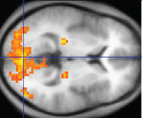
Figure \(\PageIndex{4}\): Functional Magnetic Resonance Imaging (fMRI) map. This imaging represents a transverse plane of the brain with the posterior side on the left and anterior side on the right. The orange areas represent highly active regions. In this particular example, the fMRI map shows activation of the visual cortex in response to visual stimuli. (Image credit: "1206 FMRI" by OpenStax is licensed under CC BY 4.0)
The underlying assumption is that active nervous tissue will have greater blood flow. By having the subject perform a visual task, activity all over the brain can be measured. Consider this possible experiment: the subject is told to look at a screen with a black dot in the middle (a fixation point). A photograph of a face is projected on the screen away from the center. The subject has to look at the photograph and decipher what it is. The subject has been instructed to push a button if the photograph is of someone they recognize. The photograph might be of a celebrity, so the subject would press the button, or it might be of a random person unknown to the subject, so the subject would not press the button.
In this task, visual sensory areas would be active, integrating areas would be active, motor areas responsible for moving the eyes would be active, and motor areas for pressing the button with a finger would be active. Those areas are distributed all around the brain and the fMRI images would show activity in more than just 10 percent of the brain (some evidence suggests that about 80 percent of the brain is using energy—based on blood flow to the tissue—during well-defined tasks similar to the one suggested above). This task does not even include all of the functions the brain performs. There is no language response, the body is mostly lying still in the MRI machine, and it does not consider the autonomic functions that would be ongoing in the background.
Concept Review
Nervous tissue is composed of neurons and glial cells. The basic structure of a neuron is composed of a soma (cell body) and processes. Processes can be classified in dendrites, which receive information, and axons, which carry information. The axons are partially covered by a lipid-rich sheath called myelin. Due to the high fat content of myelin, axons covered in myelin look white. Thus, the nervous tissue can also be described as gray matter and white matter on the basis of its appearance in unstained tissue. Cell bodies of neurons cluster in regions called nuclei or ganglia, depending on their location. Axons of neurons also tend to bundle up in structures called tracts (columns) or nerves (fibers), depending on their location.
The nervous system can be separated into divisions on the basis of anatomy and physiology. The anatomical divisions are the central nervous system (CNS) and peripheral nervous system (PNS). The CNS is composed of the brain and spinal cord. In the CNS, there are localized collection of neuron cell bodies called nuclei within the gray matter and bundles of axons called tracts within the white matter. The PNS is made of collections of neuron cell bodies called ganglia, and bundle of axons called nerves. A single axon can be part of a nerve when in the PNS, and then become a tract when crossing the CNS.
Functionally, the nervous system can be divided into those regions that are responsible for sensation, those that are responsible for integration, and those that are responsible for generating responses. All of these functional areas are found in both the central and peripheral anatomy. The nervous system can also be divided on the basis of how it controls the body. The somatic nervous system (SNS) is responsible for functions that result in moving skeletal muscles. Any sensory or integrative functions that result in the movement of skeletal muscle would be considered somatic. The autonomic nervous system (ANS) is responsible for functions that affect cardiac or smooth muscle tissue, or that cause glands to produce their secretions. Autonomic functions are distributed between central and peripheral regions of the nervous system. The sensations that lead to autonomic functions can be the same sensations that are part of initiating somatic responses. Somatic and autonomic integrative functions may overlap as well. A special division of the nervous system is the enteric nervous system, which is responsible for controlling the digestive organs. Parts of the autonomic nervous system overlap with the enteric nervous system. The enteric nervous system is exclusively found in the periphery because it is the nervous tissue in the organs of the digestive system.
Review Questions
Q. Which of the following cavities contains a component of the central nervous system?
A. abdominal
B. pelvic
C. cranial
D. thoracic
- Answer
-
Answer: C
Q. Which structure predominates in the white matter of the brain?
A. myelinated axons
B. neuronal cell bodies
C. autonomic ganglia
D. bundles of dendrites from the enteric nervous system
- Answer
-
Answer: A
Q. Which part of a neuron transmits an electrical signal to a target cell?
A. dendrites
B. soma
C. cell body
D. axon
- Answer
-
Answer: D
Q. Which term describes a bundle of axons in the peripheral nervous system?
A. nucleus
B. ganglion
C. tract
D. nerve
- Answer
-
Answer: D
Q. Which functional division of the nervous system would be responsible for the physiological changes seen during exercise (e.g., increased heart rate and sweating)?
A. somatic
B. autonomic
C. enteric
D. central
- Answer
-
Answer: B
Critical Thinking Questions
Q. What responses are generated by the nervous system when you run on a treadmill? Include an example of each type of tissue that is under nervous system control.
- Answer
-
A. Running on a treadmill involves contraction of the skeletal muscles in the legs through the somatic nervous system, increase in contraction of the cardiac muscle of the heart, and the production and secretion of sweat in the skin to stay cool through the autonomic nervous system.
Q. When eating food, what anatomical and functional divisions of the nervous system are involved in the perceptual experience?
- Answer
-
A. The sensation of taste associated with eating is sensed by nerves in the periphery that are involved in sensory and somatic functions. The process of chewing is somatic motor and uses skeletal muscles, while swallowing is both somatic motor (upper first phase) and autonomic motor (lower second phase).
Glossary
- afferent
- conducting or conducted inward or toward something
- autonomic nervous system (ANS)
- functional division of the nervous system that is responsible for homeostatic reflexes that coordinate control of cardiac and smooth muscle, as well as glandular tissue
- axon
- single process of the neuron that carries an electrical signal (action potential) away from the cell body toward a target cell
- brain
- the large organ of the central nervous system composed of white and gray matter, contained within the cranium and continuous with the spinal cord
- cell body
- in neurons, that portion of the cell that contains the nucleus, as opposed to the cell processes (axons and dendrites). Also known as soma
- central nervous system (CNS)
- anatomical division of the nervous system located within the cranial and vertebral cavities, namely the brain and spinal cord
- cranial nerve
- one of twelve nerves connected to the brain that are responsible for sensory or motor functions of the head and neck
- dendrite
- one of many branch-like processes that extends from the neuron cell body and functions as a contact for incoming signals (synapses) from other neurons or sensory cells
- effector
- a bodily tissue, structure, or organ (such as a gland or muscle) that becomes active in response to stimulation
- efferent
- conducted or conducting outward or away from something
- enteric nervous system (ENS)
- neural tissue associated with the digestive system that is responsible for nervous control through autonomic connections
- ganglion
- localized collection of neuron cell bodies in the peripheral nervous system
- general sense
- any sensory system that is distributed throughout the body and incorporated into organs of multiple other systems, such as the walls of the digestive organs or the skin
- glial cell
- one of the various types of neural tissue cells responsible for maintenance of the tissue, and largely responsible for supporting neurons
- gray matter
- regions of the nervous system containing cell bodies of neurons with few or no myelinated axons; actually may be more pink or tan in color, but called gray in contrast to white matter
- integration
- nervous system function that combines sensory perceptions and higher cognitive functions (memories, learning, emotion, etc.) to produce a response
- myelin
- lipid-rich insulating substance surrounding the axons of many neurons, allowing for faster transmission of electrical signals
- nerve
- cord-like bundle of axons located in the peripheral nervous system that transmits sensory input and response output to and from the central nervous system
- neuron
- neural tissue cell that is primarily responsible for generating and propagating electrical signals into, within, and out of the nervous system
- nucleus
- in the nervous system, a localized collection of neuron cell bodies that are functionally related; a “center” of neural function
- peripheral nervous system (PNS)
- anatomical division of the nervous system that is largely outside the cranial and vertebral cavities, namely all parts except the brain and spinal cord
- process
- in cells, an extension of a cell body; in the case of neurons, this includes the axon and dendrites
- response
- nervous system function that causes a target tissue (muscle or gland) to produce an event as a consequence to stimuli
- sensation
- nervous system function that receives information from the environment and translates it into the electrical signals of nervous tissue
- soma
- in neurons, that portion of the cell that contains the nucleus, as opposed to the cell processes (axons and dendrites). Also known as cell body
- somatic nervous system (SNS)
- functional division of the nervous system that is concerned with conscious perception, voluntary movement, and skeletal muscle reflexes
- somatosensation
- general senses related to the body, usually thought of as the senses of touch, which would include pain, temperature, and proprioception
- spinal cord
- organ of the central nervous system found within the vertebral cavity and connected with the periphery through spinal nerves; mediates reflex behaviors
- special sense
- any sensory system associated with a specific organ structure, namely smell, taste, sight, hearing, and balance
- spinal nerve
- one of 31 nerves connected to the spinal cord
- stimulus
- an event in the external or internal environment that registers as activity in a sensory neuron
- tract
- bundle of axons in the central nervous system having the same function and point of origin
- visceral sense
- sense associated with the internal organs
- white matter
- regions of the nervous system containing mostly myelinated axons, making the tissue appear white because of the high lipid content of myelin
Contributors and Attributions
OpenStax Anatomy & Physiology (CC BY 4.0). Access for free at https://openstax.org/books/anatomy-and-physiology