
13.2: Touch, Taste and Smell
- Page ID
- 22343

By the end of this section, you will be able to:
- Describe the different types of sensory receptors
- Describe the somatosensory receptors
- Describe the gross and microscopic structures responsible for the special senses of taste and smell
A major role of sensory receptors is to help us learn about the environment around us, or about the state of our internal environment. Stimuli from varying sources, and of different types, are received and changed into the electrochemical signals of the nervous system. This occurs when a stimulus changes the cell membrane of a sensory neuron. The stimulus causes the sensory cell to produce an action potential that is relayed into the central nervous system (CNS), where it is integrated with other sensory information—or sometimes higher cognitive functions—to become a conscious perception of that stimulus. The central integration may then lead to a motor response.
Describing sensory function with the term sensation or perception is a deliberate distinction. Sensation is the activation of sensory receptor cells at the level of the stimulus. Perception is the central processing of sensory stimuli into a meaningful pattern. Perception is dependent on sensation, but not all sensations are perceived. Receptors are the cells or structures that detect sensations. A receptor cell is changed directly by a stimulus, such as opening of ion channels in the plasma membrane.
Sensory Receptors
Stimuli in the environment activate specialized receptor cells in the peripheral nervous system. Different types of stimuli are sensed by different types of receptor cells. Receptor cells can be classified into types on the basis of three different criteria: cell type, position, and function. Receptors can be classified structurally on the basis of cell type and their position in relation to stimuli they sense. They can also be classified functionally on the basis of the transduction of stimuli, or how the mechanical stimulus, light, or chemical changed the cell membrane electrical properties.
Structural Receptor Types
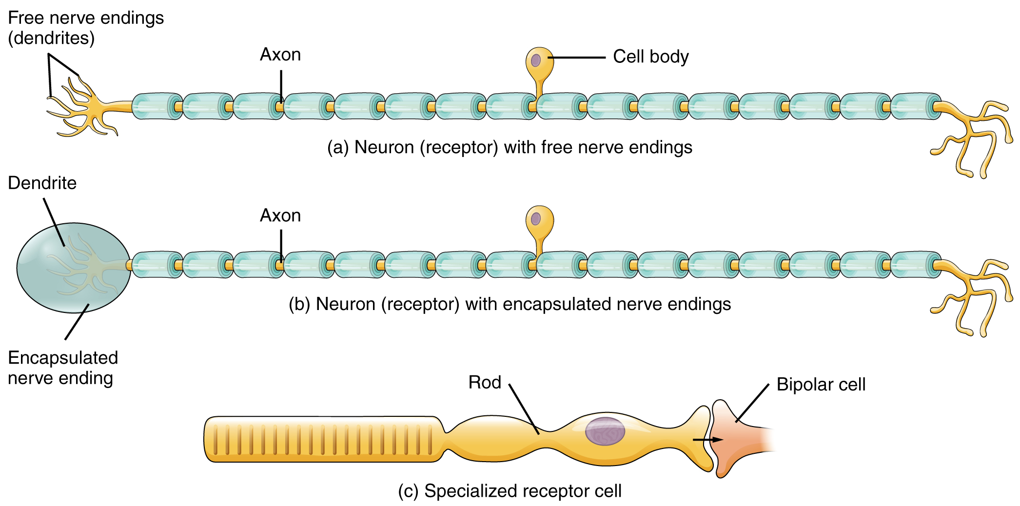
The first structural classification of sensory receptors is based on the cell type. The cells that interpret information about the environment can be either (1) a neuron that has a free nerve ending, with dendrites embedded in tissue that would receive a sensation; (2) a neuron that has an encapsulated ending in which the sensory nerve endings are covered in connective tissue that enhances their sensitivity; or (3) a specialized receptor cell, which has distinct structural components that interpret a specific type of stimulus (Figure \(\PageIndex{1}\)). The pain and temperature receptors in the dermis of the skin are examples of neurons that have free nerve endings. Also located in the dermis of the skin are lamellated corpuscles, neurons with encapsulated nerve endings that respond to pressure and touch. The cells in the retina that respond to light stimuli are an example of a specialized receptor, a photoreceptor.
Another way that receptors can be classified is based on their location relative to the stimuli. An exteroceptor is a receptor that is located near a stimulus in the external environment, such as the somatosensory receptors that are located in the skin. An interoceptor is one that interprets stimuli from internal organs and tissues, such as the receptors that sense the increase in blood pressure in the aorta or carotid sinus. Finally, a proprioceptor is a receptor located near a moving part of the body, such as a muscle, that interprets the positions of the tissues as they move.
Functional Receptor Types
A third classification of receptors is by how the receptor transduces stimuli into electrical changes. Stimuli are of three general types. Some stimuli are ions and macromolecules that affect transmembrane receptor proteins when these chemicals diffuse across the cell membrane, for example the ones found in food. Some stimuli are physical variations in the environment that affect the electrical properties of the receptor cell membrane, for example touch. Other stimuli include the electromagnetic radiation from visible light. For humans, the only electromagnetic energy that is perceived by our eyes is visible light. Some other organisms have receptors that humans lack, such as the heat sensors of snakes, the ultraviolet light sensors of bees, or magnetic receptors in migratory birds.
Receptor cells can be further categorized on the basis of the type of stimuli they transduce. Chemical stimuli can be interpreted by a chemoreceptor that interprets chemical stimuli, such as an object’s taste or smell. Osmoreceptors respond to solute concentrations of body fluids. Additionally, pain is primarily a chemical sense that interprets the presence of chemicals from tissue damage, or similar intense stimuli, through a nociceptor. Physical stimuli, such as pressure and vibration, as well as the sensation of sound and body position (balance), are interpreted through a mechanoreceptor. Another physical stimulus that has its own type of receptor is temperature, which is sensed through a thermoreceptor that is either sensitive to temperatures above (heat) or below (cold) normal body temperature.
Sensory Modalities
Ask anyone what the senses are, and they are likely to list the five major senses—taste, smell, touch, hearing, and sight. However, these are not all of the senses. The most obvious omission from this list is balance. Also, what is referred to simply as touch can be further subdivided into pressure, vibration, stretch, and hair-follicle position, on the basis of the type of mechanoreceptors that perceive these touch sensations. Other overlooked senses include temperature perception by thermoreceptors and pain perception by nociceptors.
Senses can be classified as either general or special. A general sense is one that is distributed throughout the body and has receptor cells within the structures of other organs. Mechanoreceptors in the skin, muscles, or the walls of blood vessels are examples of this type. General senses often contribute to the sense of touch, as described above, or to proprioception (awareness of body position) and kinesthesia (awareness of body movement), or to a visceral sense, which is most important to autonomic functions. A special sense is one that has a specific organ devoted to it, namely the eye, inner ear, tongue, or nose.
Each of the senses is referred to as a sensory modality. Modality refers to the way that information is encoded, which is similar to the idea of transduction. The main sensory modalities can be described on the basis of how each is transduced. The chemical senses are taste and smell. The general sense that is usually referred to as touch includes chemical sensation in the form of nociception, or pain. Pressure, vibration, muscle stretch, and the movement of hair by an external stimulus, are all sensed by mechanoreceptors. Hearing and balance are also sensed by mechanoreceptors. Finally, vision involves the activation of photoreceptors.
Listing all the different sensory modalities, which can number as many as 17, involves separating the five major senses into more specific categories, or submodalities, of the larger sense. An individual sensory modality represents the sensation of a specific type of stimulus. For example, the general sense of touch, which is known as somatosensation, can be separated into light pressure, deep pressure, vibration, itch, pain, temperature, or hair movement.
Somatosensation (Touch)
Somatosensation is considered a general sense, as opposed to the special senses discussed in this section. Somatosensation is the group of sensory modalities that are associated with touch, proprioception, and interoception. These modalities include pressure, vibration, light touch, tickle, itch, temperature, pain, proprioception, and kinesthesia. This means that its receptors are not associated with a specialized organ, but are instead spread throughout the body in a variety of organs. Many of the somatosensory receptors are located in the skin, but receptors are also found in muscles, tendons, joint capsules, ligaments, and in the walls of visceral organs.
Two types of somatosensory signals that are transduced by free nerve endings are pain and temperature. These two modalities use thermoreceptors and nociceptors to transduce temperature and pain stimuli, respectively. Temperature receptors are stimulated when local temperatures differ from body temperature. Some thermoreceptors are sensitive to just cold and others to just heat. Nociception is the sensation of potentially damaging stimuli. Mechanical, chemical, or thermal stimuli beyond a set threshold will elicit painful sensations. Stressed or damaged tissues release chemicals that activate receptor proteins in the nociceptors.
If you drag your finger across a textured surface, the skin of your finger will vibrate. Such low frequency vibrations are sensed by mechanoreceptors called Merkel cells, also known as type I cutaneous mechanoreceptors. Merkel cells are located in the stratum basale of the epidermis. Deep pressure and vibration is transduced by lamellated (Pacinian) corpuscles, which are receptors with encapsulated endings found deep in the dermis, or subcutaneous tissue. Light touch is transduced by the encapsulated endings known as tactile (Meissner) corpuscles. Follicles are also wrapped in a plexus of nerve endings known as the hair follicle plexus. These nerve endings detect the movement of hair at the surface of the skin, such as when an insect may be walking along the skin. Stretching of the skin is transduced by stretch receptors known as bulbous corpuscles. Bulbous corpuscles are also known as Ruffini corpuscles, or type II cutaneous mechanoreceptors.
Other somatosensory receptors are found in the joints and muscles. Stretch receptors monitor the stretching of tendons, muscles, and the components of joints. For example, have you ever stretched your muscles before or after exercise and noticed that you can only stretch so far before your muscles spasm back to a less stretched state? This spasm is a reflex that is initiated by stretch receptors to avoid muscle tearing. Such stretch receptors can also prevent over-contraction of a muscle. In skeletal muscle tissue, these stretch receptors are called muscle spindles. Golgi tendon organs similarly transduce the stretch levels of tendons. Bulbous corpuscles are also present in joint capsules, where they measure stretch in the components of the skeletal system within the joint. The types of nerve endings, their locations, and the stimuli they transduce are presented in Table \(\PageIndex{1}\).
| Mechanoreceptors of Somatosensation | ||
|---|---|---|
| Name | Location(s) | Stimuli |
| Free nerve endings | Dermis, cornea, tongue, joint capsules, visceral organs | Pain, temperature, mechanical deformation |
| Mechanoreceptors or Merkel's discs | Epidermal–dermal junction, mucosal membranes | Low frequency vibration (5–15 Hz) |
| Bulbous (or Ruffini's) corpuscle | Dermis, joint capsules | Stretch |
| Tactile (or Meissner's) corpuscle | Papillary dermis, especially in the fingertips and lips | Light touch, vibrations below 50 Hz |
| Lamellated (or Pacinian) corpuscle | Deep dermis, subcutaneous tissue | Deep pressure, high-frequency vibration (around 250 Hz) |
| Hair follicle plexus | Wrapped around hair follicles in the dermis | Movement of hair |
| Muscle spindle | In line with skeletal muscle fibers | Muscle contraction and stretch |
| Tendon stretch (or Golgi tendon) organ | In line with tendons | Stretch of tendon |
Gustation (Taste)
Only a few recognized submodalities exist within the sense of taste, or gustation. Until recently, only four tastes were recognized: sweet, salty, sour, and bitter. Research at the turn of the 20th century led to recognition of the fifth taste, umami, during the mid-1980s. Umami is a Japanese word that means “delicious taste,” and is often translated to mean savory. Very recent research has suggested that there may also be a sixth taste for fats, or lipids.
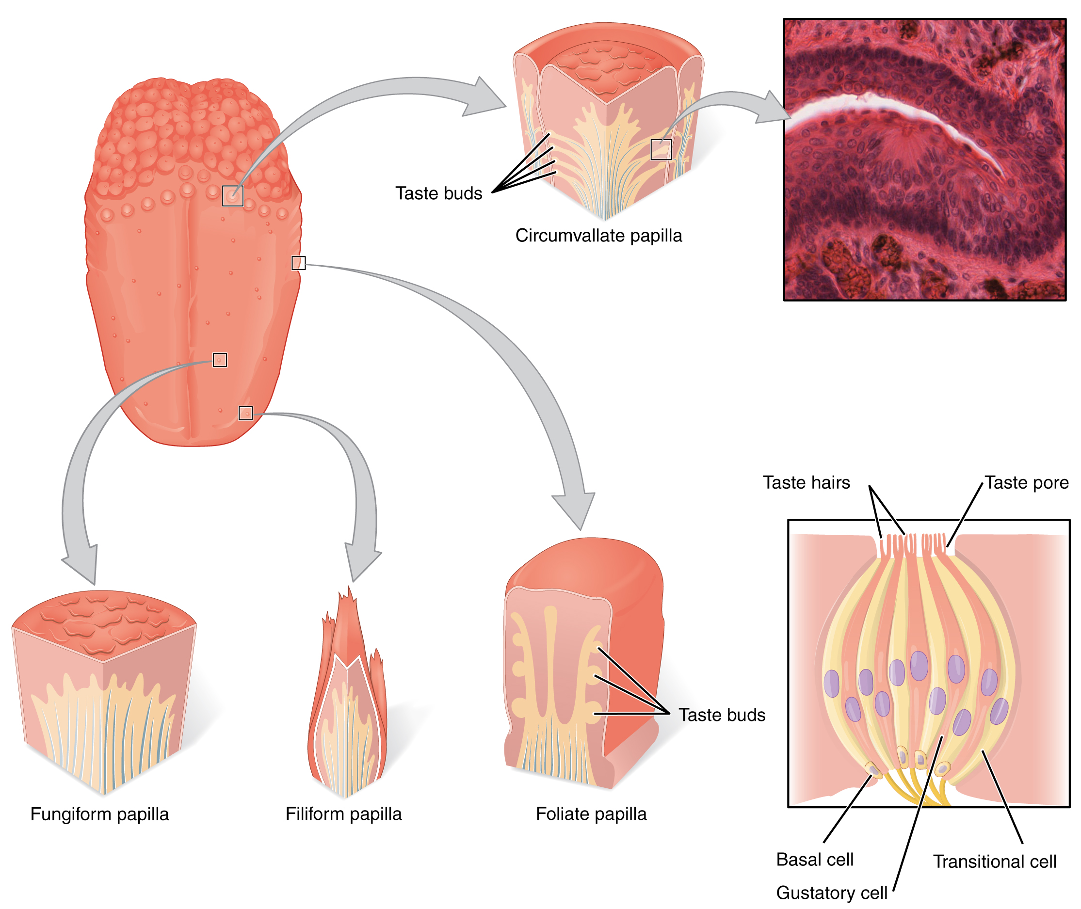
Gustation is the special sense associated with the tongue. The surface of the tongue, along with the rest of the oral cavity, is lined by a stratified squamous epithelium. Raised bumps called papillae (singular = papilla) contain the structures for gustatory transduction. There are four types of papillae, based on their appearance (Figure \(\PageIndex{2}\)): circumvallate, foliate, filiform, and fungiform. Circumvallate papillae are the largest papillae on the tongue and are located towards the posterior surface of the tongue. They contain the majority of the taste buds. Foliate papillae are located on the posterolateral region of the tongue and they are mostly used during infancy and childhood. Filiform papillae are shaped like bristles and do not contain taste buds. They are important for detecting texture and manipulating food. Fungiform papillae are located on the tip and sides of the tongues and contain few taste buds. Within the structure of the papillae are taste buds which are recessed into a taste pore and are shaped like an onion. Taste buds contain a variety of cells. Basal cells are undifferentiated stem cells located near the base of the taste bud that undergo continuous regeneration to replace the other cells of the taste buds. Transitional cells are scattered throughout the taste buds and support the other cells. Specialized gustatory receptor cells have microvilli called taste hairs and can absorb the chemicals contained within foods that are ingested. These cells release neurotransmitters based on the amount of the chemical in the food. These cells are not neurons but they are in contact with the dendrites of sensory neurons.
Once the gustatory cells are activated by the taste molecules, they release neurotransmitters onto the dendrites of sensory neurons. These neurons are part of the facial (CN VII) and glossopharyngeal (CN IX) cranial nerves, as well as a component within the vagus nerve (CN X) dedicated to the gag reflex. The facial nerve connects to taste buds in the anterior third of the tongue. The glossopharyngeal nerve connects to taste buds in the posterior two thirds of the tongue. The vagus nerve connects to taste buds in the extreme posterior of the tongue, verging on the pharynx, which are more sensitive to noxious stimuli such as bitterness. Gustatory information is then transmitted through these cranial nerves to the brain, through the thalamus, which acts as a relay station, and finally into the primary gustatory cortex of the insula. It is here that this information is processed and perception is initiated.
Novel studies have shown that gustatory cells are not only present on the tongue but also in the guts. In humans, sweet, umami and bitter taste cells are also present in the intestines and used to "taste" the ingested food and regulate its variations.
Olfaction (Smell)
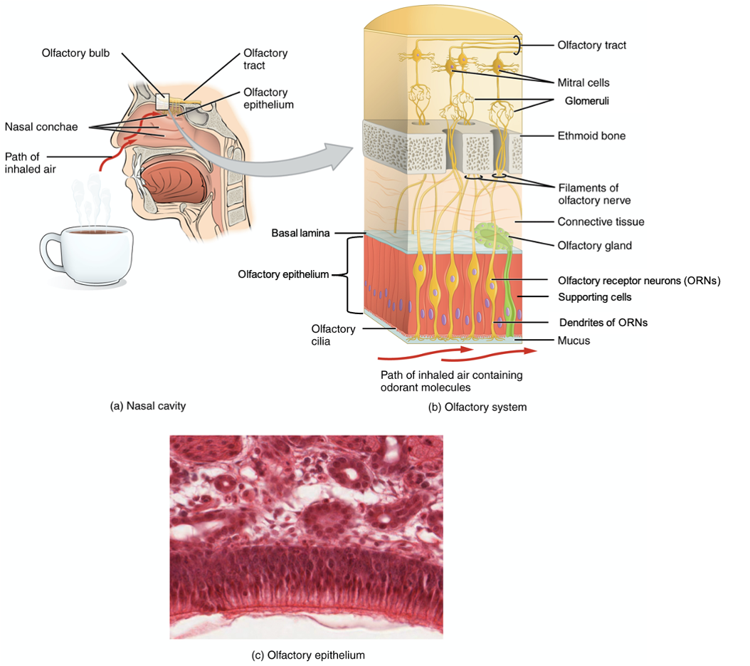
Like taste, the sense of smell, or olfaction, is also responsive to chemical stimuli. Inhaled air containing odorant molecules (smells) enters the nasal cavity and passes by the nasal conchae. The olfactory receptor neurons (ORNs) are located in a small region within the superior nasal cavity (Figure \(\PageIndex{3}\)). This region is referred to as the olfactory epithelium and also contains supporting cells and basal cells (not shown), with similar functions as the homonyms in the taste buds. Each olfactory receptor neuron has dendrites that extend from the apical surface of the epithelium into the mucus lining the cavity. Olfactory glands within the basal lamina at the interface between the olfactory epithelium and the superior connective tissue produce the mucus that lines the olfactory epithelium, The olfactory sensory neurons present nonmotile cilia called olfactory cilia (or hairs) that contain the chemoreceptor for a specific odorant molecule. As airborne molecules are inhaled through the nose, they pass over the olfactory epithelial region and dissolve into the mucus. These odorant molecules bind to proteins that keep them dissolved in the mucus and help transport them to the olfactory dendrites. The odorant–protein complex binds to a receptor protein within the cell membrane of an olfactory dendrite, producing an electrical change in the olfactory neurons.
The axon of an olfactory neuron extends from the basal surface of the epithelium towards the cribriform plate of the ethmoid bone forming the olfactory nerve (CN I). The olfactory fibers go through an olfactory foramen in the cribriform plate and converge to form spherical structures called glomeruli in the olfactory bulb, which resides on the ventral surface of the frontal lobe. Within the glomeruli, the olfactory receptor neurons synapse with mitral cells which in turn extend their axons to form the olfactory tract. From there, the axons split to travel to several brain regions. Some travel to the cerebrum, specifically to the primary olfactory cortex that is located in the inferior and medial areas of the temporal lobe. Others project to structures within the limbic system and hypothalamus, where smells become associated with long-term memory and emotional responses. This is how certain smells trigger emotional memories, such as the smell of food associated with one’s birthplace. Smell is the one sensory modality that does not synapse in the thalamus before connecting to the cerebral cortex. This intimate connection between the olfactory system and the cerebral cortex is one reason why smell can be a potent trigger of memories and emotion.
The olfactory system can recognize seven primary odors (musky, putrid, pungent, camphoraceous, ethereal, floral, pepperminty) as well as thousands of odorant molecules that produce various scents. The nasal epithelium, including the olfactory cells, can be harmed by airborne toxic chemicals. Therefore, the olfactory neurons are one of the few types of neurons that undergo mitosis and is able to replace old cells. Olfactory neurons are regularly replaced within the nasal epithelium, after which the axons of the new neurons must find their appropriate connections in the olfactory bulb. These new axons grow along the axons that are already in place in the cranial nerve.
DISORDERS OF THE...
Olfactory System: Anosmia
Blunt force trauma to the face, such as that common in many car accidents, can lead to the loss of the olfactory nerve, and subsequently, loss of the sense of smell. This condition is known as anosmia. When the frontal lobe of the brain moves relative to the ethmoid bone, the olfactory tract axons may be sheared apart. Professional fighters often experience anosmia because of repeated trauma to face and head. In addition, certain pharmaceuticals, such as antibiotics, can cause anosmia by killing all the olfactory neurons at once. If no axons are in place within the olfactory nerve, then the axons from newly formed olfactory neurons have no guide to lead them to their connections within the olfactory bulb. There are temporary causes of anosmia, as well, such as those caused by inflammatory responses related to respiratory infections or allergies.
Loss of the sense of smell can result in food tasting bland. A person with an impaired sense of smell may require additional spice and seasoning levels for food to be tasted. Anosmia may also be related to some presentations of mild depression, because the loss of enjoyment of food may lead to a general sense of despair.
The ability of olfactory neurons to replace themselves decreases with age, leading to age-related anosmia. This explains why some elderly people salt their food more than younger people do. However, this increased sodium intake can increase blood volume and blood pressure, increasing the risk of cardiovascular diseases in the elderly.
Concept Review
The cells that transduce sensory stimuli into the electrochemical signals of the nervous system are classified on the basis of structural or functional aspects of the cells. The structural classifications are either based on the anatomy of the cell that is interacting with the stimulus (free nerve endings, encapsulated endings, or specialized receptor cell), or where the cell is located relative to the stimulus (interoceptor, exteroceptor, proprioceptor). Thirdly, the functional classification is based on how the cell transduces the stimulus into a neural signal. Chemoreceptors respond to chemical stimuli and are the basis for olfaction and gustation. Related to chemoreceptors are osmoreceptors and nociceptors for fluid balance and pain reception, respectively. Mechanoreceptors respond to mechanical stimuli and are the basis for most aspects of somatosensation, as well as being the basis of audition and equilibrium in the inner ear. Thermoreceptors are sensitive to temperature changes, and photoreceptors are sensitive to light energy.
The senses are somatosensation (sensations associated with the skin and body), gustation (taste), olfaction (smell), audition (hearing), equilibrium (balance), and vision. With the exception of somatosensation, this list represents the special senses, or those systems of the body that are associated with specific organs such as the tongue or eye. The general senses can be divided into somatosensation, which is commonly considered touch, but includes tactile, pressure, vibration, temperature, and pain perception. The general senses also include the visceral senses, which are separate from the somatic nervous system function in that they do not normally rise to the level of conscious perception. The special senses are all primarily part of the somatic nervous system in that they are consciously perceived through cerebral processes, though some special senses contribute to autonomic function.
Somatosensation belongs to the general senses, which are those sensory structures that are distributed throughout the body and in the walls of various organs. Gustation and olfaction belong to the special senses. Gustation transduction is achieved by raised bumps on the tongue called papillae that contain taste buds, which in turn contain specialized gustatory receptor cells for chemical stimuli (”tastes”). Olfactory receptor neurons (unipolar) located in olfactory epithelium respond to chemical stimuli (“odorants”) of the nasal cavity. Their dendrites extend from the apical surface of the epithelium into the mucus lining the cavity and have receptors for odorants. Axons extend into the cribriform plate of the ethmoid bone to reach the olfactory bulb, forming the olfactory nerve (CN I). Once the olfactory nerve crosses the CNS, it becomes the olfactory tract and sends axons to several brain regions. The axons do not synapse onto the thalamus (unlike the other special senses).
Review Questions
Q. What type of receptor cell is responsible for transducing pain stimuli?
A. mechanoreceptor
B. nociceptor
C. osmoreceptor
D. photoreceptor
- Answer
-
B
Q. Which of these cranial nerves is part of the gustatory system?
A. olfactory
B. trochlear
C. trigeminal
D. facial
- Answer
-
D
Glossary
- basal cell
- undifferentiated stem cells located in the taste buds and olfactory epithelium
- chemoreceptor
- sensory receptor cell that is sensitive to chemical stimuli, such as in taste, smell, or pain
- encapsulated ending
- configuration of a sensory receptor neuron with dendrites surrounded by specialized structures to aid in transduction of a particular type of sensation, such as the lamellated corpuscles in the deep dermis and subcutaneous tissue
- exteroceptor
- sensory receptor that is positioned to interpret stimuli from the external environment, such as photoreceptors in the eye or somatosensory receptors in the skin
- free nerve ending
- configuration of a sensory receptor neuron with dendrites in the connective tissue of the organ, such as in the dermis of the skin, that are most often sensitive to chemical, thermal, and mechanical stimuli
- general sense
- any sensory system that is distributed throughout the body and incorporated into organs of multiple other systems, such as the walls of the digestive organs or the skin
- glomeruli
- spherical structures located in the olfactory bulb
- gustation
- sense of taste
- gustatory receptor cells
- sensory cells in the taste bud that transduce the chemical stimuli of gustation
- insula
- small region of the cerebral cortex located deep within the lateral sulcus
- interoceptor
- sensory receptor that is positioned to interpret stimuli from internal organs, such as stretch receptors in the wall of blood vessels
- kinesthesia
- sense of body movement based on sensations in skeletal muscles, tendons, joints, and the skin
- mechanoreceptor
- receptor cell that transduces mechanical stimuli into an electrochemical signal
- nociceptor
- receptor cell that senses pain stimuli
- odorant molecules
- volatile chemicals that bind to receptor proteins in olfactory neurons to stimulate the sense of smell
- olfaction
- sense of smell
- olfactory bulb
- central target of the first cranial nerve; located on the ventral surface of the frontal lobe in the cerebrum
- olfactory cilia
- hairlike structures arising from the dendrites of an olfactory receptor neuron
- olfactory epithelium
- region of the nasal epithelium where olfactory neurons are located
- olfactory gland
- gland of the olfactory epithelium that secretes mucus into the nasal cavity
- olfactory nerve
- first cranial nerve; responsible for the sense of smell
- olfactory receptor neuron
- receptor cell of the olfactory system, sensitive to the chemical stimuli of smell, the axons of which compose the first cranial nerve
- olfactory tract
- central bundle of axons deriving from the mitral cells of the olfactory bulb
- osmoreceptor
- receptor cell that senses differences in the concentrations of bodily fluids on the basis of osmotic pressure
- papilla
- for gustation, a bump-like projection on the surface of the tongue that contains taste buds
- photoreceptor
- receptor cell specialized to respond to light stimuli
- primary gustatory cortex
- region of the cerebral cortex within the insula responsible for the perception of taste
- primary olfactory cortex
- region of the cerebral cortex within the temporal lobe responsible for the perception of smell
- proprioception
- sense of position of the body in space based on sensations in skeletal muscles, tendons, joints, and the skin
- proprioceptor
- receptor cell that senses changes in the position and kinesthetic aspects of the body
- receptor cell
- cell that transduces environmental stimuli into neural signals
- sensory modality
- a particular system for interpreting and perceiving environmental stimuli by the nervous system
- somatosensation
- general sense associated with modalities lumped together as touch
- special sense
- any sensory system associated with a specific organ structure, namely smell, taste, sight, hearing, and balance
- submodality
- specific sense within a broader major sense such as sweet as a part of the sense of taste, or color as a part of vision
- supporting cells
- cells that support the olfactory receptor neurons
- taste buds
- structures within a papilla on the tongue that contain gustatory receptor cells
- taste hairs
- Hairlike projections of gustatory cells of taste buds
- taste pore
- opening of a taste bud
- thalamus
- major region of the diencephalon that is responsible for relaying information between the cerebrum and the hindbrain, spinal cord, and periphery
- thermoreceptor
- sensory receptor specialized for temperature stimuli
- transduction
- process of changing an environmental stimulus into the electrochemical signals of the nervous system
- transitional cells
- cells that support the gustatory receptor cells
- umami
- taste submodality for sensitivity to the concentration of amino acids; also called the savory sense
- visceral sense
- sense associated with the internal organs
Contributors and Attributions
OpenStax Anatomy & Physiology (CC BY 4.0). Access for free at https://openstax.org/books/anatomy-and-physiology