
14.2: Divisions of the Autonomic Nervous System
- Page ID
- 22348

By the end of this section, you will be able to:
- Compare and contrast the somatic and autonomic nervous systems
- Describe the functional differences between the sympathetic and parasympathetic divisions
- Outline the anatomical differences between the two divisions of the ANS
- Describe the preganglionic neurons, ganglia, nerves and pathways of the two divisions of the ANS
Comparison between the Somatic and Autonomic Nervous System
The nervous system can be divided into two functional parts: the somatic nervous system (SNS) and the autonomic nervous system (ANS). The major difference between these two systems is based on whether you are conscious of its process. The somatic nervous system consciously detects sensory stimuli from the special senses, skin and proprioceptors. The autonomic nervous system reflexively responds to visceral sensory stimuli, such as levels of carbon dioxide concentration in the blood or stretch caused by blood pressure, that you are not consciously aware of. Moreover, the motor efferent branches of these two systems innervate different target effectors. While the somatic motor neurons innervate and cause contraction of skeletal muscles, autonomic motor neurons innervate and control cardiac and smooth muscle, as well as glandular tissue. Thus, the motor response of the somatic nervous system is voluntary while the one of the autonomic nervous system is involuntary.
Another major difference between these two systems lies within the number of lower motor neurons that are involved in the response. In the somatic nervous system, a single lower somatic motor neuron of the brainstem or spinal cord extends from the CNS towards a skeletal muscle through a cranial or spinal nerve, respectively. These somatic motor neurons have large myelinated axons that release acetylcholine (ACh) at neuromuscular junctions. In comparison, the autonomic nervous system is composed of a chain of two lower motor neurons. The cell body of the first of the two ANS motor neurons is located in the brainstem or spinal cord and is called a preganglionic neuron. The axon of the preganglionic neuron extends outside of the CNS through cranial or spinal nerves forming a preganglionic fiber. This fiber projects to an autonomic ganglion of the peripheral nervous system. Preganglionic neurons have small myelinated axons that release acetylcholine (ACh) to excite a second motor neuron. The second motor neuron is called a ganglionic neuron. The cell body of the ganglionic neuron resides within the autonomic ganglion and its axon extends to an effector (cardiac muscle, smooth muscle, or gland) forming a postganglionic fiber. Ganglionic neurons have small unmyelinated axons that release either acetylcholine (ACh) or norepinephrine (NE) to either excite or inhibit an effector, depending on the type of receptors present on the effector. Since preganglionic and postganglionic axons are small or unmyelinated, the propagation of autonomic electrical impulses is slower compared to the somatic motor axons.
Divisions of the Autonomic Nervous System
The autonomic nervous system regulates many of the internal organs through a balance of two aspects, or divisions. The two divisions of the autonomic nervous system are the sympathetic division and the parasympathetic division. From a functional point of view, the sympathetic system is associated with the fight-or-flight response, while the parasympathetic activity is referred to by the epithet rest-and-digest. Homeostasis is the balance between the two divisions since one system complements the other. For example, the parasympathetic division will be more active when you need to conserve energy and replenish nutrient stores. The sympathetic division will activate when during exercise, stress or emergency situations. Many autonomic target effectors have dual innervation by both divisions of the autonomic nervous system, which determines their activity. For example, the heart receives connections from both the sympathetic and parasympathetic divisions: one causes heart rate to increase, whereas the other causes heart rate to decrease. From an anatomical point of view, both divisions use preganglionic and ganglionic neurons to innervate cardiac muscle, smooth muscle and glands. However, the location of preganglionic neurons within the CNS is different between the two divisions. Moreover, the location of the ganglia as well as the length of preganglionic and postganglionic axons differ in the two divisions. In the following sections, you will examine the functional and anatomical features of the sympathetic and parasympathetic divisions.
Sympathetic Division
To respond to a threat—to fight or to run away—the sympathetic system causes diverse effects as many different effector organs are activated together for a common purpose. More oxygen needs to be inhaled and delivered to skeletal muscle. The respiratory, cardiovascular, and musculoskeletal systems are all activated together. Additionally, sweating keeps the excess heat that comes from muscle contraction from causing the body to overheat. The digestive system shuts down so that blood is not absorbing nutrients when it should be delivering oxygen to skeletal muscles. To coordinate all these responses, the connections in the sympathetic system diverge from a limited region of the central nervous system (CNS) to a wide array of ganglia that project to the many effector organs simultaneously. The complex set of structures that compose the output of the sympathetic system make it possible for these disparate effectors to come together in a coordinated, systemic change.
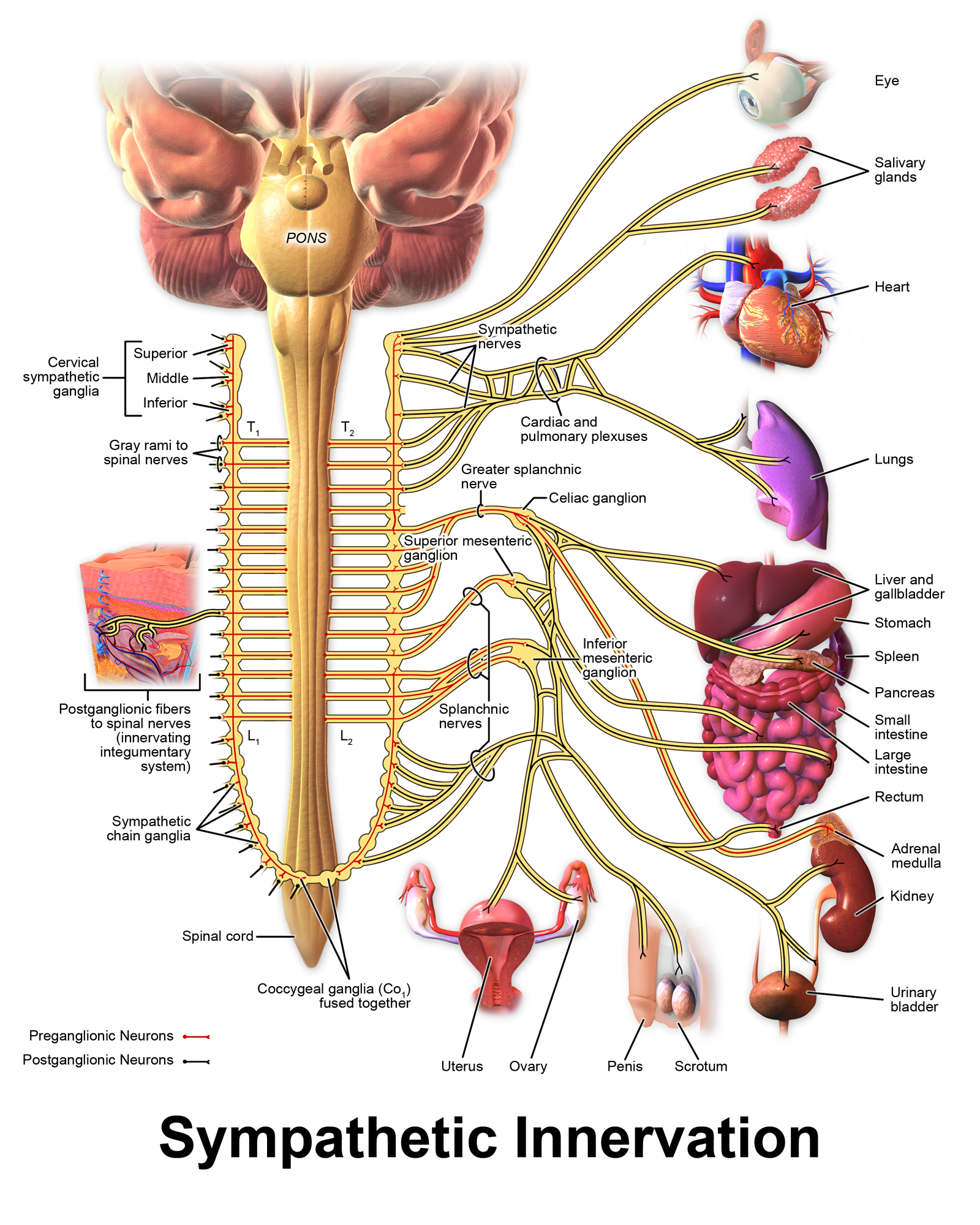
The sympathetic division of the autonomic nervous system influences various organ systems of the body through connections emerging from the first thoracic (T1) and second lumbar (L2) spinal segments (Figure \(\PageIndex{1}\)). It is referred to as the thoracolumbar system to reflect this anatomical basis. Sympathetic preganglionic neurons are located in the lateral horns of any of these spinal regions and project to ganglia adjacent to the vertebral column through the ventral roots of the spinal cord.
Sympathetic Neurons, Ganglia and Nerves
The majority of ganglia of the sympathetic system belong to a network of sympathetic chain (or trunk) ganglia that runs lateral to the vertebral column and anterior to the paired spinal nerves (Figure \(\PageIndex{1}\)). For this reason, these ganglia can also be called paravertebral ganglia. The ganglia appear as a series of clusters of neurons linked by ascending and descending axonal bridges called sympathetic trunks. There are typically 23 ganglia in the sympathetic chain on either side of the spinal column. Three correspond to the cervical region, 12 are in the thoracic region, four are in the lumbar region, and four correspond to the sacral region. The thoracic and lumbar sympathetic preganglionic fibers travel sequentially through ventral roots, spinal nerves and bundles of myelinated axons called white rami communicantes (singular = ramus communicans) to reach the correspondent paravertebral ganglia (Figure \(\PageIndex{2}\)). Here preganglionic sympathetic fibers either synapse with ganglionic neurons or often pass on through the sympathetic chain ganglion into one of its emerging nerves to synapse with ganglionic neurons elsewhere. Postganglionic fibers then travel through additional nerves to their destination in one of the organs. Many of the fibers from the postganglionic neurons in the sympathetic chain ganglia pass back into the spinal nerves through gray rami communicantes composed of unmyelinated axons and carry sympathetic information through the spinal nerves.
The cervical and sacral paravertebral ganglia are not connected to the spinal cord directly through the spinal nerves, but through sympathetic trunks. Among the cervical ganglia, the superior cervical ganglion contains ganglionic neurons that innervate structures of the head and neck such the dilator pupillae and superior tarsal muscles of the eye, the lacrimal gland, mucous membranes of the nose, palate and mouth, and salivary glands. The middle and inferior cervical ganglia contain ganglionic neurons that innervate neck and thoracic organs such as the larynx, trachea, pharynx, smooth muscle of arteries and heart.
Preganglionic sympathetic axons extending from T5-L2 do not synapse in a sympathetic chain ganglion and instead continue through the chain anteriorly towards the abdominal and pelvic organs (Figure \(\PageIndex{1}\)). These axons form splanchnic nerves and typically terminate in three autonomic ganglia called prevertebral (or collateral) ganglia. They are referred to as prevertebral because they are anterior to the vertebral column and descending aorta. The prevertebral ganglia are associated with controlling organs in the abdominal cavity, and are also considered part of the enteric nervous system. The greater splanchnic nerves originate from T5-T9 spinal nerves and synapse into the prevertebral celiac ganglia. Postganglionic axons from these ganglia innervate stomach, abdominal blood vessels, liver, gallbladder, part of the pancreas and small intestine. The lesser splanchnic nerves originate from T9-T11 spinal nerves and project to prevertebral superior mesenteric ganglia. Postganglionic axons from these ganglia innervate the large intestine. The least splanchnic nerves that extend from T12 spinal nerves project to and terminate in the prevertebral renal ganglia (not shown here). Postganglionic axons from these ganglia innervate the remainder of the pancreas and small intestine, the proximal part of the large intestine, the kidneys and proximal ureters. Lumbar splanchnic nerves that extend from L1-L2 spinal nerves terminate to the inferior mesenteric ganglia. Postganglionic axons from these ganglia project to and innervate the distal part of the large intestine, rectum, kidneys, urinary bladder, gonads and external genitalia. In addition to the above splanchnic nerves, there are also small sacral splanchnic nerves that originate from the sacral sympathetic ganglia that are not directly connected to the spinal cord and terminate into urinary and reproductive organs.
The neurons of the sympathetic autonomic ganglia are multipolar in shape, with dendrites radiating out around the cell body where synapses from the spinal cord neurons are made. Because the sympathetic ganglia are adjacent to the vertebral column, preganglionic sympathetic fibers are relatively short, and they are myelinated. Compared with the preganglionic fibers, postganglionic sympathetic fibers are long because of the relatively greater distance from the ganglion to the target effector. These postganglionic fibers are unmyelinated. A diagram that shows the connections of the sympathetic system is somewhat like a circuit diagram that shows the electrical connections between different receptacles and devices. In Figure \(\PageIndex{1}\), the “circuits” of the sympathetic system are intentionally simplified.
Sympathetic Pathways
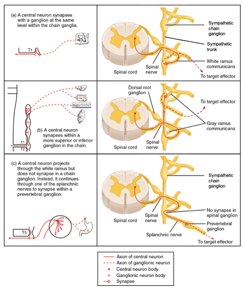
A sympathetic preganglionic axon leaving the lateral horn of the thoracolumbar spinal cord enters the sympathetic chain ganglia, where it branches toward 10-20 targets. To continue with the analogy of the circuit diagram, there are four different types of “junctions” that connect the sympathetic preganglionic axons with their effectors. The type of pathway is determined by the location and type of target effector organ being innervated. In all cases, the preganglionic axon extends into the spinal nerve at the same level as its spinal cord segment. Then it can then either (a) synapse in the paravertebral ganglion and carry information through the spinal nerve at the same level (spinal nerve pathway), (b) ascend to a more superior or descend to a more inferior paravertebral ganglion, synapse there and carry information through sympathetic nerves (sympathetic nerve pathway), (c) descend to a prevertebral (collateral) ganglion, synapse there and carry information through a splanchnic nerve (splanchnic nerve pathway) or (d) project directly to the adrenal medulla (adrenal medulla pathway). All of these branches mean that one preganglionic neuron can influence different regions of the sympathetic system very broadly, by acting on widely distributed organs. In the following paragraphs, you will examine these four different pathways. Except for the adrenal medulla pathway, these connections are represented in Figure \(\PageIndex{2}\).
The spinal nerve pathway is the most direct connection. The sympathetic preganglionic nerve projects to the sympathetic chain ganglion at the same level as the target effector. The myelinated preganglionic fiber extending from the lateral horns of the spinal cord projects to the sympathetic chain ganglion through the ventral root and spinal nerve. Through the white ramus communicans, the fiber reaches and synapses with the ganglionic neuron in the sympathetic chain ganglion. The postganglionic fiber then projects to the target effector via the gray ramus communicans, which is formed by unmyelinated axons. An example of this type is spinal nerve T1 that synapses with the T1 sympathetic chain ganglion to innervate the skin (Figure \(\PageIndex{2}\).a). Indeed, this pathway generally innervates integumentary structures such as sweat glands, arrector pili muscles, and blood vessels of the skin in the neck, torso and limbs.
The postganglionic sympathetic nerve pathway occurs when the target effectors are located superior or inferior to the spinal segment at which the sympathetic preganglionic fiber emerges. With respect to the “wiring” involved, the synapse with the ganglionic neuron occurs at sympathetic chain ganglia superior or inferior to the location of the preganglionic neuron. In order to do this, the preganglionic fiber travels through sympathetic trunks to reach the superior or inferior sympathetic chain ganglion. The postganglionic fiber does not leave the ganglion through the gray ramus communicans. Instead, it extends away from the ganglion through a sympathetic nerve. An example of this is spinal nerve T1 that innervates the eye. The spinal nerve tracks up through the sympathetic trunks until it reaches the superior cervical ganglion, where it synapses with the ganglionic neuron and projects to the eye through a sympathetic nerve (Figure \(\PageIndex{2}\).b). This pathway innervates viscera of head (sweat glands, arrector pili muscles, blood vessels of the skin; dilator pupillae, tarsal muscle and gland of the eye; salivary glands) and neck, and thoracic organs such as esophagus, heart, lungs, thoracic blood vessels.
Not all axons from the central neurons terminate in the sympathetic chain ganglia. The splanchnic nerve pathway include branches from the ventral nerve root that continue through the sympathetic chain ganglion and on to one of the prevertebral (collateral) ganglia as the greater splanchnic nerve or lesser splanchnic nerve. For example, the greater splanchnic nerve at the level of T5 synapses with a prevertebral (collateral) ganglion outside the sympathetic chain before making the connection to the postganglionic nerves that innervate the stomach (Figure \(\PageIndex{2}\).c). This pathway innervates abdominopelvic organs such as stomach, intestines, kidneys, ureters, urinary bladder and reproductive organs.
There is one additional way that preganglionic sympathetic fibers can control their effector organs and it is through the adrenal medulla pathway. However, in this pathway the preganglionic fiber does not terminate in a ganglion but instead projects to the adrenal medulla, the interior portion of the adrenal gland (Figure \(\PageIndex{1}\)). These axons are still referred to as preganglionic fibers, but the target is not a ganglion per se. The cells in the adrenal medulla that are contacted by the preganglionic fibers are called chromaffin cells. These cells are neurosecretory cells that develop from the neural crest along with the sympathetic chain ganglia. These cells in the adrenal medulla release epinephrine and norepinephrine into the bloodstream, rather than using axons to communicate with target structures. This hormonal component means that the sympathetic chemical signal can spread throughout the body very quickly and affect many organ systems at once. At the same time, these hormones remain in the bloodstream longer than neurotransmitters, prolonging the sympathetic effects.
Parasympathetic Division
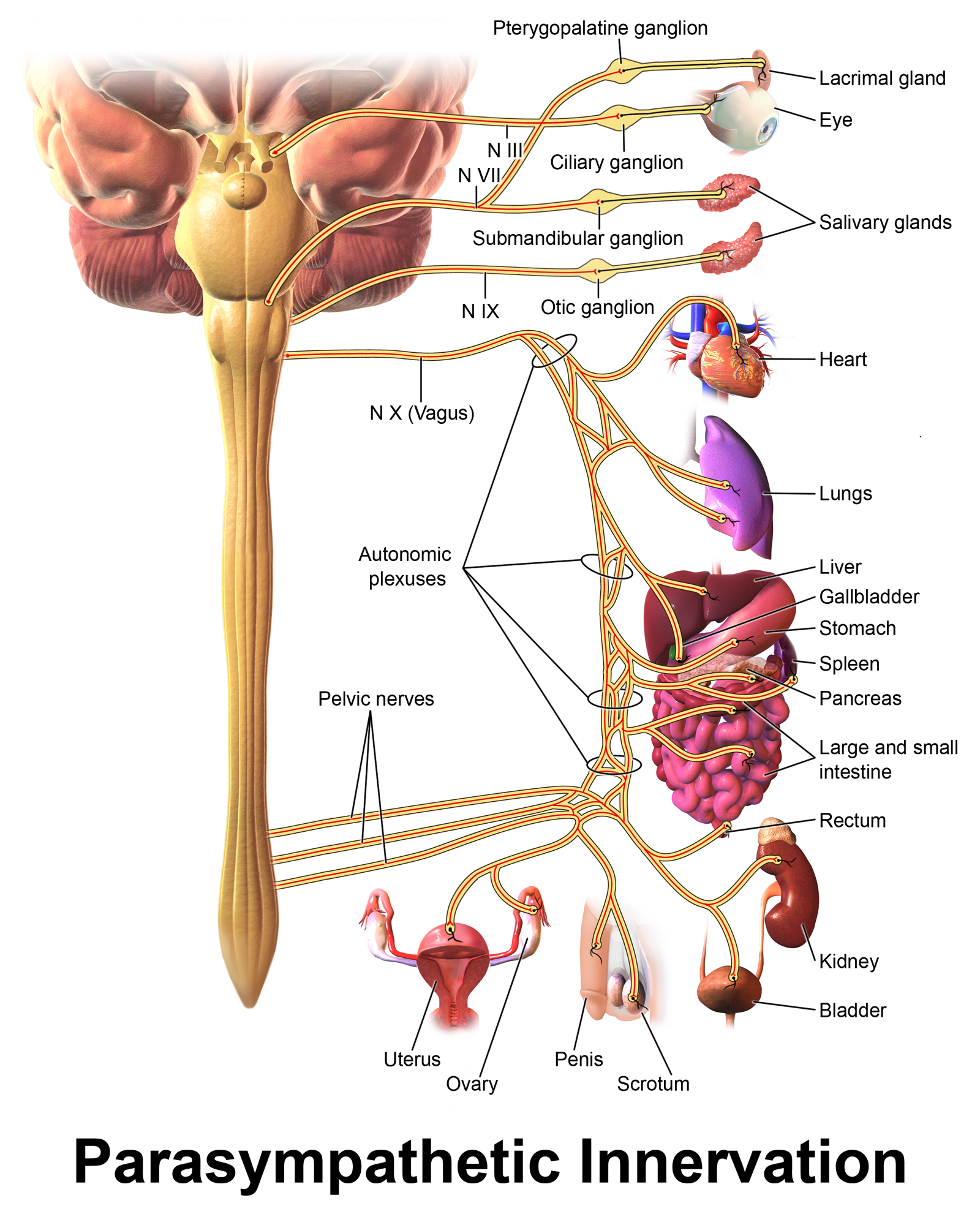
The parasympathetic division of the autonomic nervous system is named because its central neurons are located away from (para- = “apart from”) the thoracolumbar region of the spinal cord which is dedicated to the sympathetic division. Indeed, the parasympathetic system can also be referred to as the craniosacral system because the preganglionic neurons are located in nuclei of the brainstem and the lateral horn of the sacral spinal cord (S2 to S4) (\PageIndex{3}\)).
Parasympathetic Neurons, Ganglia and Nerves
The connections, or “circuits,” of the parasympathetic division are similar to the general layout of the sympathetic division with a few specific differences. The parasympathetic preganglionic fibers from the cranial region travel in cranial nerves, whereas parasympathetic preganglionic fibers from the sacral region travel in spinal nerves. The targets of these fibers are terminal ganglia, which are located near the target effector, and intramural ganglia, which are found within the walls of the target organ. Terminal ganglia receive input from cranial nerves or sacral spinal nerves. The terminal ganglia that receive input from cranial nerves are found in the head and neck, as well as the thoracic and upper abdominal cavities, whereas the terminal ganglia that receive sacral input are in the lower abdominal and pelvic cavities. The postganglionic fiber projects from the terminal ganglia a short distance to the target effector, or to the specific target tissue within the organ.
The cranial nerves associated with the parasympathetic system are the oculomotor nerve (CN III), facial nerve (VII), glossopharyngeal nerve (CN IX) and vagus nerve (CN X). These nerves generate from particular nuclei of the brainstem. Nuclei in the midbrain are part of the oculomotor complex, and parasympathetic axons from those neurons travel in the oculomotor nerve (CN III) with the somatic motor fibers that innervate the extraocular muscles. The parasympathetic preganglionic fibers within the oculomotor nerve terminate in the ciliary ganglion, which is located in the posterior orbit. The postganglionic parasympathetic fibers then project to the sphincter pupillae and ciliary muscle of the iris to control the size of the pupil and the shape of the lens.
Parasympathetic preganglionic fibers exits the pons and travel through the facial nerve (CN VII) to control the secretions of the lacrimal apparatus, nasal epithelium and salivary glands. Two branches exit the facial nerve. The first branch terminates at the pterygopalatine ganglion. Postganglionic fibers from this ganglion extend to the lacrimal gland and glands of the nasal cavity, oral cavity, and palate. The second branch terminates at the submandibular ganglion. Postganglionic fibers from this ganglion project to submandibular and sublingual salivary glands.
Parasympathetic preganglionic fibers exits the medulla oblongata and travel through the glossopharyngeal nerve (CN IX) to the otic ganglion. Postganglionic fibers from this ganglion terminate to the parotid salivary glands.
The majority of parasympathetic preganglionic axons travel through the vagus nerve (CN X) that innervates thoracic and abdominal organs as well as the gonads (ovaries and testes). Autonomic parasympathetic neurons in the medulla oblongata project through the vagus nerve to the terminal and intramural ganglia of target effectors such as heart, airways, esophagus, stomach, liver, gallbladder, pancreas, small and large intestine, kidneys, ureters, and gonads.
The remaining parasympathetic preganglionic axons originate from neurons of the lateral horns of the S2-S4 segments of the spinal cord. These axons form the pelvic splanchnic nerves that project to terminal or intramural ganglia of abdominal and pelvic organs. The main target effectors are the distal portion of the large intestine, rectum, urinary bladder, and most of reproductive organs.
Comparing the relative lengths of axons in the parasympathetic system, the preganglionic fibers are long and the postganglionic fibers are short because the ganglia are close to—and sometimes within—the target effectors. Parasympathetic preganglionic axons tend to have fewer than 4 branches. The lack of divergent branches in parasympathetic preganglionic axons prevents a systemic response and facilitates discrete and localized effects on one group of organs at a time.
Everyday Connections
Fight or Flight? What About Fright and Freeze?
The original usage of the epithet “fight or flight” comes from a scientist named Walter Cannon who worked at Harvard in 1915. The concept of homeostasis and the functioning of the sympathetic system had been introduced in France in the previous century. Cannon expanded the idea, and introduced the idea that an animal responds to a threat by preparing to stand and fight or run away. The nature of this response was thoroughly explained in a book on the physiology of pain, hunger, fear, and rage.
When students learn about the sympathetic system and the fight-or-flight response, they often stop and wonder about other responses. If you were faced with a lioness running toward you as pictured at the beginning of this chapter, would you run or would you stand your ground? Some people would say that they would freeze and not know what to do. So isn’t there really more to what the autonomic system does than fight, flight, rest, or digest. What about fear and paralysis in the face of a threat?
The common epithet of “fight or flight” is being enlarged to be “fight, flight, or fright” or even “fight, flight, fright, or freeze.” Cannon’s original contribution was a catchy phrase to express some of what the nervous system does in response to a threat, but it is incomplete. The sympathetic system is responsible for the physiological responses to emotional states. The name “sympathetic” can be said to mean that (sym- = “together”; -pathos = “pain,” “suffering,” or “emotion”).
Concept Review
The nervous system can be divided into two functional parts: the somatic nervous system (SNS) and the autonomic nervous system (ANS). The differences between these two systems lie on multiple features: the conscious level of their processes, their targets, the number of lower motor neurons involved and the neurotransmitters used. The autonomic nervous system reflexively responds to visceral sensory stimuli, such as levels of carbon dioxide concentration in the blood or stretch caused by blood pressure, that you are not consciously aware of and involuntarily controls cardiac and smooth muscle, as well as glandular tissue. The autonomic nervous system has a chain of two lower autonomic motor neurons. The first neuron is called a preganglionic neuron and resides in the brainstem or lateral horns of the spinal cord. This neuron releases ACh to a second neuron called a ganglionic neuron that is located in ganglia. The axons of ganglionic neurons are called postganglionic fibers. These nerves extend to target effectors and release either ACh or norepinephrine (NE).
The primary responsibilities of the autonomic nervous system are to regulate homeostatic mechanisms in the body. The way we respond to the world around us, to manage the internal environment on the basis of the external environment, is divided between two parts of the autonomic nervous system. The sympathetic division responds to threats and produces a readiness to confront the threat or to run away: the fight-or-flight response. The parasympathetic division plays the opposite role. When the external environment does not present any immediate danger, a restful mode descends on the body, and the digestive system is more active.
The sympathetic output of the nervous system originates out of the lateral horn of the thoracolumbar spinal cord. An axon from one of these central neurons projects by way of the ventral spinal nerve root, spinal nerve and white rami communicantes to a sympathetic chain (paravertebral) ganglion. The preganglionic fibers can synapse on ganglionic neurons here or extend to one of the prevertebral (collateral) ganglia via the splanchnic nerves (splanchnic nerve pathway). Postganglionic fibers of sympathetic chain ganglia can either return to the spinal nerve through the gray rami communicantes (spinal nerve pathway) or extend away from the ganglion through a sympathetic nerve (postganglionic sympathetic pathway). The sympathetic system also has a specialized preganglionic connection to the adrenal medulla that causes epinephrine and norepinephrine to be released into the bloodstream rather than exciting a neuron that contacts an organ directly (adrenal medulla pathway).
The parasympathetic output is based in the brainstem and sacral spinal cord. Neurons from particular nuclei in the brainstem carry parasympathetic information through four cranial nerves: oculomotor nerve (CN III), facial nerve (VII), glossopharyngeal nerve (CN IX) and vagus nerve (CN X). In the sacral spinal cord, preganglionic neurons of the lateral horn project out through pelvic splanchnic nerves. Cranial and sacral preganglionic fibers extend to terminal and intramural ganglia located close to or within the wall of target effectors. The postganglionic fibers of the ganglionic neurons then contact the target tissues within the organ to induce rest-and-digest responses. Due to the fact that parasympathetic ganglia are either close to or within the target organ, parasympathetic preganglionic axons are longer and postganglionic axons are shorter, compared to the sympathetic division.
Review Questions
Q. Which of these physiological changes would not be considered part of the sympathetic fight-or-flight response?
A. increased heart rate
B. increased sweating
C. dilated pupils
D. increased stomach motility
- Answer
-
D
Q. Which type of fiber could be considered the longest?
A. preganglionic parasympathetic
B. preganglionic sympathetic
C. postganglionic parasympathetic
D. postganglionic sympathetic
- Answer
-
A
Q. Which of these cranial nerves contains preganglionic parasympathetic fibers?
A. optic, CN II
B. facial, CN VII
C. trigeminal, CN V
D. hypoglossal, CN XII
- Answer
-
B
Q. Which of the following is not a target of a sympathetic preganglionic fiber?
A. intermural ganglion
B. prevertebral (collateral) ganglion
C. adrenal gland
D. chain ganglion
- Answer
-
A
Critical Thinking Questions
Q. In the context of a lioness hunting on the savannah, why would the sympathetic system not activate the digestive system?
A. Whereas energy is needed for running away from the threat, blood needs to be sent to the skeletal muscles for oxygen supply. The additional fuel, in the form of carbohydrates, probably wouldn’t improve the ability to escape the threat as much as the diversion of oxygen-rich blood would hinder it.
Glossary
- acetylcholine
- neurotransmitter that binds at a motor end-plate to trigger contraction
- adrenal medulla
- interior portion of the adrenal (or suprarenal) gland that releases epinephrine and norepinephrine into the bloodstream as hormones
- adrenal medulla pathway
- sympathetic pathway where preganglionic fibers innervate directly the adrenal medulla
- autonomic ganglion
- localized collection of autonomic neurons in the peripheral nervous system
- autonomic nervous system (ANS)
- functional division of the nervous system that is responsible for homeostatic reflexes that coordinate control of cardiac and smooth muscle, as well as glandular tissue
- celiac ganglion
- one of the prevertebral (collateral) ganglia of the sympathetic system that projects to the digestive system
- chromaffin cells
- neuroendocrine cells of the adrenal medulla that release epinephrine and norepinephrine into the bloodstream as part of sympathetic system activity
- ciliary ganglion
- one of the terminal ganglia of the parasympathetic system, located in the posterior orbit, axons from which project to the iris
- collateral ganglia
- ganglia outside of the sympathetic chain that are targets of sympathetic preganglionic fibers, which are the celiac, inferior mesenteric, and superior mesenteric ganglia; sometimes referred to as prevertebral ganglia
- craniosacral system
- alternate name for the parasympathetic division of the autonomic nervous system that is based on the anatomical location of central neurons in brain-stem nuclei and the lateral horn of the sacral spinal cord; also referred to as craniosacral outflow
- epinephrine
- signaling molecule released from the adrenal medulla into the bloodstream as part of the sympathetic response
- facial nerve (CN VII)
- seventh cranial nerve; responsible for contraction of the facial muscles and for part of the sense of taste, as well as causing saliva production
- fight-or-flight response
- set of responses induced by sympathetic activity that lead to either fleeing a threat or standing up to it, which in the modern world is often associated with anxious feelings
- ganglionic neuron
- specifically refers to the cell body of a neuron in the autonomic system that is located in a ganglion
- glossopharyngeal nerve (CN IX)
- ninth cranial nerve; responsible for contraction of muscles in the tongue and throat and for part of the sense of taste, as well as causing saliva production
- gray rami communicantes
- (singular = ramus communicans) unmyelinated structures that provide a short connection from a sympathetic chain ganglion to the spinal nerve that contains the postganglionic sympathetic fiber
- greater splanchnic nerve
- nerve that contains fibers from T5-T9 central sympathetic neurons that do not synapse in the chain ganglia but project onto the celiac ganglion
- inferior cervical ganglion
- the most inferior of the three paravertebral ganglia of the cervical portion of the sympathetic trunk
- inferior mesenteric ganglion
- one of the collateral ganglia of the sympathetic system that projects to the digestive system
- intramural ganglia
- terminal ganglia of the parasympathetic system that are found within the walls of the target effector
- least splanchnic nerve
- nerve that contains fibers from T12 central sympathetic neurons that do not synapse in the chain ganglia but project onto the renal ganglion
- lesser splanchnic nerve
- nerve that contains fibers from T9-T11 central sympathetic neurons that do not synapse in the chain ganglia but project onto the superior mesenteric ganglion
- lumbar splanchnic nerve
- nerve that contains fibers from L1-L2 central sympathetic neurons that project to the inferior mesenteric ganglion
- middle cervical ganglion
- the middle of the three paravertebral ganglia of the cervical portion of the sympathetic trunk
- norepinephrine
- signaling molecule released as a neurotransmitter by most postganglionic sympathetic fibers as part of the sympathetic response, or as a hormone into the bloodstream from the adrenal medulla
- oculomotor nerve (CN III)
- third cranial nerve; responsible for contraction of four of the extraocular muscles, the muscle in the upper eyelid, and pupillary constriction
- otic ganglion
- one of the terminal ganglia of the parasympathetic system, located anterior to the ear, that innervates the parotid salivary gland for salivation
- parasympathetic division
- division of the autonomic nervous system responsible for restful and digestive functions
- paravertebral ganglia
- autonomic ganglia superior to the sympathetic chain ganglia
- pelvic splanchnic nerves
- nerves that contains fibers from S2-S4 parasympathetic neurons that innervate terminal or intramural ganglia of pelvic organs
- postganglionic fiber
- axon from a ganglionic neuron in the autonomic nervous system that projects to and synapses with the target effector
- postganglionic sympathetic nerve pathway
- connection between a sympathetic preganglionic neuron and its effector through a synapse onto ganglionic neurons at the level of the sympathetic chain ganglion and extension of postganglionic fibers through a sympathetic nerve
- preganglionic fiber
- axon from a central neuron in the autonomic nervous system that projects to and synapses with a ganglionic neuron; sometimes referred to as a preganglionic neuron
- preganglionic neuron
- autonomic central neuron located in the brainstem or lateral horn of the spinal cord
- prevertebral ganglia
- autonomic ganglia that are anterior to the vertebral column and functionally related to the sympathetic chain ganglia; sometimes referred to as collateral ganglia
- pterygopalatine ganglion
- one of the terminal ganglia of the parasympathetic system, located near the junction of the maxilla and palatine bones, that innervates the lacrimal gland and glands of oral and nasal cavities and palate
- rest-and-digest response
- set of functions associated with the parasympathetic system that lead to restful actions and digestion
- sacral splanchnic nerves
- nerves that contain fibers from S1-S2 central sympathetic neurons that do not synapse in the chain ganglia but project to urinary and reproductive organs
- somatic nervous system (SNS)
- functional division of the nervous system that is concerned with conscious perception, voluntary movement, and skeletal muscle reflexes
- spinal nerve pathway
- connection between a sympathetic preganglionic neuron and its effector through a synapse onto ganglionic neurons at the level of the sympathetic chain ganglion and extension of postganglionic fibers through gray rami communicantes and spinal nerves
- splanchnic nerve pathway
- connection between a sympathetic preganglionic neuron and its effector through a synapse onto ganglionic neurons at the level of a prevertebral ganglion and extension of postganglionic fibers to the target effector
- splanchnic nerves
- paired, autonomic nerves that carry sympathetic fibers, except for the pelvic splanchnic nerves which carry parasympathetic fibers.
- submandibular ganglion
- one of the terminal ganglia of the parasympathetic system, located near the angle of the mandible, that innervates the submandibular and sublingual salivary glands for salivation
- superior cervical ganglion
- one of the paravertebral ganglia of the sympathetic system that projects to the head
- superior mesenteric ganglion
- one of the prevertebral (collateral) ganglia of the sympathetic system that projects to the digestive system
- sympathetic chain (trunk) ganglia
- series of ganglia adjacent to the vertebral column that receive input from central sympathetic neurons
- sympathetic division
- division of the autonomic nervous system associated with the fight-or-flight response
- sympathetic trunks
- sympathetic fibers connecting adjacent sympathetic chain ganglia
- target effector
- organ, tissue, or gland that will respond to the control of an autonomic or somatic or endocrine signal
- terminal ganglia
- ganglia of the parasympathetic division of the autonomic system, which are located near or within the target effector, the latter also known as intramural ganglia
- thoracolumbar system
- alternate name for the sympathetic division of the autonomic nervous system that is based on the anatomical location of central neurons in the lateral horn of the thoracic and upper lumbar spinal cord
- vagus nerve (CN X)
- tenth cranial nerve; responsible for the autonomic control of organs in the thoracic and upper abdominal cavities
- white rami communicantes
- (singular = ramus communicans) myelinated structures that provide a short connection from a sympathetic chain ganglion to the spinal nerve that contains the preganglionic sympathetic fiber
Contributors and Attributions
OpenStax Anatomy & Physiology (CC BY 4.0). Access for free at https://openstax.org/books/anatomy-and-physiology