
19.3: Anatomy of Lymphatic Vessels
- Page ID
- 22391

By the end of this section, you will be able to:
- Explain the structure and function of the lymphatic vessels
- Describe the asymmetrical pattern of lymphatic drainage into the bloodstream
A significant portion of the lymphatic system is comprised of a network of lymphatic vessels routed near the blood vessels of the cardiovascular system. The lymphatic vessels function to collect and transport fluid from the tissues of the body back to the bloodstream to help maintain a consistent blood volume. They also function to collect and transport dietary lipids and some lipid-soluble vitamins from the small intestine to the bloodstream and to distribute leukocytes throughout the body for housekeeping and immune defense.
Lymphatic Vessels
The lymphatic vessels begin as open-ended capillaries, which feed into larger and larger lymphatic vessels, and eventually empty into the bloodstream by a series of ducts. The fluid contained within lymphatic vessels is called lymph (lympha means "clear water"), a watery fluid that contains lymphocytes and other types of white blood cells, as well as proteins and lipids.
Blood pressure causes leakage of fluid from the blood capillaries, resulting in the accumulation of fluid in the interstitial space—that is, spaces between individual cells in the tissues. In humans, 20 liters of plasma is released into the interstitial space of the tissues each day due to capillary filtration. Once this filtrate is out of the bloodstream and in the tissue spaces, it is referred to as interstitial fluid. Of this, 17 liters is reabsorbed directly by the blood vessels. But what happens to the remaining three liters? Lymphatic capillaries drain the excess fluid away from the tissues and carry it back to the bloodstream via the network of lymphatic vessels. Lymph is the term used to describe interstitial fluid once it has entered the lymphatic system.
This cycle of fluids from blood plasma to interstitial fluid to lymph aids in oxygen and nutrient delivery to tissues as well as waste and pathogen removal away from tissues: it ensures that the body’s cells are constantly exposed to fresh fluid.
Lymphatic Capillaries
Lymphatic capillaries, also called the terminal lymphatics, are vessels where interstitial fluid enters the lymphatic system to become lymph fluid. Located in almost every tissue in the body, these vessels are typically interlaced among the blood capillaries of the circulatory system in the soft connective tissues of the body (Figure \(\PageIndex{1}\)). Exceptions are the red bone marrow, bones, teeth, and the cornea of the eye, which do not contain lymphatic vessels.

Lymphatic capillaries are formed by one simple layer of endothelial cells and represent the open end of the system, allowing interstitial fluid to flow into them via overlapping endothelial cells that form "flaps" that function like valves (see Figure \(\PageIndex{1}\)). When interstitial fluid pressure is low, the endothelial flaps close to prevent backflow of fluid from the capillary into the tissue. As interstitial pressure increases due to an increase in excess fluid, the spaces between the endothelial cells open up, allowing the fluid to enter the lymphatic capillary. Entry of fluid into lymphatic capillaries is also enabled by the collagen fibers that anchor the capillaries to surrounding structures. As interstitial pressure increases, the fibers pull on the endothelial cell flaps, opening them further to allow easy entry of fluid.
In the small intestine, lymphatic capillaries called lacteals are critical for the transport of dietary lipids and lipid-soluble vitamins to the bloodstream. In the small intestine, dietary triglycerides combine with other lipids and proteins, and enter the lacteals to form a milky fluid called chyle. The chyle then travels through the lymphatic vessels, and drains into the bloodstream.
Larger Lymphatic Vessels, Trunks, and Ducts
Larger lymphatic vessels are similar to veins in terms of their three-tunic structure and the presence of one-way valves to prevent backflow. These one-way valves are located fairly close to one another, and each one causes a bulge in the lymphatic vessel, giving the vessels a beaded appearance (see Figure \(\PageIndex{1}\)). A major distinction between the lymphatic and cardiovascular systems in humans is that lymph is not actively pumped by the heart, but is pulled into the lymphatic capillaries by a pressure gradient (capillary action) and is forced through the vessels by similar mechanisms that help venous blood return to the heart: the contraction of skeletal muscles during body movements and the pressure gradients created by breathing. The one-way valves in lymphatic vessels keep the lymph from flowing backwards as it moves toward its destination.
Lymph is filtered for debris and pathogens in lymph nodes as it drains through lymphatic vessels. Humans have about 500–600 lymph nodes positioned at regular intervals along the length of lymphatic vessels throughout the body and clustered in some regions of the body. Examples of clustered lymph nodes include the axillary nodes near the armpit and the inguinal nodes near the groin (Figure \(\PageIndex{2}\)). Lymph nodes are covered in more detail in the next section.
In general, lymphatic vessels of the subcutaneous tissues of the skin, that is, the superficial lymphatics, follow the same routes as veins, whereas the deep lymphatic vessels of the viscera generally follow the paths of arteries. The superficial and deep lymphatics eventually merge to form larger lymphatic vessels known as lymphatic trunks. There are four pairs of lymphatic trunks, each collecting lymph from a region of either the left or right side of the body, as well as a single intestinal trunk. Lymph from the head and neck drains into the jugular trunks. Lymph from the superficial thorax and the upper limb drains into the subclavian trunks. Lymph from the deep thorax drains into the bronchomediastinal trunks. Lymph from the lower limbs and pelvis drains into the lumbar trunks.
The largest lymphatic vessels that drain into the bloodstream are the lymphatic ducts. The overall drainage of fluid into the bloodstream via the lymphatic ducts is asymmetrical. The right lymphatic duct receives lymph from only the upper right portion of the body via the right jugular, subclavian, and bronchomediastinal trunks. The right lymphatic duct drains into the right subclavian vein lateral to its junction with the right internal jugular vein. The lymph from the rest of the body drains into the thoracic duct via all the remaining lymphatic trunks. The thoracic duct itself begins just beneath the diaphragm in the cisterna chyli, a sac-like chamber that receives lymph from the lower abdomen, pelvis, and lower limbs by way of the left and right lumbar trunks and the intestinal trunk. The left jugular, subclavian, and bronchomediastinal trunks drain into the thoracic duct near its entrance into the left subclavian vein, which is lateral to the junction of the left subclavian vein and left internal jugular vein.
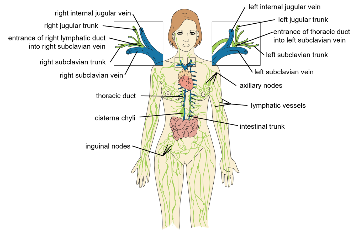 Figure \(\PageIndex{2}\): Overview of Lymphatic Vessels. Lymphatic vessels drain into lymphatic trunks. Lymphatic trunks drain into the right lymphatic duct from the upper portion of the right side of the body and into the larger thoracic duct from the upper portion of the left side of the body and both sides of the lower portion of the body. (Image Credit: “Lymphatic Vessels, Trunks, and Ducts" by Julie Jenks is a derivative of the original work by Daniel Donnelly and is licensed under CC BY 4.0)
DISORDERS OF THE....
Lymphatic System: Lymphedema
Edema is the term for localized swelling in the body, caused by fluid becoming trapped in the tissues. Lymphedema is caused by damage in the vasculature of the lymphatic system, such as by being blocked by cancer cells or destroyed by injury. When the fluid cannot flow properly to drain away from an area and be returned to the bloodstream, protein-rich interstitial fluid accumulates in the tissue spaces upstream of the damage. Figure \(\PageIndex{3}\) compares the degree of visible swelling in the left extremity impacted by each of 4 potential stages of lymphedema severity. Lymphedema may lead to serious medical consequences.
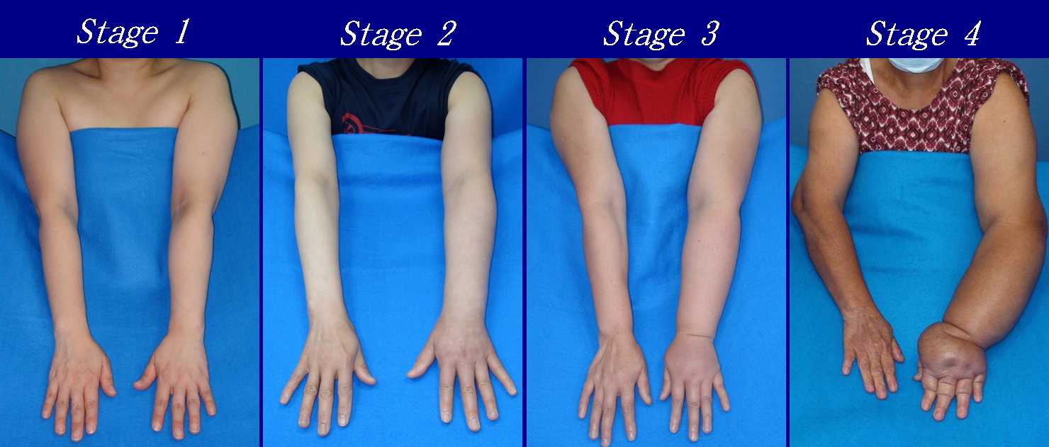 Figure \(\PageIndex{3}\): Lymphedema. When lymphatic vessels are unable to adequately return fluid to the bloodstream, the excess interstitial fluid can build up in a region of the body, typically one extremity. The resulting swelling is called lymphedema, which can be categorized into stages according to its severity. (Image credit: "Upper Limb Lymphedema" by DocHealer is licensed under CC BY-SA 4.0)
RECENT DISCOVERIES IN ANATOMY
Lymphatic Vessels near the Brain
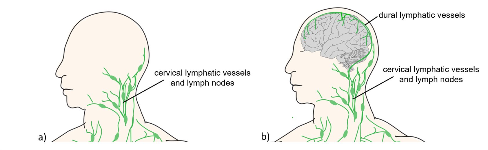
Prior to the 2015 discovery of lymphatic vessels in the dura mater surrounding the brain, maps of lymphatic vessels would depict an unexpected absence of lymphatic vessels around the brain (Figure \(\PageIndex{4}\.a)). We now understand there are lymphatic vessels embedded alongside the venous sinuses in the dura mater meningeal layer surrounding the brain (Figure \(\PageIndex{4}\.b)).
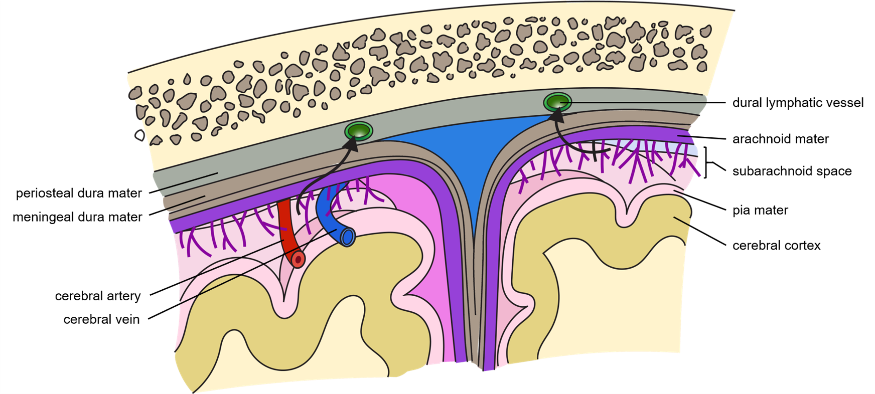
The discovery of dural lymphatic vessels adds to the understanding of several mechanisms involved in draining cerebrospinal fluid (CSF) away from the subarachnoid space around the brain. Cerebrospinal fluid supplies nervous tissue with nutrients and also collects waste products from it, so CSF is constantly secreted into spaces within and surrounding the brain and spinal cord and it is also drained away at the same rate. It has long been understood that cerebrospinal fluid drains into the dural sinuses of the bloodstream. CSF fluid can also drain into dural lymphatic vessels. Yet another mechanism of draining CSF away from the brain was described in 2012. The mechanism was dubbed the glymphatic system because while lymphatic vessels are not found deep to the dura mater around the brain, there is evidence that fluid flows along the surface of blood vessels in perivascular channels formed by astrocytes toward drainage sites (Figure \(\PageIndex{5}\)).
At the time the discovery of the glymphatic mechanism was published in 2012, it was thought that it worked to drain fluid away from the brain in the absence of nearby lymphatic vessels, but now a more dynamic picture illustrates multiple mechanisms for draining fluid away from the very metabolically active brain. The physiology of the glymphatic system mechanism of drainage is still not fully understood.
INTERACTIVE LINK
Watch this video on the role of sleep in the activity of the glymphatic system in draining fluid away from the brain. Keep in mind this video was published in 2014 prior to the discovery of the dural lymphatic vessels. What disease is associated with the accumulation of beta-amyloid proteins in the fluid around the brain caused by inadequate drainage of fluid?
- Answer
-
Inadequate drainage of fluid has been implicated in the accumulation of beta-amyloid proteins that can lead to the development of Alzheimer's disease.
Concept Review
The lymphatic vessels include a network of capillaries, vessels, trunks, and ducts that remove interstitial fluid from the tissues and return it the blood filtered of debris and pathogens by passage through lymph nodes. The lymphatics are also used to transport dietary lipids and distribute cells of the immune system.
Review Questions
Q. Which structure allows lymph from the lower right limb to enter the bloodstream?
A. thoracic duct
B. right lymphatic duct
C. right lymphatic trunk
D. left lymphatic trunk
- Answer
-
Answer: A
Critical Thinking Questions
Q. Describe the flow of lymph from its origins in interstitial fluid somewhere in the left arm to its emptying into the venous bloodstream.
- Answer
-
A. Fluid enters lymphatic capillaries when rising pressure caused by the build-up of interstitial fluid pushes open a flap where adjacent endothelial cells of a lymphatic capillary overlap. The interstitial fluid is then called lymph and it drains into larger lymphatic vessels. The lymph can only go in one direction due to one-way valves in the vessels, and it eventually drains into the left subclavian trunk. The left subclavian trunk merges with the thoracic duct that drains the lymph into the left subclavian vein near the internal jugular vein.
Glossary
- bronchomediastinal trunks
- pair of large lymphatics that collect lymph from smaller lymphatic vessels in the deep thorax
- chyle
- lipid-rich lymph inside the lymphatic capillaries of the small intestine
- cisterna chyli
- bag-like vessel that forms the beginning of the thoracic duct
- intestinal trunk
- large lymphatic that collects lymph from smaller lymphatic vessels in the abdomen
- jugular trunks
- pair of large lymphatics that collect lymph from smaller lymphatic vessels in the head and neck
- lacteal
- lymphatic capillary of the small intestine that collects milky lipid-rich chyle from the small intestine
- lumbar trunks
- pair of large lymphatics that collect lymph from smaller lymphatic vessels in the lower limbs and pelvis
- lymph
- watery fluid in a lymphatic vessel that contains leukocytes, plasma proteins, and lipids
- lymph node
- one of the bean-shaped organs found associated with the lymphatic vessels that function to filter lymph of debris and pathogens
- lymphatic capillaries
- smallest of the lymphatic vessels and the origin of lymph flow
- lymphatic system
- network of lymphatic vessels, lymph nodes, and ducts that carries lymph from the tissues and back to the bloodstream
- lymphatic trunks
- large lymphatics that collect lymph from smaller lymphatic vessels and empties into the blood via lymphatic ducts
- lymphedema
- swelling, typically in one extremity, caused by an accumulation of interstitial fluid in tissues upstream of damaged or blocked lymphatic vessels
- lymphocytes
- white blood cells characterized by a large nucleus and small rim of cytoplasm
- right lymphatic duct
- drains lymph fluid from the upper right side of body into the right subclavian vein
- subclavian trunks
- pair of large lymphatics that collect lymph from the upper limb and superficial thorax
- thoracic duct
- large duct that drains lymph from the lower limbs, left thorax, left upper limb, and the left side of the head
References
Abbott, N.J., Pizzo, M.E., Preston, J.E. et al. The role of brain barriers in fluid movement in the CNS: is there a ‘glymphatic’ system?. Acta Neuropathol 135, 387–407 (2018). [Accessed 17 Apr 2021]
Iliff JJ, Wang M, Liao Y, Plogg BA, Peng W, Gundersen GA, Benveniste H, Vates GE, Deane R, Goldman SA, Nagelhus EA, Nedergaard M. A paravascular pathway facilitates CSF flow through the brain parenchyma and the clearance of interstitial solutes, including amyloid β. Sci Transl Med. 2012 Aug 15;4(147):147ra111. doi: 10.1126/scitranslmed.3003748. PMID: 22896675; PMCID: PMC3551275. [Accessed 18 Jan 2021]
Louveau A, Smirnov I, Keyes TJ, Eccles JD, Rouhani SJ, Peske JD, Derecki NC, Castle D, Mandell JW, Lee KS, Harris TH, Kipnis J. Structural and functional features of central nervous system lymphatic vessels. Nature. 2015 Jul 16;523(7560):337-41. doi: 10.1038/nature14432. Epub 2015 Jun 1. Erratum in: Nature. 2016 May 12;533(7602):278. PMID: 26030524; PMCID: PMC4506234. [Accessed 18 Jan 2021]
Contributors and Attributions
OpenStax Anatomy & Physiology (CC BY 4.0). Access for free at https://openstax.org/books/anatomy-and-physiology