
20.2: Organs and Structures of the Respiratory System
- Page ID
- 22400

By the end of this section, you will be able to:
- List the structures that make up the respiratory system
- Compare and contrast the histology and functions of the conducting zone and the respiratory zone
The major organs of the respiratory system function primarily to provide oxygen to body tissues for cellular respiration, remove the waste product carbon dioxide, and help to maintain acid-base balance. Portions of the respiratory system are also used for non-vital functions, such as sensing odors, speech production, and for straining, such as during childbirth or coughing (Figure \(\PageIndex{1}\)).
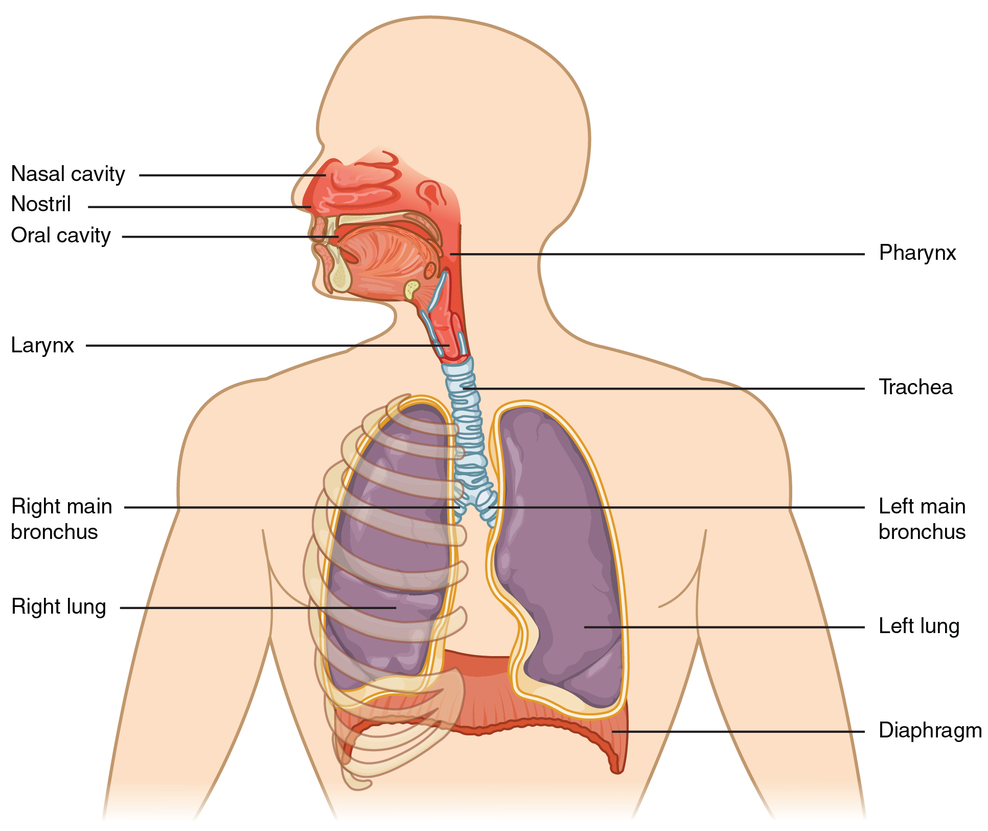
Functionally, the respiratory system can be divided into a conducting zone and a respiratory zone. The conducting zone of the respiratory system includes the organs and structures not directly involved in gas exchange. Gas exchange occurs in the respiratory zone. The conducting zone delivers air to and from the respiratory zone.
Conducting Zone
The major functions of the conducting zone are to provide a route for incoming and outgoing air, remove debris and pathogens from the incoming air, and warm and humidify the incoming air. Several structures within the conducting zone perform other functions as well. The epithelium of the nasal passages, for example, is essential to sensing odors, and the bronchial epithelium that lines the lungs can metabolize some airborne carcinogens.
The Nose and its Adjacent Structures
The primary entrance and exit for the respiratory system is through the nose. When discussing the nose, it is helpful to divide it into two major sections: the external nose, and the nasal cavity or internal nose.
The external nose consists of the surface and skeletal structures that result in the outward appearance of the nose and contribute to its numerous functions (Figure \(\PageIndex{2}\)). The root is the region of the nose located between the eyebrows. The bridge is the part of the nose that connects the root to the rest of the nose. The dorsum nasi is the ridge that runs the length of the nose. The apex is the tip of the nose. The philtrum is the concave surface that connects the apex of the nose to the upper lip.
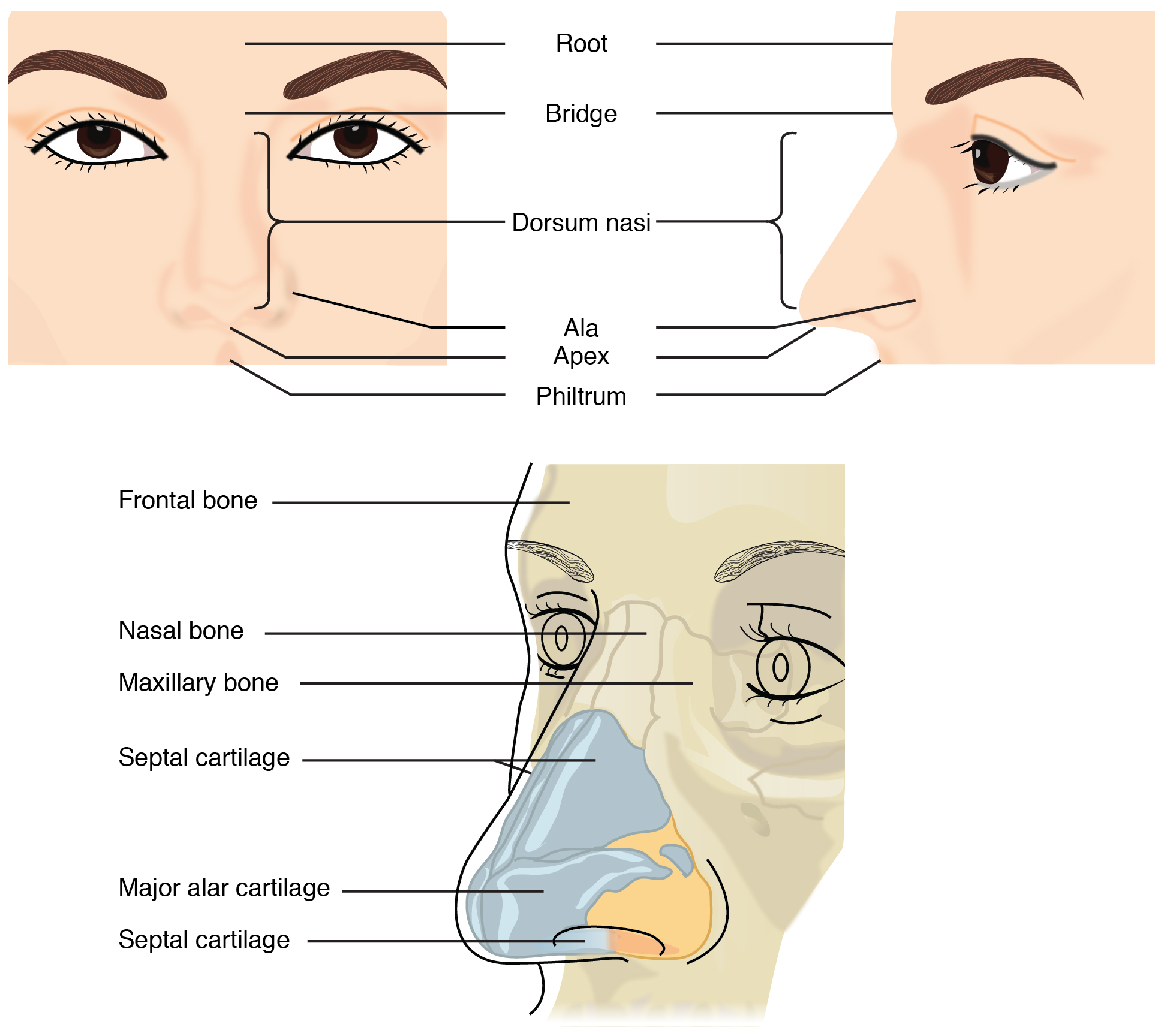
Underneath the thin skin of the nose are its skeletal features (see Figure \(\PageIndex{2}\)). While the root and bridge of the nose consist of bone, most of the protruding portion of the nose is composed of cartilage. As a result, when looking at a skull much of the nose is missing. The nasal bone is one of a pair of bones that lies under the bridge of the nose. The nasal bone articulates superiorly with the frontal bone and laterally with the maxillary bones.
On either side of the apex, the alar cartilages, composed of hyaline cartilage, form the medial portion of the external nares, the narrow openings into each nasal cavity. The alae (singular = ala) form the lateral portion of the external nares and lack cartilage. The external nares open into the nasal cavity, which is separated into left and right sections by the nasal septum. The nasal septum is formed anteriorly by a portion of the septal cartilage, the flexible hyaline cartilage you can move with your fingers that forms the majority of the dorsum nasi, and posteriorly by the perpendicular plate of the ethmoid bone superiorly (a cranial bone located just posterior to the nasal bones) and the thin vomer bone inferiorly (whose name refers to its plough shape).
Each lateral wall of the nasal cavity has three bony projections, called the superior, middle, and inferior nasal conchae (Figure \(\PageIndex{3}\)). The inferior nasal conchae are separate bones, whereas the superior and middle nasal conchae are portions of the ethmoid bone. Several bones that help form the walls of the nasal cavity have hollowed air-containing spaces called the paranasal sinuses. The increased surface area of both the nasal cavities and the sinuses contribute to the richness of the tone of sounds produced, as evidenced by the change in your voice when you plug your nose or are congested. Each paranasal sinus is named for its associated bone: frontal sinus, maxillary sinus, sphenoidal sinus, and ethmoidal sinuses. The hollowed sinuses also lighten the weight of the skull.
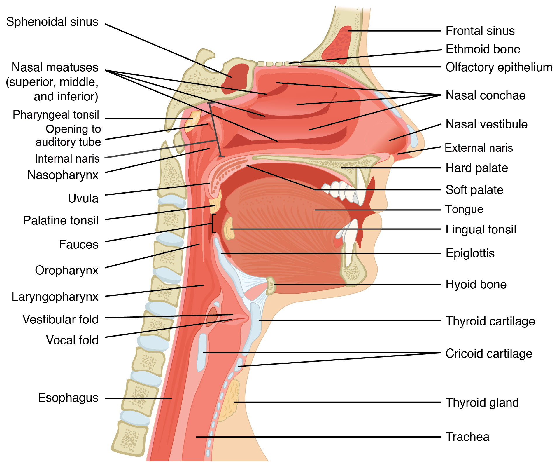 Figure \(\PageIndex{3}\): Upper Airway. A sagittal section of the upper airway and surrounding structures is illustrated. Air passes through the external naris and nasal vestibule as it enters the nasal cavity, which has raised nasal conchae and depressed meatuses that increase its surface area. The nasal cavity narrows at the internal naris that meets the nasopharynx. The frontal and sphenoidal sinus are shown as hollowed out spaces within the frontal and sphenoid bones, respectively. Air then passes by the tonsils as it moves through the nasopharynx above the uvula, the oropharynx posterior to the oral cavity, and the laryngopharynx before entering the larynx superior to the trachea. (Image credit: "Upper Airway" by Julie Jenks is licensed under CC BY 4.0/A derivative from the original work)
The external nares and nasal vestibule at the anterior portion of each nasal cavity are composed of skin: lined with stratified squamous epithelium and contain sebaceous glands and hair follicles embedded in the dermis that serve to prevent the passage of large debris, such as dirt, through the nasal cavity.
The conchae, meatuses, and paranasal sinuses are lined by a mucous membrane or mucosa called the respiratory epithelium composed of ciliated pseudostratified columnar epithelium (Figure \(\PageIndex{4}\) shows the same epithelium lining in the trachea). Mucous spelled with an "o" is the adjective form of the word, while mucus spelled without an "o" is the noun form used to describe the secretion produced by mucous membranes. The respiratory epithelium contains goblet cells, one of the specialized, columnar epithelial cells that produce mucus to trap debris. The cilia of the respiratory epithelium help remove the mucus and debris from the nasal cavity with a constant beating motion, sweeping materials towards the throat to be swallowed. Interestingly, cold air slows the movement of the cilia, resulting in accumulation of mucus that may in turn lead to a runny nose during cold weather. The tissue-covered nasal conchae and meatuses within the nasal cavities create turbulent airflow and provide an increased surface area to aid in filtering, warming, and humidifying air as it enters the body. Capillaries located just beneath the nasal epithelium warm the air by convection. Serous and mucus-producing cells also secrete the lysozyme enzyme and proteins called defensins, which have antibacterial properties. Immune cells that patrol the connective tissue deep to the respiratory epithelium provide additional protection.
A specialized olfactory epithelium used to detect odors (olfaction is the sense of smell) is found at the superior surface of the nasal cavity in the area of the olfactory foramina of the ethmoid bone and is covered in more detail in the chapter that includes the special senses of the nervous system.
The floor of the nasal cavity is composed of the palate. The hard palate at the anterior region of the nasal cavity is composed of mucosa covering bone, while the soft palate at the posterior portion of the nasal cavity consists of mucosa covering muscle tissue. Inhaled air moves deeper into the upper airway when it leaves the nasal cavities via the internal nares, also known as posterior nasal apertures, narrowings at the back of each nasal cavity, and moves into the pharynx.
Pharynx
The pharynx is a tube formed by skeletal muscle and lined by mucous membrane that is continuous with that of the nasal cavities (see Figure \(\PageIndex{3}\)). The pharynx is divided into three major regions: the nasopharynx, the oropharynx, and the laryngopharynx (Figure \(\PageIndex{5}\)).
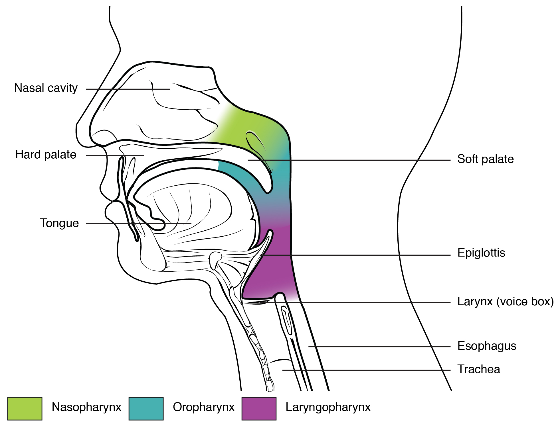
The nasopharynx is posterior to the conchae of the nasal cavity, and it is meant to serve only as an airway. At the top of the nasopharynx is the pharyngeal tonsil, also called the adenoid, an aggregate of lymphoid reticular tissue. The function of the pharyngeal tonsil is not well understood, but it contains a rich supply of lymphocytes and is covered with ciliated epithelium that traps and destroys invading pathogens that enter during inhalation. The pharyngeal tonsil is large in children, but interestingly, tends to regress with age and may even disappear. The uvula is a small bulbous, teardrop-shaped structure located at the apex of the soft palate. Both the uvula and soft palate move like a pendulum during swallowing, swinging upward to close off the nasopharynx to prevent ingested materials from entering the nasal cavity. In addition, auditory (Eustachian) tubes that connect to each middle ear cavity open into the nasopharynx. This connection is why colds may lead to ear infections.
Unlike the nasopharynx which is a passageway for air only, the oropharynx is a passageway for both air and food. The oropharynx is bordered superiorly by the nasopharynx and anteriorly by the oral cavity. The fauces is the opening at the connection between the oral cavity and the oropharynx (see Figure \(\PageIndex{3}\)). As the nasopharynx becomes the oropharynx, the epithelium changes from ciliated pseudostratified columnar epithelium to a stratified squamous epithelium. The oropharynx contains two distinct sets of tonsils, the palatine and lingual tonsils. A palatine tonsil is one of a pair of structures located laterally in the oropharynx in the area of the fauces. The lingual tonsil is located at the base of the tongue. Similar to the pharyngeal tonsil, the palatine and lingual tonsils are composed of lymphoid tissue, and trap and destroy pathogens entering the body through the oral or nasal cavities.
The laryngopharynx is inferior to the oropharynx. It continues the route for ingested material and air until its inferior end, where the digestive and respiratory systems diverge. It is posterior to the open epiglottis, allowing air to move in and out of the larynx. When you swallow ingested material, the epiglottis closes and the material moves into the esophagus. The stratified squamous epithelium of the oropharynx is continuous with the laryngopharynx.
Larynx
The larynx is a cartilaginous structure inferior to the laryngopharynx that connects the pharynx to the trachea and helps regulate the volume of air that enters and leaves the lungs, protects the respiratory passages from ingested materials, and produces sound (Figure \(\PageIndex{6}\)). The structure of the larynx is formed by several pieces of cartilage. With the exception of the epiglottis, the cartilages of the larynx are comprised of hyaline cartilage. Three large cartilage pieces—the thyroid cartilage (anterior), epiglottis (superior), and cricoid cartilage (inferior)—form the major structure of the larynx, which is also known as the voice box. The thyroid cartilage is the largest piece of cartilage that makes up the larynx and functions to form a protective shield across the anterior of the larynx. The thyroid cartilage consists of the laryngeal prominence, or “Adam’s apple,” which tends to be more prominent in males. The thick cricoid cartilage forms a continuous ring around the larynx to hold the airway open as pressure changes during ventilation. It has a taller posterior region and a shorter anterior region inferior to the thyroid cartilage. The cricoid cartilage is connected to the thyroid cartilage by the cricothyroid ligament across the midline anteriorly and is connected to the most superior cartilage of the trachea by the cricotracheal ligament. Three smaller, paired cartilages—the arytenoids, corniculates, and cuneiforms—attach to the epiglottis and the vocal cords as well as the muscle that helps move the vocal cords to produce sound.
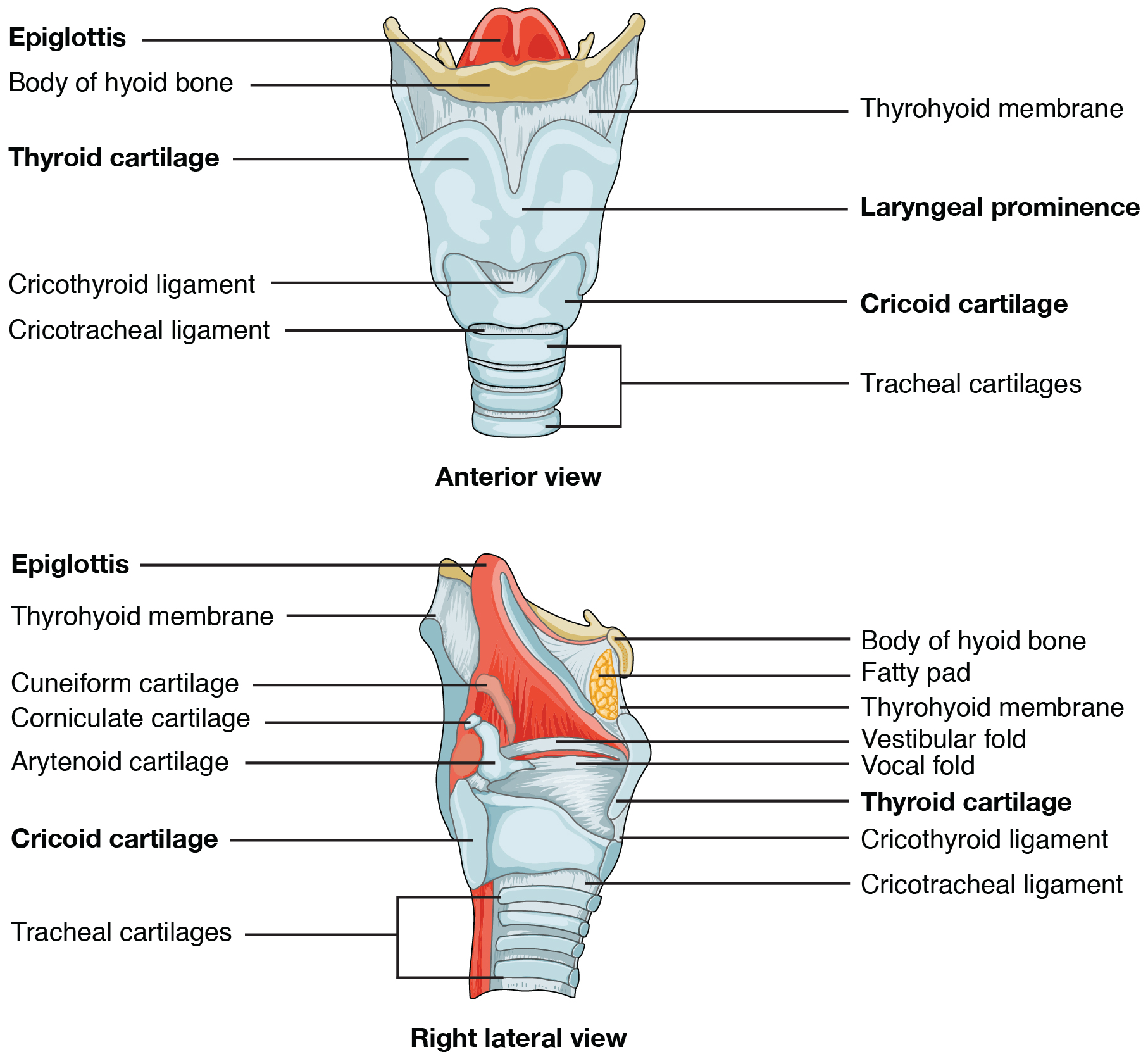
The epiglottis, attached to the thyroid cartilage by a ligament, is a very flexible piece of elastic cartilage that is tethered to the body of the hyoid bone and remains open unless you are swallowing. The hyoid bone is also connected to the thyroid cartilage via the thyrohyoid membrane (see Figure \(\PageIndex{6}\)). A cushioning pad of adipose connective tissue is positioned between the thyrohyoid membrane and the epiglottis. When the hyoid bone moves during swallowing, the pharynx and larynx are lifted upward, allowing the pharynx to expand and the epiglottis to swing downward to cover the glottis. These movements produce a larger area for food to pass through, while preventing ingested food and beverages from entering the trachea.
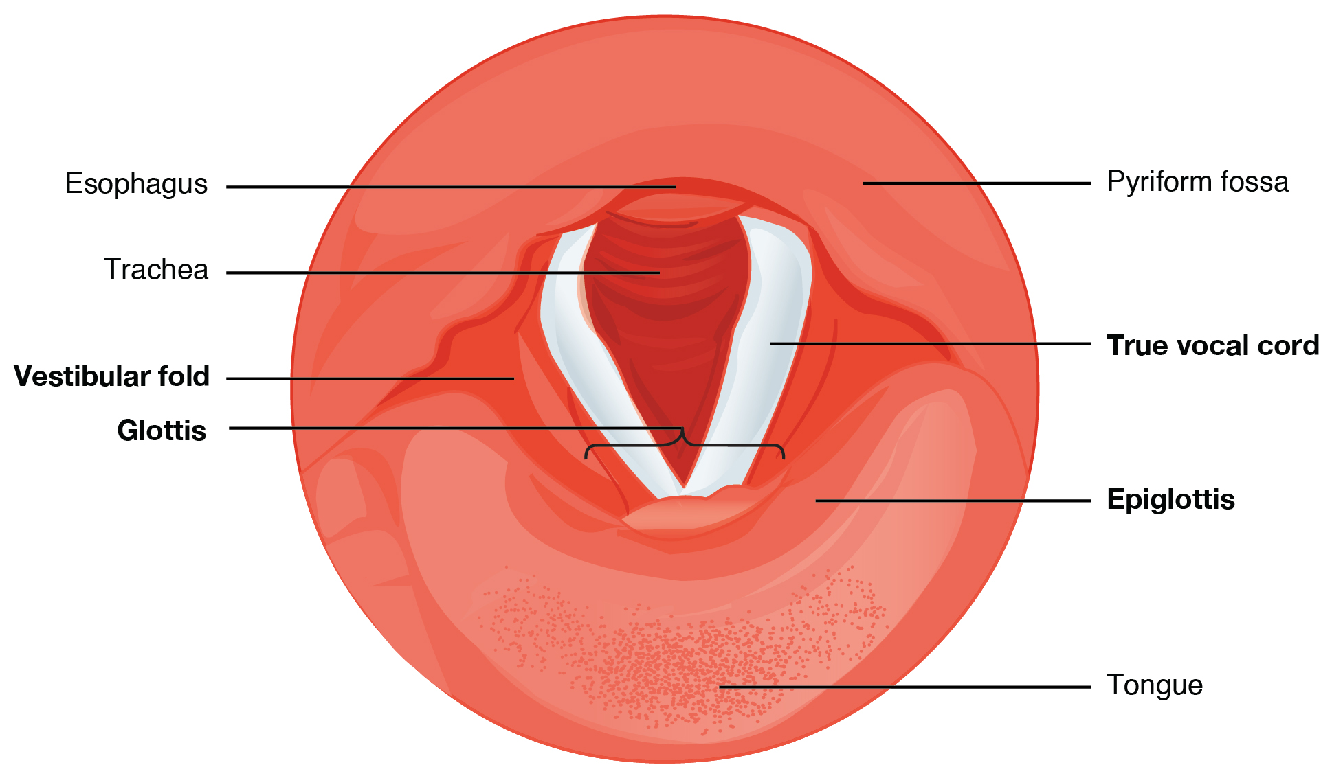
The glottis is composed of the vestibular folds, the true vocal cords, and the space between these folds through which air passes to and from the trachea (Figure \(\PageIndex{7}\)). A vestibular fold, or false vocal cord, is one of a pair of folded sections of mucous membrane that sit superior to the vocal folds and may function to protect the vocal folds and amplify sound. A true vocal cord (or vocal fold) is one of the white, membranous folds attached by muscle to the thyroid and arytenoid cartilages of the larynx on their outer edges. The inner edges of the true vocal cords contain an elastic vocal ligament covered in epithelial tissue that vibrates as air moves across it to produce sound. The size of the membranous folds of the true vocal cords differs between individuals, producing voices with different pitch ranges. Folds in males tend to be larger than those in females, which create a deeper voice. Movements of the muscles attaching the folds to the thyroid and arytenoid cartilages reposition the vocal folds to adjust the pitch of sounds produced.
Continuous with the laryngopharynx, the superior portion of the larynx is lined with stratified squamous epithelium, transitioning into ciliated pseudostratified columnar epithelium that contains goblet cells. Similar to the nasal cavity and nasopharynx, this specialized epithelium produces mucus to trap debris and pathogens as they enter the trachea. Beginning in this region and continuing throughout much of the conducting zone, cilia beat the mucus upward towards the laryngopharynx, where it can be swallowed down the esophagus into the acidic environment of the stomach that functions to kill pathogens trapped in the mucus. The action of the cilia moving the mucus upward to be swallowed is referred to as the mucous escalator.
Trachea
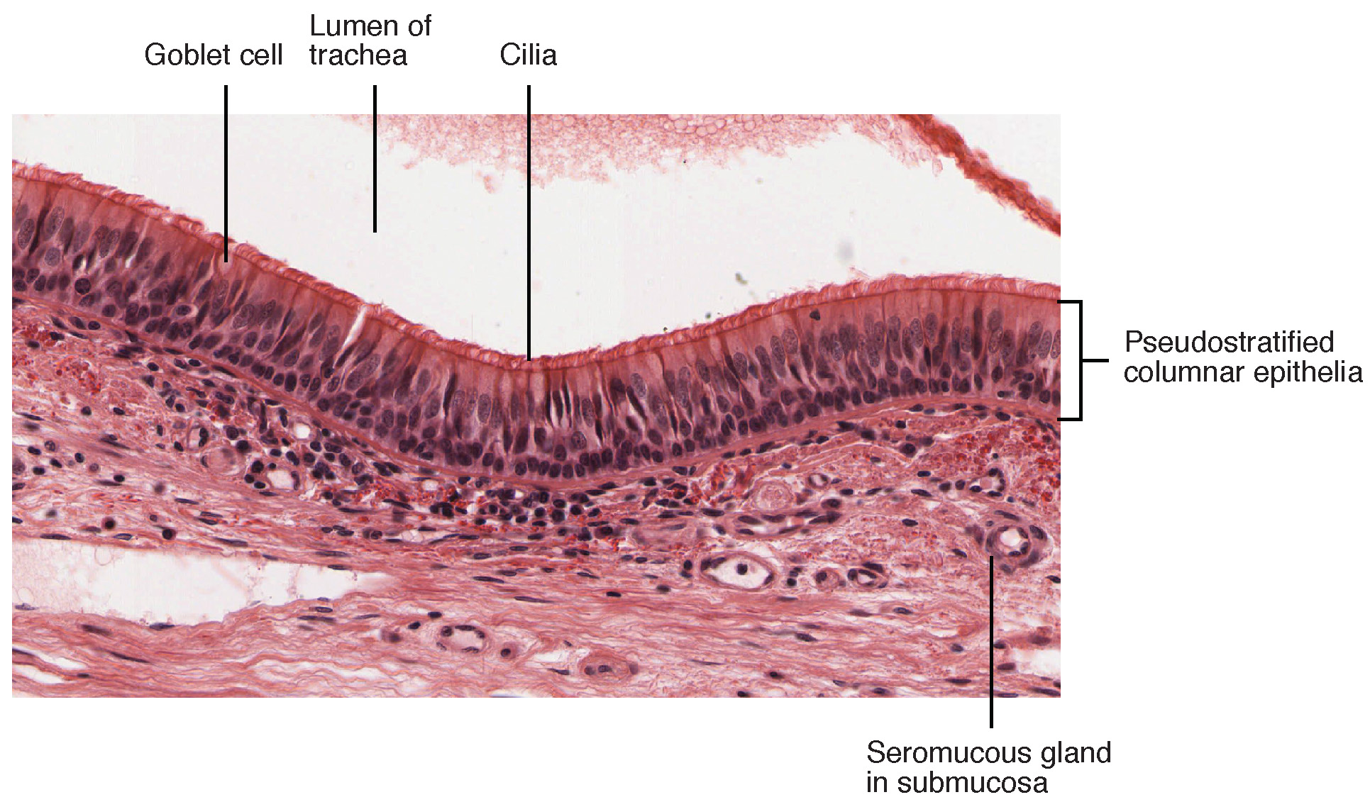
The trachea (windpipe) extends from the larynx toward the lungs (Figure \(\PageIndex{8}\)). The mucosal layer of the wall of the trachea is lined with ciliated pseudostratified columnar epithelium featuring mucus-secreting goblet cells and covering a lamina propria of areolar connective tissue. The submucosa contains dense irregular connective tissue and houses seromucous glands that secrete a lubricating mucus that shares properties with serous fluid. The wall of the trachea contains 16 to 20 stacked, C-shaped pieces of hyaline cartilage that are positioned horizontally with the opening in the C at the back of the trachea. The incomplete rings of cartilage provide structural support that prevent the trachea from collapsing and protect it. The cartilages are connected to one another by dense connective tissue. The fibroelastic membrane consists of the trachealis muscle, made of smooth muscle, and elastic connective tissue. It is a flexible membrane that spans the gap across the the C-shaped cartilages at the posterior of the trachea, allowing the trachea to stretch and expand slightly during inhalation and exhalation. Its flexibility also allows the trachea to accommodate ingested materials passing through the esophagus that borders the trachea posteriorly. In addition, the smooth muscle of the trachealis muscle can be contracted to force air through the trachea during exhalation. The superficial wrapping of the trachea is an adventitia of dense irregular connective tissue.
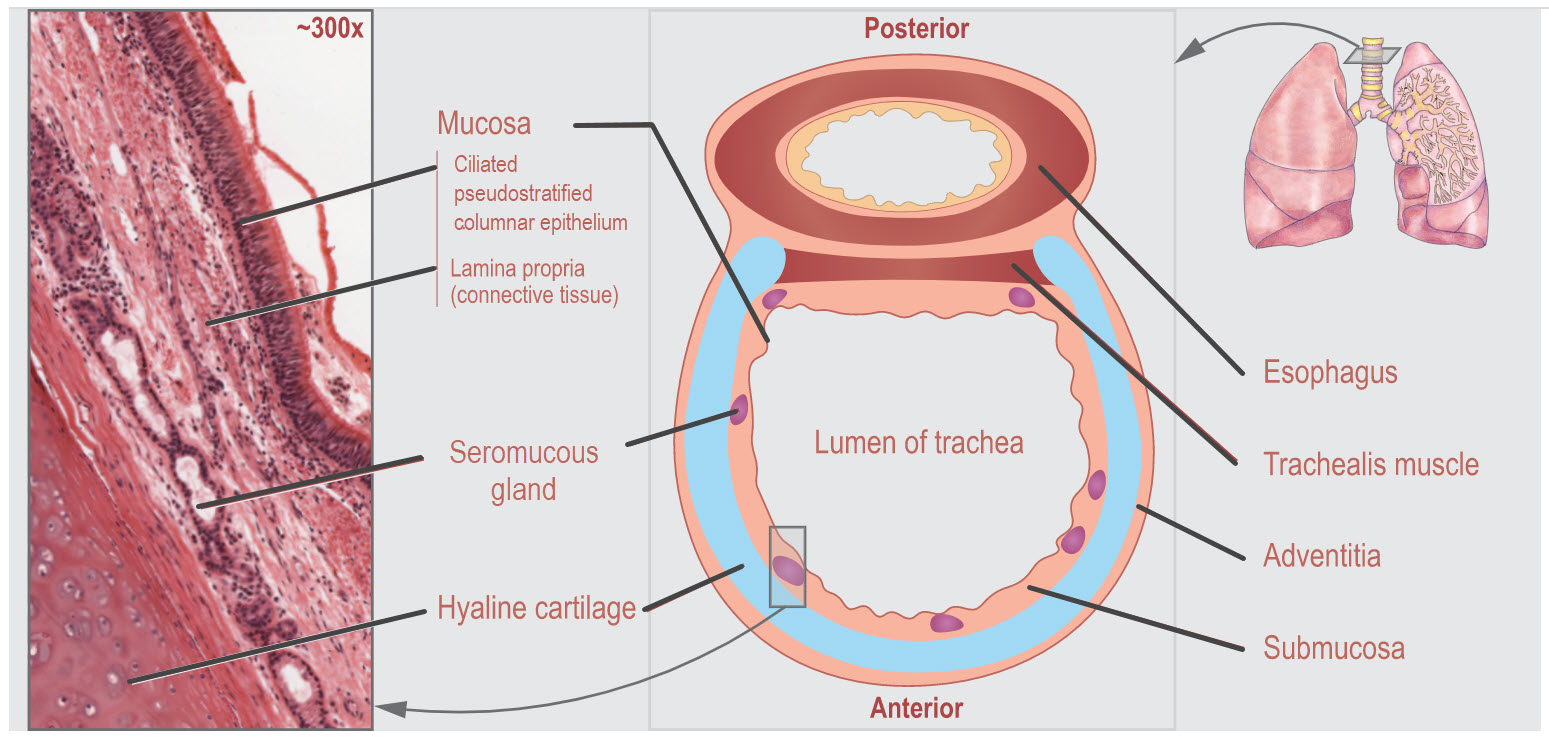 Figure \(\PageIndex{8}\): Trachea. The mucosa lines the hollow lumen of the trachea and is comprised of ciliated pseudostratified columnar epithelium that contains goblet cells covering a lamina propria of connective tissue. The submucosa houses seromucous glands. The C-shaped rings of hyaline cartilage are positioned deep to the mucosa with the opening at the posterior, which is spanned by the trachealis muscle and connective tissue. The superficial layer of the trachea is adventitia. The esophagus is positioned posterior to the trachea. LM × 1220. (Image credit: "Trachea" by Julie Jenks is licensed under CC BY 4.0/A derivative from the original work/Micrograph provided by the Regents of University of Michigan Medical School © 2012)
Bronchial Tree
The trachea branches into the right and left primary (main) bronchi (singular = bronchus) at the carina. The carina is a raised structure that contains specialized nervous tissue that induces violent coughing if a foreign body, such as food, is present. These bronchi are also lined by ciliated pseudostratified columnar epithelium containing mucus-producing goblet cells (Figure \(\PageIndex{9}\)). Rings of cartilage, similar to those of the trachea, support the structure of the bronchi and prevent their collapse. The primary bronchi branch to the secondary (lobar) bronchi that deliver air to the individual lobes of each lung. The secondary bronchi branch to the tertiary (segmental) bronchi that deliver air to the bronchopulmonary segments that comprise each lobe. The lobes and bronchopulmonary segments of the lungs will be covered in more detail in the next section.
The bronchial tree (or respiratory tree) is the collective term used for these highly branching bronchi, since they resemble branches of an upside-down tree. The main function of the bronchi, like other conducting zone structures, is to provide a passageway for air to move into and out of each lung. In addition, the mucous membrane traps debris and pathogens, cilia continue to work as a mucous escalator moving mucus up toward the laryngopharynx to be swallowed. Hyaline cartilage is featured within the wall of all the bronchi to reinforce the airways and help them stay open through pressure changes, but its quantity decreases with each branch point. The C-shaped rings of cartilage in the trachea become irregular plates of cartilage in the bronchi that are smaller and more sporadic in the tertiary bronchi than in the primary bronchi. A layer of smooth muscle is present in the bronchi that may bronchoconstrict, make the lumen diameter smaller by contracting the smooth muscle, or bronchodilate, make the lumen diameter larger by relaxing the smooth muscle, to adjust air flow in each passage. The quantity of smooth muscle in the wall relative to the lumen diameter increases with each branch point, meaning that the capability to adjust air flow is greater in the tertiary bronchi than in the primary bronchi.
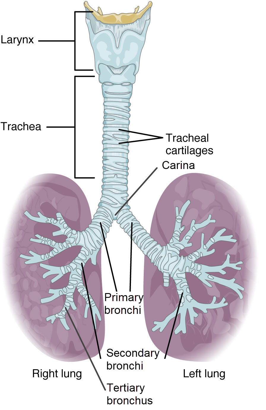 Figure \(\PageIndex{9}\): Bronchial Tree. The tracheal tube is formed by stacked, C-shaped pieces of hyaline cartilage. The trachea branches at the carina to the primary bronchi that deliver air to each lung. The primary bronchi branch into the secondary bronchi that deliver air to the lobes of each lung. The secondary bronchi branch into the tertiary bronchi that deliver air into the bronchopulmonary segments of each lobe. (Image credit: "Bronchial Tree" by Julie Jenks is licensed under CC BY 4.0 / A derivative from the original work)
Bronchi continue to branch from the tertiary bronchi, getting smaller and smaller. Bronchioles, which are 1 mm in diameter or less, further branch until they become the tiny terminal bronchioles, which lead to the structures of gas exchange. There are approximately 30,000 terminal bronchioles in each lung. As they branch, the epithelium of the bronchioles transitions from ciliated pseudostratified columnar epithelium to a thinner simple columnar epithelium to an even thinner simple cuboidal epithelium in the terminal bronchioles. Cilia and mucus-producing cells are sporadic in the largest bronchioles and then disappear as the bronchioles get smaller. The walls of the bronchioles do not contain cartilage like those of the bronchi, but they feature a significant layer of smooth muscle to change their diameter to adjust air flow.
Respiratory Zone
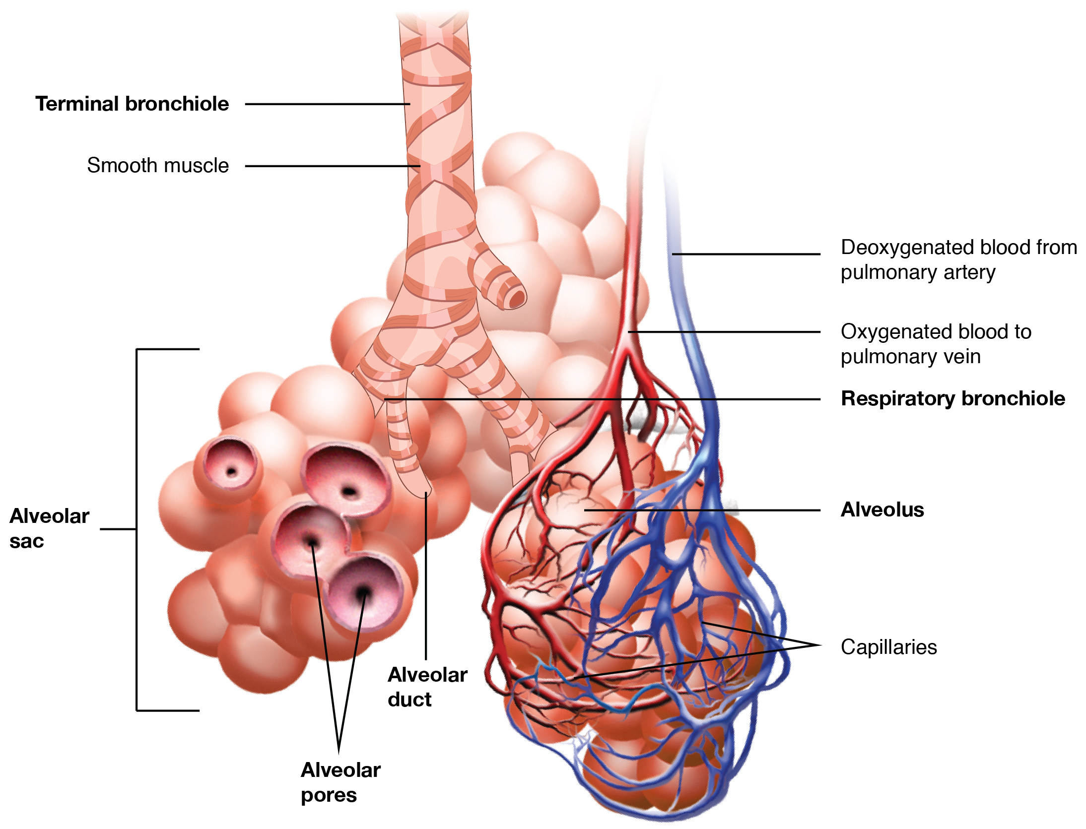
In contrast to the conducting zone, the respiratory zone includes structures that are directly involved in gas exchange. The respiratory zone begins where the terminal bronchioles join a respiratory bronchiole, the smallest type of bronchiole (Figure \(\PageIndex{10}\)), which then leads to an alveolar duct that opens into a cluster of alveoli. Respiratory bronchioles are lined with simple cuboidal epithelium.
Alveoli
An alveolar duct is a tube whose wall contains smooth muscle and connective tissue and is lined with simple cuboidal epithelium. Each alveolar duct opens into a cluster of alveoli. An alveolus is one of the many small, grape-like sacs that are attached to the alveolar ducts. There are an average of about 480 million alveoli in the lungs, yielding an incredibly large surface area for gas exchange.
An alveolar sac is a cluster of many individual alveoli that are responsible for gas exchange. Each alveolar sac is surrounded by a capillary bed with a network of capillaries that distribute several capillaries to route along the outside of each alveolus in the alveolar sac (Figure \(\PageIndex{11}\)). Deoxygenated blood is delivered to the capillary bed via an arteriole branching from the pulmonary artery and oxygenated blood is collected by a venule that drains to the pulmonary vein. An alveolus is approximately 200 μm in diameter with elastic walls that allow the alveolus to stretch during air intake like a balloon inflating, which greatly increases the surface area available for gas exchange, and then return to a smaller size to assist with expiration. Alveoli are connected to their neighbors by alveolar pores, which help maintain equal air pressure throughout the alveoli and lung.
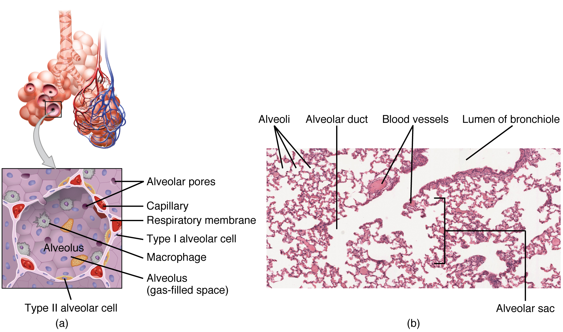
The simple squamous epithelium of the alveolar wall consists of three major cell types: type I alveolar cells, type II alveolar cells, and alveolar macrophages (see Figure \(\PageIndex{11}\)). Type I alveolar cells (aka type I pneumocytes or squamous alveolar cells) are squamous epithelial cells of the alveoli, which constitute up to 97 percent of the alveolar surface area. These cells are about 25 nm (there are a thousand nanometers in a millimeter!) thick and are highly permeable to gases. Type II alveolar cells (aka type II pneumocytes or great alveolar cells) are interspersed among the type I cells and secrete pulmonary surfactant, a substance composed of phospholipids and proteins that reduces the surface tension of the alveoli. Like tiny balloons, the alveoli expand when they fill with air during an inhale and deflate during an exhale. The reduced surface tension from the surfactant prevents the walls of each alveolus from sticking closed during and after exhaling. Type II alveolar cells also play an important role in initiating repair of an alveolus is damaged. Roaming around the alveolar wall is the alveolar macrophage, a phagocytic cell of the immune system that removes debris and pathogens that have reached the alveoli.
The simple squamous epithelium formed by type I alveolar cells is attached to a thin, elastic basement membrane. This epithelium is extremely thin and borders the endothelial membrane of capillaries. Taken together, the alveoli and capillary membranes form a respiratory membrane that is approximately 0.5 mm thick. The respiratory membrane allows gases to cross by simple diffusion, allowing oxygen to be picked up by the blood for transport and CO2 to be released into the air of the alveoli to be excreted during the exhale.
DISORDERS OF THE...
Respiratory System: Asthma
Asthma is common condition that affects the lungs in both adults and children. Approximately 8.2 percent of adults (18.7 million) and 9.4 percent of children (7 million) in the United States suffer from asthma. In addition, asthma is the most frequent cause of hospitalization in children.
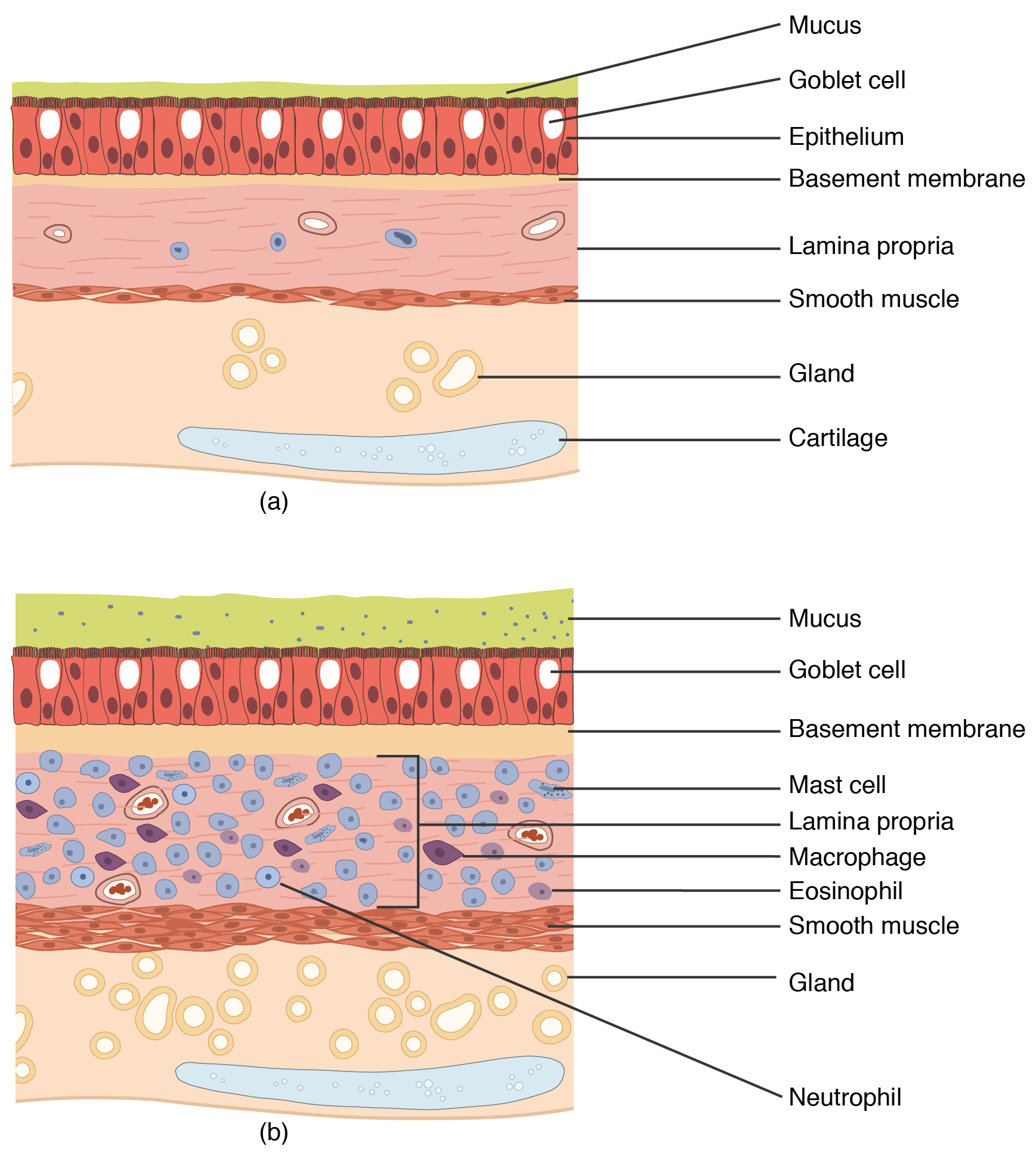
Asthma is a chronic disease characterized by inflammation and edema of the airway, and bronchospasms (that is, constriction of the bronchioles), which can inhibit air from entering the lungs. In addition, excessive mucus secretion can occur, which further contributes to airway occlusion (Figure \(\PageIndex{12}\)). Cells of the immune system, such as eosinophils and macrophages, may also be involved in infiltrating the walls of the bronchi and bronchioles.
Bronchospasms occur periodically and lead to an “asthma attack.” An attack may be triggered by environmental factors such as dust, pollen, pet hair, or dander, changes in the weather, mold, tobacco smoke, and respiratory infections, or by exercise and stress.
Symptoms of an asthma attack involve coughing, shortness of breath, wheezing, and tightness of the chest. Symptoms of a severe asthma attack that requires immediate medical attention would include difficulty breathing that results in blue (cyanotic) lips or face, confusion, drowsiness, a rapid pulse, sweating, and severe anxiety. The severity of the condition, frequency of attacks, and identified triggers influence the type of medication that an individual may require. Longer-term treatments are used for those with more severe asthma. Short-term, fast-acting drugs that are used to treat an asthma attack are typically administered via an inhaler. For young children or individuals who have difficulty using an inhaler, asthma medications can be administered via a nebulizer.
In many cases, the underlying cause of the condition is unknown. However, recent research has demonstrated that certain viruses, such as human rhinovirus C (HRVC), and the bacteria Mycoplasma pneumoniae and Chlamydia pneumoniae that are contracted in infancy or early childhood, may contribute to the development of many cases of asthma.
Concept Review
The respiratory system is responsible for obtaining oxygen and getting rid of carbon dioxide, and aiding in speech production and in sensing odors. From a functional perspective, the respiratory system can be divided into two major areas: the conducting zone and the respiratory zone. The conducting zone consists of all of the structures that provide passageways for air to travel into and out of the lungs: the nasal cavity, pharynx, larynx, trachea, bronchi, and most bronchioles. The nasal passages contain the conchae and meatuses that expand the surface area of the cavity, which helps to warm and humidify incoming air, while removing debris and pathogens. The pharynx is composed of three major sections: the nasopharynx, which is continuous with the nasal cavity; the oropharynx, which borders the nasopharynx and the oral cavity; and the laryngopharynx, which connects the oropharynx to the larynx and esophagus.
The larynx is a cartilaginous structure involved in passing air to and from the trachea. The epiglottis closes during swallowing to prevent ingested materials from passing to the trachea. The true vocal cords within the glottis of the larynx function in the production of sound. The trachea delivers air to and from the bronchial tree, a pathway of branching tubes with several levels of bronchi and tiny bronchioles that conduct air to the respiratory zone. The respiratory zone includes the structures of the lung that are directly involved in gas exchange: the respiratory bronchioles and alveoli. The lining of the conducting zone is composed mostly of ciliated pseudostratified columnar epithelium with goblet cells. The mucus secreted by goblet cells traps pathogens and debris, whereas beating cilia move the mucus superiorly via the mucous escalator toward the throat, where it is swallowed. As the bronchioles become smaller and smaller, and nearer the alveoli, the epithelium thins and is simple squamous epithelium in the alveoli. The endothelium of the surrounding capillaries, together with the alveolar epithelium, forms the respiratory membrane. This is a blood-air barrier through which gas exchange occurs by simple diffusion.
Review Questions
Q. Which of the following anatomical structures is not part of the conducting zone?
A. pharynx
B. nasal cavity
C. alveoli
D. bronchi
- Answer
-
C
Q. What is the function of the conchae in the nasal cavity?
A. increase surface area
B. exchange gases
C. maintain surface tension
D. maintain air pressure
- Answer
-
A
Q. The fauces connects which of the following structures to the oropharynx?
A. nasopharynx
B. laryngopharynx
C. nasal cavity
D. oral cavity
- Answer
-
D
Q. Which of the following are structural features of the trachea?
A. C-shaped cartilage
B. smooth muscle fibers
C. cilia
D. all of the above
- Answer
-
D
Q. Which of the following structures is not part of the conducting zone?
A. trachea
B. bronchi
C. terminal bronchioles
D. respiratory bronchioles
- Answer
-
D
Q. What is the role of alveolar macrophages?
A. to secrete pulmonary surfactant
B. to secrete antimicrobial proteins
C. to remove pathogens and debris
D. to facilitate gas exchange
- Answer
-
C
Critical Thinking Questions
Q. Describe the three regions of the pharynx and their functions.
- Answer
-
The pharynx has three major regions. The first region is the nasopharynx, which is connected to the posterior nasal cavity. The second region is the oropharynx, which is continuous with the nasopharynx and is connected to the oral cavity at the fauces. The laryngopharynx is connected to the oropharynx and the esophagus and trachea. Both the oropharynx and laryngopharynx are passageways for air and food and drink, while the nasopharynx is designed only for air. The epithelium of the nasopharynx is a ciliated pseudostratified columnar epithelium with mucus-secreting goblet cells to help trap debris and pathogens from air, whereas the oropharynx and laryngopharynx are lined with a stratified squamous epithelium for extra protection from abrasion and exposures related to swallowing ingested food and drink.
Q. If a person sustains an injury to the epiglottis, what would be the functional consequence?
- Answer
-
The epiglottis is a region of the larynx that is important during the swallowing of food or drink. As a person swallows, the pharynx moves upward and the epiglottis closes over the trachea, preventing food or drink from entering the trachea. If a person’s epiglottis were injured, this mechanism would be impaired. As a result, the person may have problems with food or drink entering the trachea, and possibly, the lungs. Over time, this may cause infections such as pneumonia to set in.
Q. Compare and contrast the conducting and respiratory zones.
- Answer
-
The conducting zone of the respiratory system includes the organs and structures that are not directly involved in gas exchange, but perform other duties such as providing a passageway for air, trapping and removing debris and pathogens, and warming and humidifying incoming air. Such structures include the nasal cavity, pharynx, larynx, trachea, and most of the bronchial tree. The respiratory zone includes all the organs and structures that are directly involved in gas exchange, including the respiratory bronchioles, alveolar ducts, and alveoli.
References
Bizzintino J, Lee WM, Laing IA, Vang F, Pappas T, Zhang G, Martin AC, Khoo SK, Cox DW, Geelhoed GC, et al. Association between human rhinovirus C and severity of acute asthma in children. Eur Respir J [Internet]. 2011 ; 37(5):1037–1042. [cited 2013 Mar 22]
Kumar V, Ramzi S, Robbins SL. Robbins. Basic Pathology. 7th ed. Philadelphia (PA): Elsevier Ltd; 2005.
Martin RJ, Kraft M, Chu HW, Berns, EA, Cassell GH. A link between chronic asthma and chronic infection. J Allergy Clin Immunol [Internet]. 2001; 107(4):595-601. [cited 2013 Mar 22]
Ochs, Matthias, et al. “The Number of Alveoli in the Human Lung.” American Journal of Respiratory and Critical Care Medicine, vol. 169, no. 1, 2004, pp. 120–124., doi:10.1164/rccm.200308-1107oc. [Accessed 26 Apr 2021].
Glossary
- ala
- (plural = alae) small, flaring structure of a nostril that forms the lateral side of the external naris
- alar cartilage
- cartilage that supports the apex of the nose and helps shape the nares; it is connected to the septal cartilage and connective tissue of the alae
- alveolar duct
- small tube that leads from the terminal bronchiole to the respiratory bronchiole and is the point of attachment for alveoli
- alveolar macrophage
- immune system cell of the alveolus that removes debris and pathogens
- alveolar pore
- opening that allows airflow between neighboring alveoli
- alveolar sac
- cluster of alveoli
- alveolus
- small, grape-like sac that performs gas exchange in the lungs
- apex
- tip of the external nose
- bronchial tree
- collective name for the multiple branches of the bronchi and bronchioles of the respiratory system
- bridge
- portion of the external nose that lies in the area of the nasal bones
- bronchiole
- branch of bronchi that are 1 mm or less in diameter and terminate at alveolar sacs
- bronchoconstriction
- contraction of the smooth muscle in the wall of a bronchus or bronchiole to reduce the volume of the lumen and adjust air flow
- bronchodilation
- relaxation of the smooth muscle in the wall of a bronchus or bronchiole to increase the volume of the lumen and adjust air flow
- bronchus
- tube connected to the trachea that branches into many subsidiaries and provides a passageway for air to enter and leave the lungs
- carina
- raised structure at the divergence of the left and right main bronchi from the trachea that induces coughing if a foreign body (such as food) is detected
- conducting zone
- region of the respiratory system that includes the organs and structures that provide passageways for air and are not directly involved in gas exchange
- cricoid cartilage
- portion of the larynx composed of a ring of cartilage with a wide posterior region and a thinner anterior region; attached to the esophagus
- dorsum nasi
- intermediate portion of the external nose that connects the bridge to the apex and is supported by the nasal bone
- epiglottis
- leaf-shaped piece of elastic cartilage that is a portion of the larynx that swings to close the trachea during swallowing
- external naris
- (plural = external nares) opening of the nostril to the nasal cavity
- external nose
- region of the nose that is easily visible to others
- fauces
- portion of the posterior oral cavity that connects the oral cavity to the oropharynx
- fibroelastic membrane
- specialized membrane that connects the ends of the C-shape cartilage in the trachea; contains smooth muscle fibers
- glottis
- opening between the vocal folds through which air passes when producing speech
- laryngeal prominence
- region where the two lamina of the thyroid cartilage join, forming a protrusion known as “Adam’s apple”
- laryngopharynx
- portion of the pharynx bordered by the oropharynx superiorly and esophagus and trachea inferiorly; serves as a route for both air and food
- larynx
- cartilaginous structure that produces the voice, prevents food and beverages from entering the trachea, and regulates the volume of air that enters and leaves the lungs
- lingual tonsil
- lymphoid tissue located at the base of the tongue
- meatus
- one of three recesses (superior, middle, and inferior) in the nasal cavity attached to the conchae that increase the surface area of the nasal cavity
- nasal bone
- bone of the skull that lies under the root and bridge of the nose and is connected to the frontal and maxillary bones
- nasal septum
- wall composed of bone and cartilage that separates the left and right nasal cavities
- nasopharynx
- portion of the pharynx flanked by the conchae and oropharynx that serves as an airway
- oropharynx
- portion of the pharynx flanked by the nasopharynx, oral cavity, and laryngopharynx that is a passageway for both air and food
- palatine tonsil
- one of the paired structures composed of lymphoid tissue located anterior to the uvula at the roof of isthmus of the fauces
- paranasal sinus
- one of the cavities within the skull that is connected to the conchae that serve to warm and humidify incoming air, produce mucus, and lighten the weight of the skull; consists of frontal, maxillary, sphenoidal, and ethmoidal sinuses
- pharyngeal tonsil
- structure composed of lymphoid tissue located in the nasopharynx; also known as the adenoid
- pharynx
- region of the conducting zone that forms a tube of skeletal muscle lined with respiratory epithelium; located between the nasal conchae and the esophagus and trachea
- philtrum
- concave surface of the face that connects the apex of the nose to the top lip
- primary bronchus (main bronchus)
- passageway of the conducting zone branching directly off the trachea that delivers air to each lung
- pulmonary surfactant
- substance composed of phospholipids and proteins that reduces the surface tension of the alveoli; made by type II alveolar cells
- respiratory bronchiole
- specific type of bronchiole that leads to alveolar sacs
- respiratory epithelium
- ciliated lining of much of the conducting zone that is specialized to remove debris and pathogens, and produce mucus
- respiratory membrane
- alveolar and capillary wall together, which form an air-blood barrier that facilitates the simple diffusion of gases
- respiratory zone
- includes structures of the respiratory system that are directly involved in gas exchange
- root
- region of the external nose between the eyebrows
- secondary bronchus (lobar bronchus)
- passageway of the conducting zone branching directly off the primary bronchi that delivers air to a lobe of either lung
- septal cartilage
- flexible hyaline cartilage that forms the anterior portion of the nasal septum
- tertiary bronchus (segmental bronchus)
- passageway of the conducting zone branching directly off the secondary bronchi that delivers air to a bronchopulmonary segment within a lobe of either lung
- thyroid cartilage
- largest piece of cartilage that makes up the larynx and consists of two lamina
- trachea
- tube composed of cartilaginous rings and supporting tissue that connects the lung bronchi and the larynx; provides a route for air to enter and exit the lung
- trachealis muscle
- smooth muscle located in the fibroelastic membrane of the trachea
- true vocal cord
- one of the pair of folded, white membranes that have a free inner edge that oscillates as air passes through to produce sound
- type I alveolar cell
- squamous epithelial cells that are the major cell type in the alveolar wall; highly permeable to gases
- type II alveolar cell
- cuboidal epithelial cells that are the minor cell type in the alveolar wall; secrete pulmonary surfactant
- vestibular fold
- part of the folded region of the glottis composed of mucous membrane; supports the epiglottis during swallowing
Contributors and Attributions
OpenStax Anatomy & Physiology (CC BY 4.0). Access for free at https://openstax.org/books/anatomy-and-physiology