
21.7: Accessory Organs in Digestion- Liver, Pancreas, and Gallbladder
- Page ID
- 22414

By the end of the section, you will be able to:
- State the main digestive roles of the liver, pancreas, and gallbladder
- Identify three main features of liver histology that are critical to its function
- Discuss the composition and function of bile
- Identify the major types of enzymes and buffers present in pancreatic juice
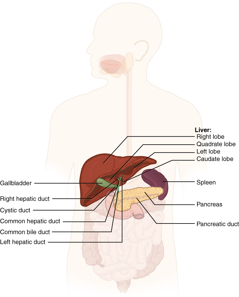
Chemical digestion in the small intestine relies on the activities of three accessory digestive organs: the liver, pancreas, and gallbladder (Figure 21.7.1). The digestive role of the liver is to produce bile and export it to the duodenum. The gallbladder primarily stores, concentrates, and releases bile. The pancreas produces pancreatic juice, which contains digestive enzymes and bicarbonate ions, and delivers it to the duodenum.
Liver
The liver is the largest gland in the body, weighing about three pounds in an adult. It is also one of the most important organs. In addition to being an accessory digestive organ, it plays a number of roles in metabolism and regulation. The liver lies inferior to the diaphragm in the right upper quadrant of the abdominal cavity and receives protection from the surrounding ribs.
The liver is divided into two primary lobes: a large right lobe and a much smaller left lobe. In the right lobe, some anatomists also identify an inferior quadrate lobe and a posterior caudate lobe, which are defined by internal features. The liver is connected to the abdominal wall and diaphragm by five peritoneal folds referred to as ligaments. These are the falciform ligament, the coronary ligament, two lateral ligaments, and the ligamentum teres hepatis. The falciform ligament and ligamentum teres hepatis are actually remnants of the umbilical vein, and separate the right and left lobes anteriorly. The lesser omentum tethers the liver to the lesser curvature of the stomach.
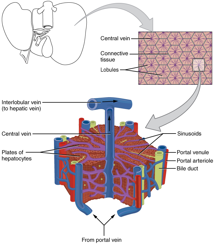
The porta hepatis (“gate to the liver”) is where the hepatic artery and hepatic portal vein enter the liver. These two vessels, along with the common hepatic duct, run behind the lateral border of the lesser omentum on the way to their destinations. As shown in Figure 21.7.2, the hepatic artery delivers oxygenated blood from the heart to the liver. The hepatic portal vein delivers partially deoxygenated blood containing nutrients absorbed from the small intestine and actually supplies more oxygen to the liver than do the much smaller hepatic arteries. In addition to nutrients, drugs and toxins are also absorbed. After processing the bloodborne nutrients and toxins, the liver releases nutrients needed by other cells back into the blood, which drains into the central vein and then through the hepatic vein to the inferior vena cava. With this hepatic portal circulation, all blood from the alimentary canal passes through the liver. This largely explains why the liver is the most common site for the metastasis of cancers that originate in the alimentary canal.
Histology
The liver has three main components: hepatocytes, bile canaliculi, and hepatic sinusoids. A hepatocyte is the liver’s main cell type, accounting for around 80 percent of the liver's volume. These cells play a role in a wide variety of secretory, metabolic, and endocrine functions. Plates of hepatocytes called hepatic laminae radiate outward from the central vein in each hepatic lobule. The liver lobule is a hexagon shape with portal triads on each corner at the perimeter of the hexagon. The portal triad is a distinctive arrangement consisting of three basic structures: a bile duct, a small branch of the hepatic artery (portal arteriole) bringing in oxygenated blood and a branch of the hepatic portal vein (portal venule) delivering nutrient rich blood. The bile duct carries freshly made bile away from hepatocytes toward the gallbladder and duodenum.
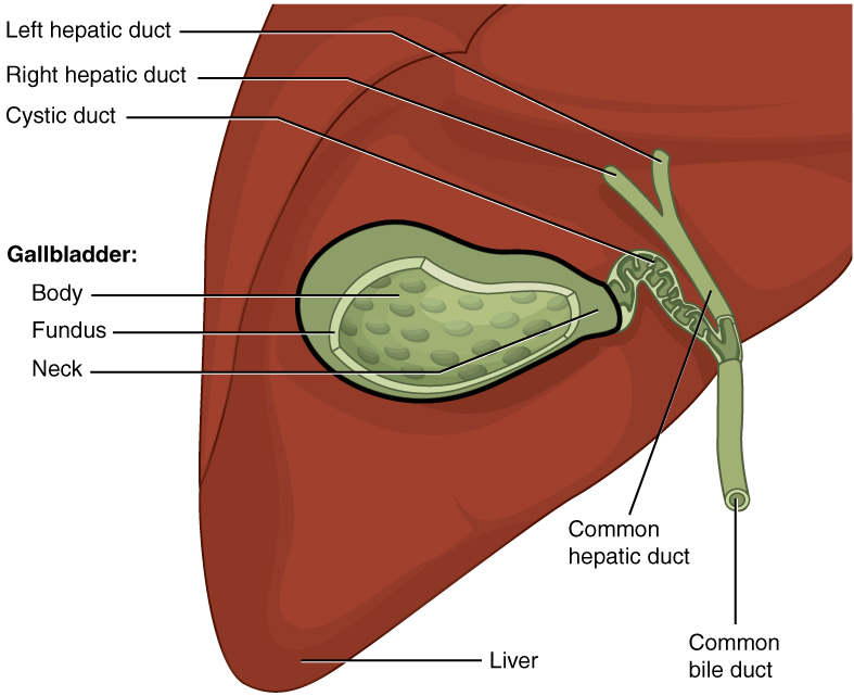
Between adjacent hepatocytes, grooves in the cell membranes provide room for each bile canaliculus (plural = canaliculi). These small ducts accumulate the bile produced by hepatocytes. From the hepatocyes, bile flows first into bile ductules and then into bile ducts. The bile ducts unite to form the larger right and left hepatic ducts, which themselves merge and exit the liver as the common hepatic duct (Figure 21.7.4). This duct then joins with the cystic duct from the gallbladder, forming the common bile duct through which bile flows into the small intestine.
A hepatic sinusoid is an open, porous blood space formed by fenestrated capillaries from nutrient-rich hepatic portal veins and oxygen-rich hepatic arteries. Hepatocytes are tightly packed around the fenestrated endothelium of these spaces, giving them easy access to the blood. From their central position, hepatocytes process the nutrients, toxins, and waste materials carried by the blood. Materials such as bilirubin are processed and excreted into the bile canaliculi. Other materials including proteins, lipids, and carbohydrates are processed and secreted into the sinusoids or just stored in the cells until called upon. The hepatic sinusoids combine and send blood to a central vein. Blood then flows through a hepatic vein into the inferior vena cava. This means that blood and bile flow in opposite directions. The hepatic sinusoids also contain star-shaped reticuloendothelial cells (Kupffer cells), phagocytes that remove dead red and white blood cells, bacteria, and other foreign material that enter the sinusoids.
Bile
Recall that lipids are hydrophobic, that is, they do not dissolve in water. Thus, before they can be digested in the watery environment of the small intestine, large lipid globules must be broken down into smaller lipid globules, a process called emulsification. Bile is a mixture secreted by the liver to accomplish the emulsification of lipids in the small intestine.
Hepatocytes secrete about one liter of bile each day. A yellow-brown or yellow-green alkaline solution (pH 7.6 to 8.6), bile is a mixture of water, bile salts, bile pigments (including bilirubin), phospholipids (such as lecithin), electrolytes, cholesterol, and triglycerides. Some molecules in bile have regions attracted to water molecules and regions attracted to lipids. This results in the large lipid globules being pulled apart into many tiny lipid fragments of about 1 µm in diameter. This change dramatically increases the surface area available for lipid-digesting enzyme activity. This is the same way dish soap works on fats mixed with water.
Bile salts act as emulsifying agents, explained above, so they are also important for the absorption of digested lipids. While most constituents of bile are eliminated in feces, bile salts are reclaimed by the enterohepatic circulation. Once bile salts reach the ileum, they are absorbed and returned to the liver in the hepatic portal blood. The hepatocytes then excrete the bile salts into newly formed bile. Thus, this precious resource is recycled.
Bilirubin, the main bile pigment, is a waste product produced when the spleen removes old or damaged red blood cells from the circulation. These breakdown products, including proteins, iron, and toxic bilirubin, are transported to the liver via the splenic vein of the hepatic portal system. In the liver, proteins and iron are recycled, whereas bilirubin is excreted in the bile. It accounts for the green color of bile. Bilirubin is eventually transformed by intestinal bacteria into stercobilin, a brown pigment that gives your stool its characteristic color! In some disease states, bile does not enter the intestine, resulting in white (‘acholic’) stool with a high fat content, since virtually no fats are broken down or absorbed.
Hepatocytes work non-stop, but bile production increases when fatty chyme enters the duodenum and stimulates the secretion of the gut hormone secretin. Between meals, bile is produced but conserved. The valve-like hepatopancreatic ampulla closes, allowing bile to divert to the gallbladder, where it is concentrated and stored until the next meal.
Pancreas
The soft, oblong, glandular pancreas lies transversely in the retroperitoneum behind the stomach. Its head is nestled into the “c-shaped” curvature of the duodenum with the body extending to the left about 15.2 cm (6 in) and ending as a tapering tail in the hilum of the spleen. It performs a curious mix of exocrine (secreting digestive enzymes) and endocrine (releasing hormones into the blood) functions (Figure 21.7.3). Histology of the pancreas reveals round clusters of acinar cells connected to the pancreatic duct and the bigger clusters of pancreatic islets.
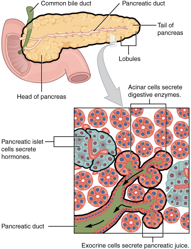
The exocrine part of the pancreas arises as little grape-like cell clusters, each called an acinus (plural = acini), located at the terminal ends of pancreatic ducts. These acinar cells secrete enzyme-rich pancreatic juice into tiny merging ducts that form two dominant ducts. The larger duct fuses with the common bile duct (carrying bile from the liver and gallbladder) just before entering the duodenum via a common opening (the hepatopancreatic ampulla). The smooth muscle sphincter of the hepatopancreatic ampulla controls the release of pancreatic juice and bile into the small intestine. The second and smaller pancreatic duct, the accessory duct (duct of Santorini), present in approximately 30% of the population, runs from the pancreas directly into the duodenum, approximately 1 inch above the hepatopancreatic ampulla. When present, it is a persistent remnant of pancreatic development.
Scattered through the sea of exocrine acini are small islands of endocrine cells, the islets of Langerhans. These vital cells produce the hormones pancreatic polypeptide, insulin, glucagon, and somatostatin. These hormones play roles in regulating several metabolic functions.
Pancreatic Juice
The pancreas produces over a liter of pancreatic juice each day. Unlike bile, it is clear and composed mostly of water along with some salts, sodium bicarbonate, and several digestive enzymes. Sodium bicarbonate is responsible for the slight alkalinity of pancreatic juice (pH 7.1 to 8.2), which serves to buffer the acidic gastric juice in chyme, inactivate pepsin from the stomach, and create an optimal environment for the activity of pH-sensitive digestive enzymes in the small intestine. Pancreatic enzymes are active in the digestion of sugars, proteins, and fats.
The pancreas produces protein-digesting enzymes in their inactive forms. These enzymes are activated in the duodenum. If produced in an active form, they would digest the pancreas (which is exactly what occurs in the disease, pancreatitis). The intestinal brush border enzyme enteropeptidase stimulates the activation of an enzyme that starts the cascade of other enzymes to digest proteins.
The enzymes that digest starch (amylase), fat (lipase), and nucleic acids (nuclease) are secreted in their active forms, since they do not attack the pancreas as do the protein-digesting enzymes.
Pancreatic Secretion
Regulation of pancreatic secretion is the job of hormones and the parasympathetic nervous system. The entry of acidic chyme into the duodenum and the presence of proteins and fats in the duodenum stimulates the release of multiple hormones of digestion. Parasympathetic regulation occurs through the vagus nerve, cranial nerve X, mainly during the cephalic and gastric phases of gastric secretion. Usually, the pancreas secretes just enough bicarbonate to counterbalance the amount of HCl produced in the stomach.
Gallbladder
Figure 21.7.4 is a drawing showing the anterior view of the gallbladder, part of the liver superior to the gallbladder, and the duct connecting the liver to gallbladder, eventually becoming the common bile duct. The gallbladder is 8–10 cm (~3–4 in) long and is nested in a shallow area on the posterior aspect of the right lobe of the liver. This muscular sac stores, concentrates, and, when stimulated, propels the bile into the duodenum via the common bile duct. The gallblader can accumulate gall stones over time, resulting in removal of the gallbladder with the gall stones in some people. It is divided into three regions. The fundus is the widest portion and tapers medially into the body, which in turn narrows to become the neck. The neck angles slightly superiorly as it approaches the hepatic duct. The cystic duct is 1–2 cm (less than 1 in) long and turns inferiorly as it bridges the neck and hepatic duct.
The simple columnar epithelium of the gallbladder mucosa is organized in rugae, similar to those of the stomach. There is no submucosa in the gallbladder wall. The wall’s middle, muscular coat is made of smooth muscle fibers. When these fibers contract, the gallbladder’s contents are ejected through the cystic duct and into the bile duct (Figure 21.7.4). Visceral peritoneum reflected from the liver capsule holds the gallbladder against the liver and forms the outer coat of the gallbladder. The gallbladder's mucosa absorbs water and ions from bile, concentrating it by up to 10-fold.
Concept Review
Chemical digestion in the small intestine cannot occur without the help of the liver and pancreas. The liver produces bile and delivers it to the common hepatic duct. Bile contains bile salts and phospholipids, which emulsify large lipid globules into tiny lipid droplets, a necessary step in lipid digestion and absorption. The gallbladder stores and concentrates bile, releasing it when it is needed by the small intestine.
The pancreas produces the enzyme- and bicarbonate-rich pancreatic juice and delivers it to the small intestine through ducts. Pancreatic juice buffers the acidic gastric juice in chyme, inactivates pepsin from the stomach, and enables the optimal functioning of digestive enzymes in the small intestine.
Review Questions
Q. Which of these statements about bile is true?
A. It is brown.
B. It is stored in the liver.
C. It is synthesized in the gallbladder.
D. Bile salts are recycled.
- Answer
-
Answer: D
Q. Pancreas ________.
A. is anterior to the stomach.
B. has only a body and tail.
C. has a pancreatic duct to carry pancreatic juice to duodenum.
D. merges with the cystic duct.
- Answer
-
Answer: C
Critical Thinking Questions
Q. When will gall stones be dangerous?
- Answer
-
A. The pancreas secretes protein-digesting enzymes and empties them into the duodenum via the pancreatic duct. The pancreatic duct merges with the common bile duct prior to entering into the duodenum. A gall stone blocking this merging point can be dangerous because it leads to pancreatitis since the pancreatic enzymes are not emptied into the duodenum.
Q. Describe the location of hepatocytes in the liver and how this arrangement enhances their function.
- Answer
-
A. The hepatocytes are the main cell type of the liver. They process, store, and release nutrients into the blood. Radiating out from the central vein, they are tightly packed around the hepatic sinusoids, allowing the hepatocytes easy access to the blood flowing through the sinusoids.
Glossary
- accessory duct
- (also, duct of Santorini) duct that runs from the pancreas into the duodenum
- acinus
- cluster of glandular epithelial cells in the pancreas that secretes pancreatic juice in the pancreas
- bile
- alkaline solution produced by the liver and important for the emulsification of lipids
- bile canaliculus
- small duct between hepatocytes that collects bile
- bilirubin
- main bile pigment, which is responsible for the brown color of feces
- central vein
- vein that receives blood from hepatic sinusoids
- common bile duct
- structure formed by the union of the common hepatic duct and the gallbladder’s cystic duct
- common hepatic duct
- duct formed by the merger of the two hepatic ducts
- cystic duct
- duct through which bile drains and enters the gallbladder
- enterohepatic circulation
- recycling mechanism that conserves bile salts
- enteropeptidase
- intestinal brush-border enzyme that activates trypsinogen to trypsin
- gallbladder
- accessory digestive organ that stores and concentrates bile
- hepatic artery
- artery that supplies oxygenated blood to the liver
- hepatic lobule
- hexagonal-shaped structure composed of hepatocytes that radiate outward from a central vein
- hepatic portal vein
- vein that supplies deoxygenated nutrient-rich blood to the liver
- hepatic sinusoid
- blood capillaries between rows of hepatocytes that receive blood from the hepatic portal vein and the branches of the hepatic artery
- hepatic vein
- vein that drains into the inferior vena cava
- hepatocytes
- major functional cells of the liver
- liver
- largest gland in the body whose main digestive function is the production of bile
- pancreas
- accessory digestive organ that secretes pancreatic juice
- pancreatic juice
- secretion of the pancreas containing digestive enzymes and bicarbonate
- porta hepatis
- “gateway to the liver” where the hepatic artery and hepatic portal vein enter the liver
- portal triad
- bile duct, hepatic artery branch, and hepatic portal vein branch
- reticuloendothelial cell
- (also, Kupffer cell) phagocyte in hepatic sinusoids that filters out material from venous blood from the alimentary canal
Contributors and Attributions
OpenStax Anatomy & Physiology (CC BY 4.0). Access for free at https://openstax.org/books/anatomy-and-physiology