
23.1: Introduction to the Reproductive System
- Page ID
- 22429

Chapter Learning Objectives:
- Describe the anatomy of the male and female reproductive systems, including their accessory structures
- Explain the role of hypothalamic and pituitary hormones in male and female reproductive function
- Trace the path of a sperm cell from its initial production through fertilization of an oocyte
- Explain the events in the ovary prior to ovulation
- Describe the development and maturation of the sex organs and the emergence of secondary sex characteristics during puberty
Small, uncoordinated, and slick with amniotic fluid, a newborn encounters the world outside of her mother’s womb. We do not often consider that a child’s birth is proof of the healthy functioning of both her mother’s and father’s reproductive systems. Moreover, her parents’ endocrine systems had to secrete the appropriate regulating hormones to induce the production and release of unique male and female gametes, reproductive cells containing the parents’ genetic material (one set of 23 chromosomes). Her parent’s reproductive behavior had to facilitate the transfer of male gametes—the sperm—to the female reproductive tract at just the right time to encounter the female gamete, an oocyte (egg). Finally, combination of the gametes (fertilization) had to occur, followed by implantation and development. In this chapter, you will explore the male and female reproductive systems, whose healthy functioning can culminate in the powerful sound of a newborn’s first cry.
Brief Overview
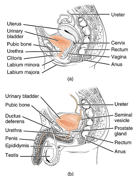
Before diving into the details, let's cover some basic common terms. Unique for its role in human reproduction, a gamete is a specialized sex cell carrying 23 chromosomes—one half the number in body cells. Gonads are the organs that make the gametes. Once the gametes are made, they travel in tubular structures. These structures are often the site for permanent sterilization procedures because if the gametes of the opposite sex cannot meet, then fertilization will not be possible. (Table 23.1.1) Comparing the midsagittal view of the female and male pelvis, we see multiple differences (Figure 23.1.2). Discussions on the female reproductive anatomy will be covered in section 23.3. Discussions on the male reproductive anatomy will be covered in section 23.2.
Table 23.1.1 Male and Female Reproductive Systems
| Term | Male | Female |
| Gonads | Testes | Ovaries |
| Gametes | Sperms | Ova (ovum, singular, also known as eggs) |
| Tubes/Pathways | Vas deferens or Ductus deferens | Oviducts or Fallopian tubes |
| Permanent Sterilization Procedure | Vasectomy | Tubal ligation |
Contributors and Attributions
OpenStax Anatomy & Physiology (CC BY 4.0). Access for free at https://openstax.org/books/anatomy-and-physiology