
8: The Axial Muscles
- Page ID
- 12528
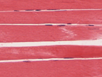
Skeletal muscle is found attached to bones. It consists of long multinucleate fibers. The fibers run the entire length of the muscle they come from and so are usually too long to have their ends visible when viewed under the microscope. The fibers are relatively wide and very long, but unbranched. Fibers are not individual cells, but are formed from the fusion of thousands of precursor cells. This is why they are so long and why individual fibers are multinucleate (a single fiber has many nuclei). The nuclei are usually up against the edge of the fiber. There are striations in skeletal muscle. These are alternating dark and light bands perpendicular to the edge of the fiber that are present all along the fiber.
The muscles of the head and neck
Figure 8.1 lists the muscles of the head and neck that you will need to know. A single platysma muscle is only shown in the lateral view of the head muscles in Figure 8.1. There are two platysma muscles, one on each side of the neck. Each is a broad sheet of a muscle that covers most of the anterior neck on that side of the body. The other anterior neck muscles are below them, and most models have the platysma muscles cut away to show the deeper muscles. The platysma muscles help pull down the lower jaw (mandible.)
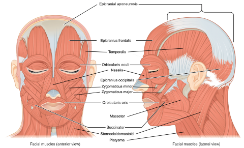
Figure 8.1. The muscles of the head.
Under the platysma are two sternocleidomastoid muscles. One on each side of the neck. These muscles have two origins, one on the sternum and the other on the clavicle. They insert on the mastoid process of the temporal bone. They can flex or extend the head, or can rotate the towards the shoulders.
The epicranius muscle is also very broad and covers most of the top of the head. The epicranius muscle includes a middle section which is all aponeurosis. The actual muscle tissue is only found over the forehead (the portion of the muscle called the epicranius frontalis; sometimes called the frontal belly of the epicranius) and the back of the head (the portion of the muscle called the epicranius occipitalis; sometimes called the occipital belly of the epicranius).
The buccinator muscles, one on each side of the face, compress the cheeks when contracted. The name is from the Latin for trumpet, which requires blowing air out of the cheeks to play, and also reflects the anatomical adjective for the cheek, buccal.
The two masseter muscles are also on each side of the face. They close the jaw when contracted. Its name is derived from the same Greek root as mastication, which means to chew.
The zygomaticus major muscles and the zygomaticus minor muscles are found on each side of the face both have their origins on the zygomatic bone. They both can change the shape of the mouth by elevating it.
LAB 8 EXERCISE 8-1
The following are muscles of facial expression. For each, give its location and describe its action when it contracts. Complete figure 8.1 above by adding in any muscles found in the table below.
.2 The following are muscles of mastication. For each, give its location and describe its action when it contracts.
|
Muscle |
Location |
Action when contracted |
|---|---|---|
|
Masseter |
||
|
Temporalis |
||
|
Medial pterygoid |
||
|
Lateral pterygoid |
LICENSES AND ATTRIBUTIONS
CC LICENSED CONTENT, ORIGINAL
- The following are muscles of facial expression. For each, give its location and describe its action when it contracts. Complete figure 8.1 above by adding in any muscles found in the table below.
Muscle
Location
Action when contracted
Epicranius frontalis
Epicranius occipitalis
Orbicularis oculi
Zygomaticus major
Zygomaticus minor
Buccinator
Orbicularis oris
Platysma
Levator labii sup.
Depressor labii inf.
Levator anguli oris
Depressor anguli oris
- The following are muscles of mastication. For each, give its location and describe its action when it contracts.
A&P Labs. Authored by: Ross Whitwam. Provided by: Mississippi University for Women. Located at: http://www.muw.edu. License: CC BY-SA: Attribution-ShareAlike
CC LICENSED CONTENT, SPECIFIC ATTRIBUTION
Figure 8-1. The muscles of the head.. Authored by: OpenStax College. Located
at: https://cnx.org/resources/9b369a7466..._the_Muscles_o f_Facial_Expressions.jpg. License: CC BY-SA: Attribution-ShareAlike
The muscles of the trunk
Information
Figures 8.2 and 8.3 shows many of the muscles of the body’s trunk that you need to know, as well as some of the muscles of the arms and legs you will learn about in the next lab.
The deltoid muscles are the triangular muscles over each shoulder.
Some of the trunk muscles have been given nicknames by gym rats. For instance, the pecs are the pectoralis major muscles at each breast.
Lats are the latissimus dorsi muscle that covers most of the lower back with its lateral fibers.
The upper back is covered by the large trapezius muscle that is almost diamond-shaped as it extends from the neck, out to the shoulders, then tapers in midway down the back.
Obliques are the external oblique muscle whose fibers angle down as it covers both sides of the abdominal region. The external oblique muscle has two sets of fibers, which cover the left and right abdomen, that are connected by a wide aponeurosis sheet in the center of the abdomen. In most muscle models that aponeurosis sheet is cut away to reveal the rectus abdominis muscles below it.
What gym rats call the core muscles are three layers of muscle that sit over the abdomen. These layers are shown in Figure 8.4. The outer layer is the external oblique muscle, with its aponeurosis covering the medial abdomen. Under the external oblique are the internal obliques on the sides of the abdomen and the rectus abdominis muscle in-between the internal obliques. The fibers of the internal obliques run up at an angle, opposite in direction to the fiber angle of the external obliques. The rectus abdominis muscle is also known as the abs. The deepest layer has the transverse abdominis muscle, whose fibers run laterally. Its fibers are concentrated at the sides of the abdomen and, like the external oblique, has an aponeurosis covering the medial abdomen under the rectus abdominis.
Extending from the back and wrapping around the sides of the rib cage is the serratus anterior muscle. This muscle’s anterior edges are serrated like the teeth of a saw because this muscle’s origins are on ribs 1 through 8 and each serration is the attachment point to another rib. This muscle is shown in Figure 8.5.
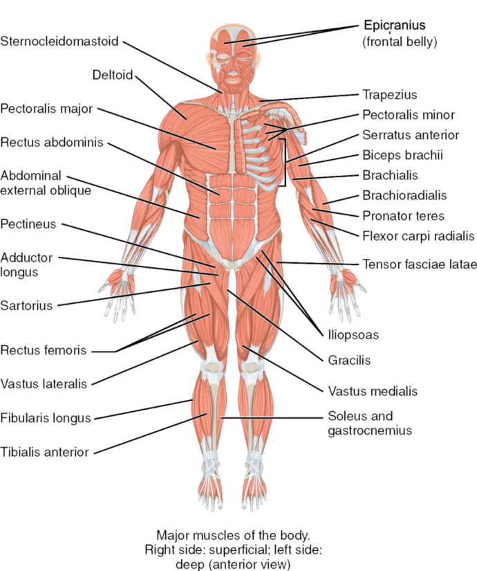
Figure 8.2. The major muscles of the body, anterior view. Anatomical right shows superficial muscles. Anatomical left shows deep muscles.
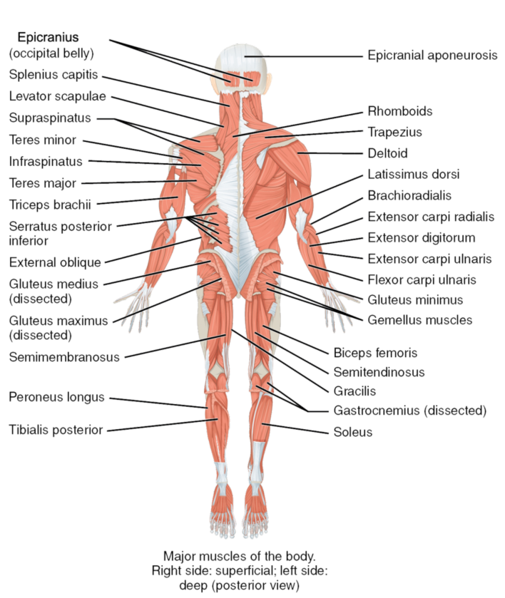
Figure 8.3. The major muscles of the body, posterior view. Anatomical right shows superficial muscles. Anatomical left shows deep muscles.
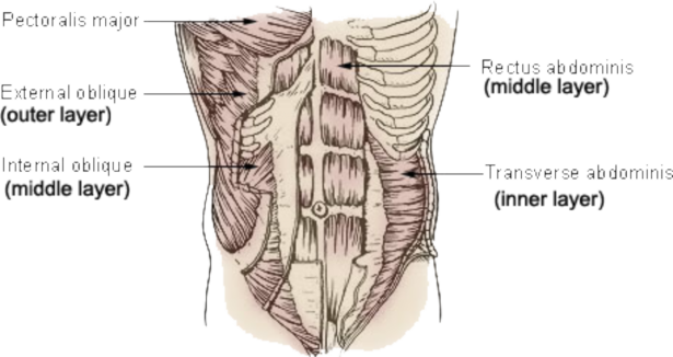
Figure 8.4. The three layers of muscles in the abdomen.
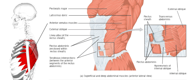
Figure 8.5. The external muscles of the body, lateral view.
LAB 8 EXERCISE 8-2
- The following are muscles that move the pectoral girdle. For each, give its location and describe its action when it contracts.
Muscle
Location
Action when contracted
Trapezius
Pectoralis minor
Serratus
anterior
- The following are muscles that move the arm. For each, give its location and describe its action when it contracts.
Muscle
Location
Action when contracted
Pectoralis major
Latissimus dorsi
Deltoid
- The following are muscles of the abdominal wall. For each, give its location and describe its action when it contracts.
Muscle
Location
Action when contracted
Rectus abdominis
External oblique
Internal oblique
Transversus abdominis

- Label the indicated facial muscles in Figure 8.6.
1
2
3
5
5
6
8
7
10
12
11
9
Figure 8.6. Facial muscles.
LICENSES AND ATTRIBUTIONS
CC LICENSED CONTENT, ORIGINAL
- A&P Labs. Authored by: Ross Whitwam. Provided by: Mississippi University for Women. Located at: http://www.muw.edu. License: CC BY-SA: Attribution-ShareAlike
CC LICENSED CONTENT, SHARED PREVIOUSLY
- Figure 8-5. The external muscles of the body, lateral view.. Authored by: This image was made out of, or made from, content published in a BodyParts3D/Anatomography web site. . Provided by: BodyParts3D, u00a9 The Database Center for Life Science licensed under CC Attribution-Share Alike 2.1 Japan.. Located
at: commons.wikimedia.org/wiki/F...es_lateral.png. License: CC BY-SA: Attribution- ShareAlike
CC LICENSED CONTENT, SPECIFIC ATTRIBUTION
- Figure 8-2. The major muscles of the body, anterior view. Anatomical right shows superficial muscles. Anatomical left shows deep muscles.. Authored by: OpenStax College. Located
at: https://cnx.org/resources/8a3b1231f3...s_of_Muscles.j pg. License: CC BY-SA: Attribution-ShareAlike
- Figure 8-3. The major muscles of the body, posterior view. Anatomical right shows superficial muscles. Anatomical left shows deep muscles.. Authored by: OpenStax College. Located
at: https://cnx.org/resources/8a3b1231f3...s_of_Muscles.j pg. License: CC BY-SA: Attribution-ShareAlike
- Figure 8-5. The external muscles of the body, lateral view.. Authored by: OpenStax College. Located
at: https://cnx.org/resources/33fa36d780...he_Abdomen.jpg. License: CC BY-SA: Attribution-ShareAlike
- Figure 8-6. Facial muscles.. Authored by: Patrick J. Lynch. Located
at: commons.wikimedia.org/wiki/F...ad_anatomy.jpg. License: CC BY-SA: Attribution-ShareAlike
PUBLIC DOMAIN CONTENT
- Figure 8-4. The three layers of muscles in the abdomen.. Authored by: Arcadian. Located
at: commons.wikimedia.org/wiki/F...nk_muscles.jpg. License: Public Domain: No Known Copyright
LAB 8 EXERCISE 8-3

3
1
A
3 Layers of connective tissue
Identify the following: epimysium * perimysium * endomysium * muscle fascicle * muscle fiber
Identify the following: origin * insertion *
B
extensor * flexor
1
2
3
4
5
2
4

Note: make some flashcards for studying the
insertions, origins and actions!
C
List the defining visual characteristics of this muscle and draw arrows to features on the photograph that illustrate each characteristic.

- Obtain a slide of skeletal muscle tissue from the slide box.
- View the slide on an appropriate objective.
- Fill out the blanks next to your drawing.
In the circle below, draw a representative sample of key features you identified, taking care to correctly and clearly draw their true shapes and directions. Draw your structures proportionately to their size in your microscope’s field of view.

Type of muscle tissue:

LAB 8 EXERCISE 8-4
IDENTIFY MUSCLES OF THE HEAD & FACE
1
3
6
7
2
4
5


*connective tissue
insertion, not a muscle
LAB 8 EXERCISE 8-5
IDENTIFY THE MUSCLES OF THE TORSO
1
4
5
2
6
7
3
8
LAB 8 EXERCISE 8-6
IDENTIFY THE MUSCLES OF OR NEAR THE BACK
1
4
5
6
2
7
3
8
MODELS: Head & Neck and Torso
Name
Action
Origin
Insertion
Muscles of facial expression
Frontalis (epicranius)
Raises eyebrows , wrinkles forehead
Frontal Bone
Skin of the brow
Occipitalis (epicranius)
Pull scalp posteriorly
Occipital bone
Aponeurosis connecting to frontalis
Orbicularis oris
Closes mouth
Maxillae and Mandible
Lips
Zygomaticus (major/minor)
smile
Zygomatic Bone
mouth
Orbicularis oculi
Closes eye
Margin of Orbit
Eyelid
Masseter
Elevates mandible
Zygomatic Arch
Mandible
Temporalis
Elevates mandible
Temporal Bone
Mandible
Buccinator
Presses cheek inward
Maxillae and Mandible
orbicularis oris
Muscles of the head, vertebral column and abdominal wall
Splenius capitis
extend + laterally flex head
upper spine
Temporal & occipital
Sternocleidomastoid
Flexes + rotates neck (also elevates ribs)
Sternum & Clavicle
Mastoid Process
Scalenes
Flexes neck (also elevates ribs)
Cervical Vertebrae
1st Two Ribs
Rectus abdominus
Flexes vertebral column , compresses abdomen
Pubis
Lower Ribs and Xiphoid
External oblique
Flexes + rotates vertebral column,
compresses abdomen
Lower Ribs
Linea alba and Ilium
Internal oblique
Flexes + rotates vertebral column, compresses abdomen
Lumbar Vertebrae & Ilium
Lower Ribs, Linea alba, Sternum
Transverse abdominus
Compresses abdomen
Lower Ribs, Ilium, Lumbar
Vertebrae
Linea Alba & Pubis
Erector spinae group
Extends vertebral column
Ilium, Sacrum, Ribs, Vertebrae
Ribs, Vertebrae, Base of Skull
Levator scapulae
Extends neck (also elevates scapula)
Cervical Vertebrae
Scapula
Name
Action
Origin
Insertion
Thoracic & shoulder muscles
Pectoralis major
Flexes, adducts + medially rotates arm at shoulder
Sternum & Clavicle
Humerus
Pectoralis minor
Elevates ribs (also moves scapula anterior and inferior)
Ribs
Scapula
External intercostals
Elevates ribs
Inferior Rib
Superior rib
Internal intercostals
Depresses ribs
Superior rib
Inferior Rib
Diaphragm
Increases thoracic volume
Xiphoid Process, Ribs, Lumbar Vertebrae
Central Tendinous Sheet
Arm movers
Serratus anterior
Moves and fixes scapula anteriorly
Ribs
Scapula
Trapezius
Elevates, retracts, depresses+ rotates scapula upward (also extends neck)
Occipital Bone & Thoracic Vertebrae
Scapula
Rhomboids (major & minor)
Elevates + adducts scapula
Cervical & Thoracic
Vertebrae
Scapula
Latissimus dorsi
Extends, adducts + medially rotates arm at shoulder
Thoracic, Lumbar Vertebrae, Ribs
Humerus
Deltoid
Abducts arm at shoulder (also anterior fibers flex + posterior fibers extend arm at shoulder)
Clavicle & Scapula
Humerus
Teres major
Medially rotate arm
Scapula
Humerus
Rotator cuff (SITS)
Infraspinatus
Laterally rotates shoulder
Scapula
Humerus
Supraspinatus
Abducts shoulder
Scapula
Humerus
Subscapularis
Medially rotates shoulder
Scapula
Humerus
Teres minor
Laterally rotates shoulder
Scapula
Humerus
- A&P Labs. Authored by: Ross Whitwam. Provided by: Mississippi University for Women. Located at: http://www.muw.edu. License: CC BY-SA: Attribution-ShareAlike