
1.14: Muscular System
- Page ID
- 64963

- Identify the anatomy of the muscular system
- Describe the main functions of the muscular system
- Spell the medical terms of the muscular system and use correct abbreviations
- Explore common diseases, disorders, and procedures related to the muscular system
- Identify the medical specialties associated with the muscular system
Muscular System Word Parts
Click on prefixes, combining forms, and suffixes to reveal a list of word parts to memorize for the Muscular System.
Introduction to the Muscular System
When most people think of muscles, they think of the muscles that are visible just under the skin, particularly of the limbs. These are skeletal muscles, so-named because most of them move the skeleton. But there are two additional types of muscles: the smooth muscle and the cardiac muscle. The body has over 600 muscles which contribute significantly to the body’s weight.
Watch this video:

A YouTube element has been excluded from this version of the text. You can view it online here: https://pb.libretexts.org/med/?p=156
Media 14.1 Muscles, Part 2 – Organismal Level: Crash Course A&P #22 [Online video]. Copyright 2015 by CrashCourse.
Muscular System Medical Terms
Anatomy (Structures) of the Muscular System
Muscle is one of the four primary tissue types of the body, and it is made up of specialized cells called fibers. The body contains three types of muscle tissue: skeletal muscle, , cardiac muscle, and smooth muscle (see Figure 14.1). All three muscle tissues have some properties in common; they all exhibit a quality called excitability as their plasma membranes can change their electrical states (from polarized to depolarized) and send an electrical wave called an action potential along the entire length of the membrane. Fascia is fibrous connective tissue that encloses muscles.
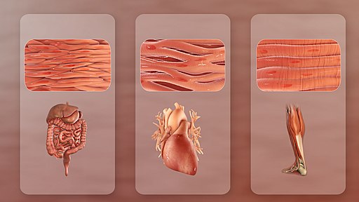
Three Types of Muscle Tissues
- Smooth – mainly associated with the walls of the internal organs. Also known as visceral muscles and are responsible for involuntary muscle movement – such as breathing, etc.
- Cardiac – heart muscle or myocardium. Its appearance is similar to a skeletal muscle and is responsible for the pumping of blood. It gives the heart beat.
- Skeletal – closely associated with the skeletal system. Also known as striated muscles and are responsible for voluntary muscle movement – such as swallowing, etc.
Smooth Muscle
Smooth muscle, so named because the cells do not have striations, is present in the walls of hollow organs like the urinary bladder, uterus, stomach, intestines, and in the walls of passageways, such as the arteries and veins of the circulatory system, and the tracts of the respiratory, urinary, and reproductive systems. Smooth muscle is also present in the eyes, where it functions to change the size of the iris and alter the shape of the lens; and in the skin where it causes hair to stand erect in response to cold temperature or fear.
Cardiac Muscle
Cardiac muscle tissue is only found in the heart. Highly coordinated contractions of cardiac muscle pump blood into the vessels of the circulatory system. Similar to skeletal muscle, cardiac muscle is striated and organized into sarcomeres, possessing the same banding organization as skeletal muscle (see Figure 14.1). Cardiac muscle fibers cells also are extensively branched and are connected to one another at their ends by intercalated discs. An intercalated disc allows the cardiac muscle cells to contract in a wave-like pattern so that the heart can work as a pump.
Skeletal Muscle
Skeletal muscles act not only to produce movement but also to stop movement, such as resisting gravity to maintain posture. Small, constant adjustments of the skeletal muscles are needed to hold a body upright or balanced in any position. Muscles also prevent excess movement of the bones and joints, maintaining skeletal stability and preventing skeletal structure damage or deformation.
Skeletal muscles are located throughout the body at the openings of internal tracts to control the movement of various substances. These muscles allow functions, such as swallowing, urination, and defecation, to be under voluntary control. Skeletal muscles also protect internal organs (particularly abdominal and pelvic organs) by acting as an external barrier or shield to external trauma and by supporting the weight of the organs.
Skeletal muscles contribute to the maintenance of homeostasis in the body by generating heat. This heat is very noticeable during exercise, when sustained muscle movement causes body temperature to rise, and in cases of extreme cold, when shivering produces random skeletal muscle contractions to generate heat.
Concept Check
- Compare and contrast the 3 types of muscles tissues.
- Where in the body do you find each of the muscle types?
Physiology (Function) of the Muscular System
The main function of the muscular system is to assist with movement. Muscles work as antagonistic pairs. As one muscle contracts, the other muscle relaxes. This contraction pulls on
the bones and assists with movement. Contraction is the shortening of the muscle fibers while relaxation lengthens the fibers. This sequence of relaxation and contraction is influenced by the nervous system.
Muscles also work to keep the posture of the body. This is done through muscle contraction where the trunk is kept straight either when sitting or standing.

A YouTube element has been excluded from this version of the text. You can view it online here: https://pb.libretexts.org/med/?p=156
Media 14.2 Review of joint movements with Dr. Mike. Copyright 2021, Dr. Matt & Dr. Mike.
ACTION |
MEANING |
| flexion | Decreasing the angle between two bones; bending a limb. |
| extension | Increasing the angle between two bones; straightening out a limb. |
| abduction | Movement away from the midline of the body |
| adduction | Movement toward the midline of the body |
| rotation | Circular movement around an axis (central point). Internal rotation is toward the center of the body and external rotation is away from the center of the body. |
| dorsiflexion | Decreasing the angle of the ankle join so that the foot moves upward, toward the knee or ceiling. This is the opposite movement of stepping on the gas pedal when driving a car. |
| plantar flexion | Motion that moves the foot downward toward the ground as when pointing the toes or stepping on the gas pedal. Plant/o means sole of the foot. |
| supination | As applied to the hand and forearm, where the elbow is bent, the act of turning the palm up. As applied to the foot, it is outward roll of the foot/ankle during normal motion. |
| pronation | As applied to the hand and forearm, where the elbow is bent, the act of turning the palm down. As applied to the foot, it is inward roll of the foot/ankle during normal motion. |
Naming of Muscles
There are many nomenclatures for naming muscles. Some of these include:
- divisions – biceps, triceps, quadriceps
- size – maximus (largest), minimus (smallest)
- shape – deltoid (triangular), trapezious (trapezoid)
- action – flexor (to flex), adductor (towards midline of body)
Muscular System Medical Abbreviations
Common Diseases and Disorders
Muscular Dystrophy
Muscular dystrophies are rare inherited diseases that weaken muscles, decreasing the individual’s mobility. There are several kinds of muscular dystrophy that affect different muscle groups and symptoms develop at different ages (Centers for Disease Control and Prevention, 2020). The most common kind of muscular dystrophy is Duchenne/Becker muscular dystrophy (DMD/BMD). This disease primarily effects boys and symptoms may include frequent falls and trouble keeping up with peers. Since all muscles are affected, the person will eventually require a wheelchair and assistance with breathing (Muscular Dystrophy Association, 2021). To learn more please visit the Centers for Disease Control and Prevention muscular dystrophy webpage.
Cerebral Palsy
Cerebral Palsy (CP) is caused by an interruption to the normal development of a person’s brain leading to weakness with muscles. Depending on the area of the brain that is affected, signs and symptoms will vary in the type and severity between individuals. Balance and coordination are often challenging due the inability to control muscles (Centers for Disease Control and Prevention, 2020). To learn more about Cerebral palsy please visit the Centers for Disease Control and Prevention.
Carpal Tunnel Syndrome
Carpal tunnel syndrome may present with pain, numbness or weakness to the hand(s) caused by pressure on the median nerve. Some causes for this pressure are work related such as keyboarding with improper body mechanics, illness such as arthritis, and even pregnancy (National Institute of Neurological Disorders and Stroke, 2020). To learn more, visit the National Institute of Neurological Disorders and Stroke webpage.
Paralysis
Paralysis is the loss of strength and control of the muscles in parts of the body. Paralysis can be localized where it affects specific areas such as the face, feet, vocal chords, etc., or it can be generalized where it affects a larger area of the body. There are various types of generalized paralysis, including:
- Paresis – a partial paralysis wherein there is still some control of the muscles
- Paraplegia – paralysis that affects both legs and lower part of the body.
- Quadriplegia – affects both arms, both legs and sometimes from the neck down
- Hemiplegia – affects one side of the body. For example, the arm and leg on the same side of the body (Cleveland Clinic, 2017)
To learn more about paralysis, please visit the Cleveland Clinic’s Paralysis information web page.
Sprain and Strain
A sprain is an injury to a joint whereby a ligament is stretched or torn.
A strain is an injury to a muscle whereby a tendon is stretched or torn.
Diagnostic Procedures
Electromyography (EMG) is a procedure that assesses the function of nerve cells that control muscles. Electrodes, either attached to the skin or inserted into the muscle, allow for the recording of electrical impulses. EMG can identify functional problems with the peripheral nerves, muscles, or with the signals between the nerves and the muscles. This is just one test in a series of tests that assist in the diagnosis of neuromuscular disorders (Mayo Clinic Staff, 2019). To learn more, please visit the Mayo Clinic’s Electromyography webpage.
Magnetic Resonance Imaging (MRI) (Fig 14.4) is a test that uses radio frequency waves and a magnetic field to produce clear images that aid in the diagnosis of a wide range of conditions (National Institute of Biomedical Imaging and Bioengineering, n.d.). Leung (2017) notes that there has been increased clinical use in using MRI for the treatment and monitoring of muscular disorders due to the high-quality MRI images that distinguish skeletal muscles from fat (para. 4). To learn more, please visit the National Institute of Biomedical Imaging and Bioengineering MRI webpage.
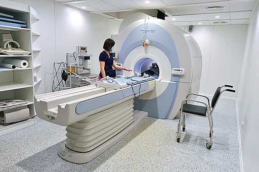
Figure 14.4. Vantage Atlas MRI scanner from Toshiba. From Mos.ru, CC BY 4.0 via Wikimedia Commons
Range of Motion Testing is a diagnostic procedures used to determine the amount of movement around a specific joint.
Medical Terminology in Context
Medical Specialties Related to Muscular System
Orthopedic Surgeon
Orthopedic Surgeons are medical doctors who complete specialized training in the prevention, diagnosis, treatment and surgery of disorders and diseases related to the musculoskeletal systems (American Academy of Orthopaedic Surgeons, 2021). For more details please visit the American Academy of Orthopaedic Surgeons’s page on Orthopaedic Surgeons.
Neurologist
Neurologists are medical doctors who complete an additional specialized training in the prevention, diagnosis, and treatment of disorders and conditions related to the brain, spinal cord, nerves and muscles (American Academy of Neurology, 2021). For more details visit the American Academy of Neurology’s webpage.
Kinesiologist
Kinesiologists are regulated health-care professionals with a degree in kinesiology or related discipline. Kinesiologists work in a variety of settings that assist people with pain management, injury prevention, and health promotion through biomechanics (American Kinesiology Association, 2021). To explore career options for kinesiologists, visit the American Kinesiology Association’s Career Center webpage. To learn more about kinesiologists and careers in this field, visit the Zippia Kinesiologist Overview webpage.
Occupational Therapist
Occupational therapist helps people across the lifespan to do the things they want and need to do through the therapeutic use of daily activities (occupations). Occupational therapy practitioners enable people of all ages to live life to its fullest by helping them promote health, and prevent—or live better with—injury, illness, or disability. To learn more please visit the American Occupational Therapy Association webpage.
Certified Occupational Therapist Assistant
Certified occupational therapist assistants and aides help patients develop, recover, improve, as well as maintain the skills needed for daily living and working. To learn more please visit the Occupational Outlook Handbook: Occupational Therapy Assistants and Aides.
Physical Therapist
Physical therapists help people to maximize their quality of life. They work with people of all ages and abilities, and in a variety of settings. They help people rehabilitate from devastating injuries, manage chronic conditions, avoid surgery and prescription drugs, and create healthy habits. To learn more please visit: American Physical Therapy Association
Physical Therapist Assistant
Assist physical therapists in providing physical therapy treatments and procedures. May, in accordance with state laws, assist in the development of treatment plans, carry out routine functions, document the progress of treatment, and modify specific treatments in accordance with patient status and within the scope of treatment plans established by a physical therapist. Generally requires formal training. To learn more please visit: Physical Therapist Assistants
Test Yourself
References
American Academy of Neurology. (2021). What is a neurologist?https://www.aan.com/tools-and-resources/medical-students/careers-in-neurology/what-is-a-neurologist/
American Academy of Orthopaedic Surgeons. (2021). Orthopaedic Surgeons: Restoring mobility and keeping our nation in motion. https://www.aaos.org/about/what-is-an-orthopaedic-surgeon/
American Kinesiology Association. (2021). Career Center. https://www.americankinesiology.org/SubPages/Pages/Careers%20In%20Kinesiology#rn
American Occupational Therapy Association. (2021).What is Occupational Therapy. https://www.aota.org/conference-events/otmonth/what-is-ot.aspx
American Physical Therapy Association. (2021). Careers in Physical Therapy. https://www.apta.org/your-career/careers-in-physical-therapy
Centers for Disease Control and Prevention. (2020). What is cerebral palsy? CDC. https://www.cdc.gov/ncbddd/cp/facts.html
Centers for Disease Control and Prevention. (2020). What is muscular dystrophy? CDC. https://www.cdc.gov/ncbddd/musculardystrophy/facts.html
[CrashCourse]. (2015, July 15). Muscles, part 2 – organismal level: Crash course A&P #22 [Video]. YouTube. https://youtu.be/I80Xx7pA9hQ
Leung, J. (2016, November 25). Magnetic resonance imaging patterns of muscle involvement in genetic muscle diseases: a systematic review. Journal of Neurology, 264(7), 1320-1333. https://dx.doi.org/10.1007%2Fs00415-016-8350-6
Mayo Clinic Staff. (2019, May 21). Electromyography (EMG). Mayo Clinic Patient Care and Information. https://www.mayoclinic.org/tests-procedures/emg/about/pac-20393913
Muscular Dystrophy Association. (2021). Duchenne Muscular Dystrophy (DMD). https://www.mda.org/disease/duchenne-muscular-dystrophy
National Institute of Biomedical Imaging and Bioengineering. (n.d.) Magnetic Resonance Imaging (MRI). National Institutes of Health. https://www.nibib.nih.gov/science-education/science-topics/magnetic-resonance-imaging-mri
National Institute of Neurological Disorders and Stroke. (2020). Carpal Tunnel Syndrome Fact Sheet. National Institutes of Health. https://www.ninds.nih.gov/Disorders/Patient-Caregiver-Education/Fact-Sheets/Carpal-Tunnel-Syndrome-Fact-Sheet
Occupational Therapy Assistants and Aides. (Sept 2021). Occupational Outlook Handbook. https://www.bls.gov/ooh/healthcare/occupational-therapy-assistants-and-aides.htm
U.S. Bureau of Labor Statistics. (2021). Occupational Therapists. https://www.bls.gov/ooh/healthcare/physical-therapists.htm#tab-1
U.S. Bureau of Labor Statistics. (2021). Physical Therapist Assistants. https://www.bls.gov/oes/current/oes312021.htm#nat
Unless otherwise indicated, this chapter contains material adapted from Anatomy and Physiology (on OpenStax), by Betts, et al. and is used under a a CC BY 4.0 international license. Download and access this book for free at https://openstax.org/books/anatomy-and-physiology/pages/1-introduction.