
18.9B: Histology of the Small Intestine
- Page ID
- 50446
The small intestine wall has four layers: the outermost serosa, muscularis, submucosa, and innermost mucosa.
- Describe the histology of the small intestine
Key Points
- The outermost layer of the intestine, the serosa, is a smooth membrane consisting of a thin layer of cells that secrete serous fluid, and a thin layer of connective tissue.
- The muscularis is a region of muscle adjacent to the submucosa membrane. It is responsible for gut movement (also called peristalsis ). It usually has two distinct layers of smooth muscle: circular and longitudinal.
- The submucosa is the layer of dense irregular connective tissue or loose connective tissue that supports the mucosa; it also joins the mucosa to the bulk of underlying smooth muscle.
- The mucosa is the innermost tissue layer of the small intestines and is a mucous membrane that secretes digestive enzymes and hormones. The intestinal villi are part of the mucosa.
- The three sections of the small intestine look similar to each other at a microscopic level, but there are some important differences. The jejunum and ileum do not have Brunner’s glands in the submucosa, while the ileum has Peyer’s patches in the mucosa, but the duodenum and jejunum do not.
Key Terms
- Brunner’s glands: Compound, tubular, submucosal glands found in that portion of the duodenum that is above the hepatopancreatic sphincter (sphincter of Oddi).
- Peyer’s patches: Patches of lymphoid tissue or lymphoid nodules on the walls of the ileum in the small intestine.
- intestinal wall: The wall of the small intestine is composed of four layers, from the outside to the inside: serosa, muscularis, submucosa, and mucosa.
The Small Intestine’s Layers
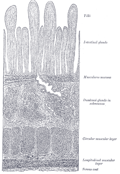
Section of duodenum: This image shows the layers of the duodenum: the serosa, muscularis, submucosa, and mucosa.
The small intestine has four tissue layers:
- The serosa is the outermost layer of the intestine. The serosa is a smooth membrane consisting of a thin layer of cells that secrete serous fluid, and a thin layer of connective tissue. Serous fluid is a lubricating fluid that reduces friction from the movement of the muscularis.
- The muscularis is a region of muscle adjacent to the submucosa membrane. It is responsible for gut movement, or peristalsis. It usually has two distinct layers of smooth muscle: circular and longitudinal.
- The submucosa is the layer of dense, irregular connective tissue or loose connective tissue that supports the mucosa, as well as joins the mucosa to the bulk of underlying smooth muscle.
- The mucosa is the innermost tissue layer of the small intestines, and is a mucous membrane that secretes digestive enzymes and hormones. The intestinal villi are part of the mucosa.
The three sections of the small intestine look similar to each other at a microscopic level, but there are some important differences. The jejunum and ileum do not have Brunner’s glands in the submucosa, while the ileum has Peyer’s patches in the mucosa, but the duodenum and jejunum do not.
Brunner’s Glands
Brunner’s glands (or duodenal glands) are compound tubular submucosal glands found in the duodenum. The main function of these glands is to produce a mucus-rich, alkaline secretion (containing bicarbonate) in order to neutralize the acidic content of chyme that is introduced into the duodenum from the stomach, and to provide an alkaline condition for optimal intestinal enzyme activity, thus enabling absorption to take place and lubricate the intestinal walls.
Peyer’s Patches
Peyer’s patches are organized lymph nodules. They are aggregations of lymphoid tissue that are found in the lowest portion of the small intestine, which differentiate the ileum from the duodenum and jejunum.
Because the lumen of the gastrointestinal tract is exposed to the external environment, much of it is populated with potentially pathogenic microorganisms. Peyer’s patches function as the immune surveillance system of the intestinal lumen and facilitate the generation of the immune response within the mucosa.
Intestinal Villi
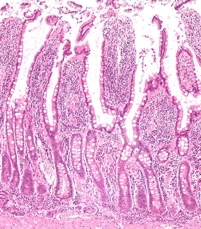
Micrograph of the small intestine: A low-magnification micrograph of small intestinal mucosa that shows villi.
Intestinal villi (singular: villus) are tiny, finger-like projections that protrude from the epithelial lining of the mucosa. Each villus is approximately 0.5–1.6 mm in length and has many microvilli (singular: microvillus), each of which are much smaller than a single villus.
Villi increase the internal surface area of the intestinal walls. This increased surface area allows for more intestinal wall area to be available for absorption. An increased absorptive area is useful because digested nutrients (including sugars and amino acids) pass into the villi, which is semi-permeable, through diffusion, which is effective only at short distances.
In other words, the increased surface area (in contact with the fluid in the lumen) decreases the average distance traveled by the nutrient molecules, so the effectiveness of diffusion increases.
The villi are connected to blood vessels that carry the nutrients away in the circulating blood.