
20.3D: Yolk Sac Development
- Page ID
- 50613
The yolk sac is vascularized and contributes nutrients to the embryo.
- Discuss the location and purpose of the yolk sac
Key Points
- The yolk sac is the earliest visible portion of the gestational sac and is situated on the ventral portion of the embryo, arising from extra-embryonic endoderm.
- The blood to and from the yolk sac is conveyed via the primitive aorta and vitelline circulation.
- At approximately the end of the fourth week, the yolk sac is connected to the primitive digestive system, which allows the yolk sac to contribute nutrients to the embryo.
Key Terms
- yolk sac: A membranous sac attached to an embryo that provides early nourishment in the form of yolk in bony fishes, sharks, reptiles, birds, and mammals. It functions as the developmental circulatory system of the human embryo before internal circulation begins.
- vitelline circulation: The system of blood flowing from the embryo to the yolk sac and back again.
- gestational sac: The gestational sac (or gestation sac) is the only available intrauterine structure that can be used to determine if an intrauterine pregnancy (IUP) exists until the embryo is identified.
- Heuser’s membrane: Also called the exocoelomic membrane, it is a short lived combination of hypoblast cells and extracellular matrix.
- mesenchyme: A type of tissue characterized by loosely associated cells that lack polarity and are surrounded by a large extracellular matrix.
The yolk sac is the first element seen in the gestational sac during pregnancy. In humans, it is usually visible at five weeks of gestation. Identifying a true gestation sac is a critical landmark, and is reliably seen in early pregnancy through ultrasound. The yolk sac, situated on the ventral aspect of the embryo, is lined by extra-embryonic endoderm, outside of which is a layer of extra-embryonic mesenchyme derived from the mesoderm.
Blood is conveyed to the wall of the sac by the primitive aorta. After circulating through a wide-meshed capillary plexus, it is returned by the vitelline veins to the tubular heart of the embryo. This vitelline circulation absorbs nutritive material from the yolk sac that is conveyed to the embryo.
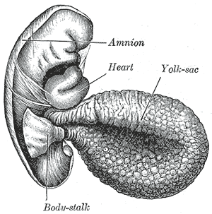
Yolk sac: The yolk sac is a membranous sac attached to the embryo that provides nourishment in the form of yolk.
At the end of the fourth week, the yolk sac has the appearance of a small, pear-shaped vesicle (umbilical vesicle) opening into the digestive tube by a long, narrow tube, the vitelline duct. The vesicle can be seen in the afterbirth as a small, somewhat oval-shaped body whose diameter varies from 1 mm to 5 mm. It is situated between the amnion and the chorion and may lie on or at a varying distance from the placenta.
As a rule, the vitelline duct undergoes complete obliteration during the seventh week. In about 2% of the cases, its proximal portion persists as a diverticulum from the small intestine (Meckel’s diverticulum), which is situated about 60 cm proximal to the ileocecal valve. It may be attached by a fibrous cord to the abdominal wall at the umbilicus. Sometimes, a narrowing of the lumen of the ileum is seen opposite the site of attachment of the duct.
The yolk sac starts forming during the second week of embryonic development, at the same time of the shaping of the amniotic sac. The hypoblast starts proliferating laterally and descending. In the meantime Heuser’s membrane, located on the opposite pole of the developing vesicle, starts its upward proliferation and meets the hypoblast.