

2.9: Cell Physiology — Body Fluids and Fluid Compartment
- Page ID
- 90502

\( \newcommand{\vecs}[1]{\overset { \scriptstyle \rightharpoonup} {\mathbf{#1}} } \)
\( \newcommand{\vecd}[1]{\overset{-\!-\!\rightharpoonup}{\vphantom{a}\smash {#1}}} \)
\( \newcommand{\dsum}{\displaystyle\sum\limits} \)
\( \newcommand{\dint}{\displaystyle\int\limits} \)
\( \newcommand{\dlim}{\displaystyle\lim\limits} \)
\( \newcommand{\id}{\mathrm{id}}\) \( \newcommand{\Span}{\mathrm{span}}\)
( \newcommand{\kernel}{\mathrm{null}\,}\) \( \newcommand{\range}{\mathrm{range}\,}\)
\( \newcommand{\RealPart}{\mathrm{Re}}\) \( \newcommand{\ImaginaryPart}{\mathrm{Im}}\)
\( \newcommand{\Argument}{\mathrm{Arg}}\) \( \newcommand{\norm}[1]{\| #1 \|}\)
\( \newcommand{\inner}[2]{\langle #1, #2 \rangle}\)
\( \newcommand{\Span}{\mathrm{span}}\)
\( \newcommand{\id}{\mathrm{id}}\)
\( \newcommand{\Span}{\mathrm{span}}\)
\( \newcommand{\kernel}{\mathrm{null}\,}\)
\( \newcommand{\range}{\mathrm{range}\,}\)
\( \newcommand{\RealPart}{\mathrm{Re}}\)
\( \newcommand{\ImaginaryPart}{\mathrm{Im}}\)
\( \newcommand{\Argument}{\mathrm{Arg}}\)
\( \newcommand{\norm}[1]{\| #1 \|}\)
\( \newcommand{\inner}[2]{\langle #1, #2 \rangle}\)
\( \newcommand{\Span}{\mathrm{span}}\) \( \newcommand{\AA}{\unicode[.8,0]{x212B}}\)
\( \newcommand{\vectorA}[1]{\vec{#1}} % arrow\)
\( \newcommand{\vectorAt}[1]{\vec{\text{#1}}} % arrow\)
\( \newcommand{\vectorB}[1]{\overset { \scriptstyle \rightharpoonup} {\mathbf{#1}} } \)
\( \newcommand{\vectorC}[1]{\textbf{#1}} \)
\( \newcommand{\vectorD}[1]{\overrightarrow{#1}} \)
\( \newcommand{\vectorDt}[1]{\overrightarrow{\text{#1}}} \)
\( \newcommand{\vectE}[1]{\overset{-\!-\!\rightharpoonup}{\vphantom{a}\smash{\mathbf {#1}}}} \)
\( \newcommand{\vecs}[1]{\overset { \scriptstyle \rightharpoonup} {\mathbf{#1}} } \)
\(\newcommand{\longvect}{\overrightarrow}\)
\( \newcommand{\vecd}[1]{\overset{-\!-\!\rightharpoonup}{\vphantom{a}\smash {#1}}} \)
\(\newcommand{\ket}[1]{\left| #1 \right>}\)
\(\newcommand{\bra}[1]{\left< #1 \right|}\)
\(\newcommand{\braket}[2]{\left< #1 \vphantom{#2} \right| \left. #2 \vphantom{#1} \right>}\)
\(\newcommand{\braopket}[3]{\left< #1 \vphantom{#2}\vphantom{#3} \right| #2 \vphantom{#1}\vphantom{#3} \left| #3 \vphantom{#1}\vphantom{#2} \right>}\)
\(\newcommand{\qmvec}[1]{\mathbf{\vec{#1}}}\)
\(\newcommand{\op}[1]{\hat{\mathbf{#1}}}\)
\(\newcommand{\expect}[1]{\langle #1 \rangle}\)
\(\newcommand{\dfn}[1]{\emph{\textbf{#1}}}\)
Cell physiology focuses on how cells maintain balance and carry out essential functions that keep the body alive and functioning.
- Explain the importance of water in the body
- Contrast the composition of the intracellular fluid with that of the extracellular fluid
- Explain the importance of protein channels in the movement of solutes
- Identify the causes and symptoms of edema
Cell physiology is the study of how cells function. This field of biology looks at the everyday activities that keep cells alive and working, like taking in nutrients, making energy, building proteins, and getting rid of waste.
Think of it as understanding how each tiny cell acts like a mini factory:
-
It has systems for communication,
-
machinery for making products,
-
and pathways for transporting materials in and out.
In A&P, learning about cell physiology helps you understand how the body works at its most basic level — and how all those small cellular processes add up to keep tissues, organs, and organ systems running smoothly.
The chemical reactions of life take place in aqueous solutions. The dissolved substances in a solution are called solutes. In the human body, solutes vary in different parts of the body, but may include proteins—including those that transport lipids, carbohydrates, and, very importantly, electrolytes. Often in medicine, a mineral dissociated from a salt that carries an electrical charge (an ion) is called an electrolyte. For instance, sodium ions (Na+) and chloride ions (Cl-) are often referred to as electrolytes.
In the body, water moves through selectively-permeable cell membranes and from one compartment of the body to another by a process called osmosis. Osmosis is basically the diffusion of water from regions of higher water concentration to regions of lower water concentration, along an osmotic gradient across a selectively-permeable membrane. As a result, water will move into and out of cells and tissues, depending on the relative concentrations of the water and solutes found there. An appropriate balance of solutes inside and outside of cells must be maintained to ensure normal function.
Body Water Content
The human body is made mostly of water. In fact, water makes up about 75% of an infant’s body, and around 50–60% of an adult’s body. As we get older, that percentage can drop to about 45%. Why does it change? Because as we grow, the amount of muscle, fat, bone, and other tissues in our body changes too.
Some parts of the body have more water than others. For example, the brain and kidneys are about 80–85% water, they are basically water-packed! On the other hand, your teeth are mostly solid, with only about 8–10% water. (See figure below).
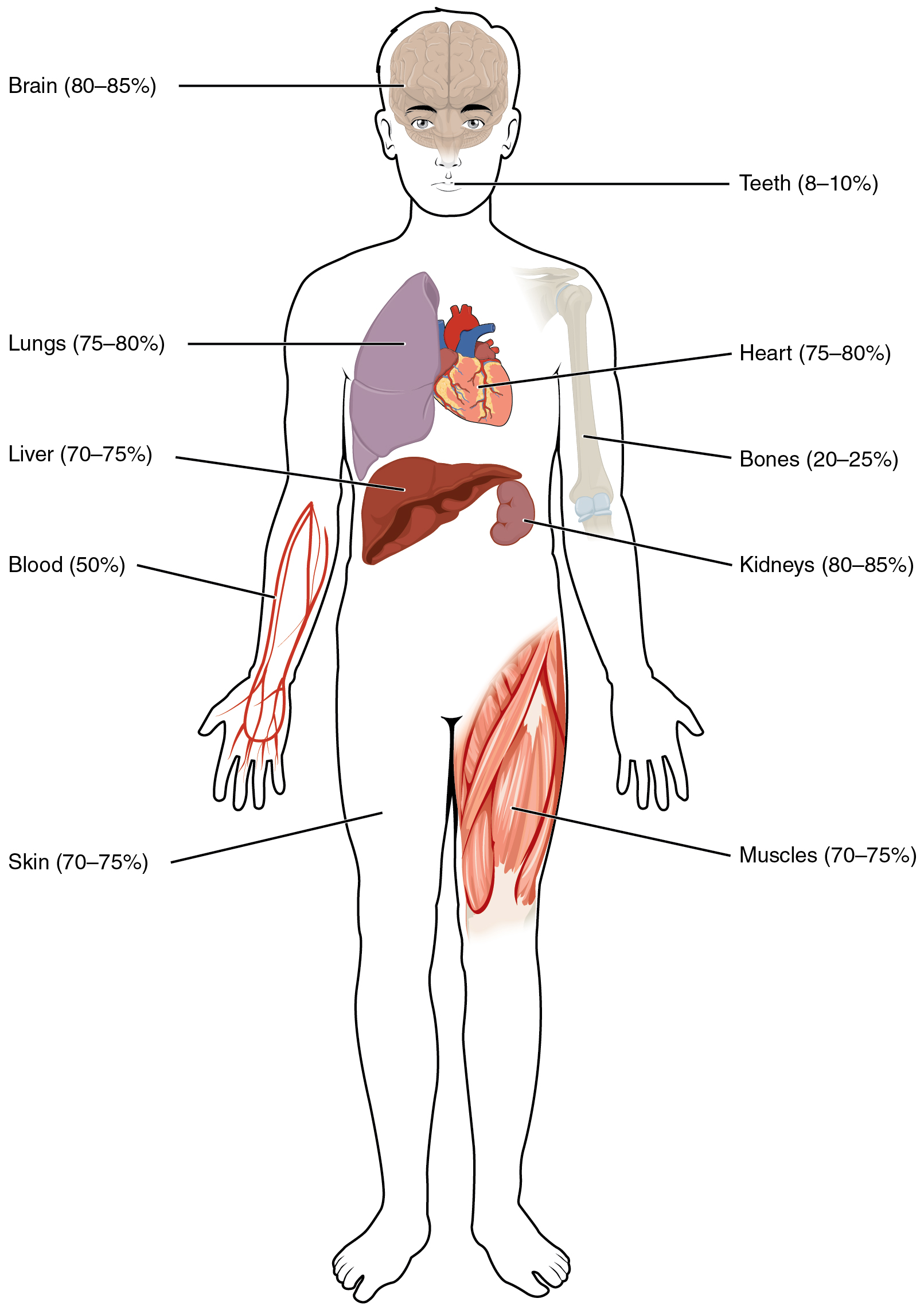
Figure \(\PageIndex{1}\): Water Content of the Body’s Organs and Tissues. Water content varies in different body organs and tissues, from as little as 8 percent in the teeth to as much as 85 percent in the brain.
Fluid Compartments
Body fluids can be discussed in terms of their specific fluid compartment, a location that is largely separate from another compartment by some form of a physical barrier. The intracellular fluid (ICF) compartment is the system that includes all fluid enclosed in cells by their plasma membranes.
Extracellular fluid (ECF) surrounds all cells in the body. Extracellular fluid has two primary constituents: the fluid component of the blood — called plasma — and the interstitial fluid (IF) that surrounds all cells not in the blood (See figure below).
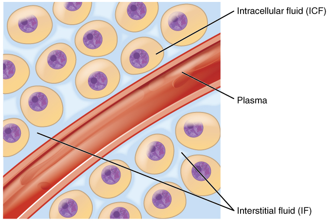
Figure \(\PageIndex{2}\): Fluid Compartments in the Human Body. The intracellular fluid (ICF) is the fluid within cells. The interstitial fluid (IF) is part of the extracellular fluid (ECF) between the cells. Blood plasma is the second part of the ECF. Materials travel between cells and the plasma in capillaries through the IF.
Intracellular Fluid
The ICF lies within cells and is the principal component of the cytosol/cytoplasm. The ICF makes up about 60 percent of the total water in the human body, and in an average-size adult male, the ICF accounts for about 25 liters (seven gallons) of fluid (Figure \(\PageIndex{3}\)). This fluid volume tends to be very stable, because the amount of water in living cells is closely regulated. If the amount of water inside a cell falls to a value that is too low, the cytosol becomes too concentrated with solutes to carry on normal cellular activities; if too much water enters a cell, the cell may burst and be destroyed.
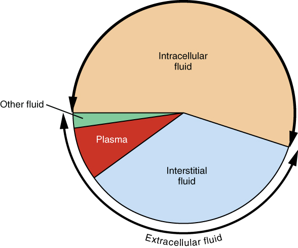
Figure \(\PageIndex{3}\): Pie Chart Showing the Proportion of Total Body Fluid in Each of the Body’s Fluid Compartments. Most of the water in the body is intracellular fluid. The second largest volume is the interstitial fluid, which surrounds cells that are not blood cells. Next is plasma, then a small proportion of "other fluid."
Extracellular Fluid
The ECF accounts for the other one-third of the body’s water content. Approximately 20 percent of the ECF is found in plasma. Plasma travels through the body in blood vessels and transports a range of materials, including blood cells, proteins (including clotting factors and antibodies), electrolytes, nutrients, gases, and wastes. Gases, nutrients, and waste materials travel between capillaries and cells through the IF. Cells are separated from the IF by a selectively permeable cell membrane that helps regulate the passage of materials between the IF and the interior of the cell.
The body has other water-based ECF. These include the cerebrospinal fluid (CSF) that bathes the brain and spinal cord, lymph, the synovial fluid in joints, the pleural fluid in the pleural cavities, the pericardial fluid in the cardiac sac, the peritoneal fluid in the peritoneal cavity, and the aqueous humor of the eye. Because these fluids are outside of cells, these fluids are also considered components of the ECF compartment.
Composition of Body Fluids
The compositions of the two components of the ECF — plasma and IF — are more similar to each other than either is to the ICF (Figure \(\PageIndex{4}\)). Blood plasma has high concentrations of sodium, chloride, bicarbonate, and protein. The IF has high concentrations of sodium, chloride, and bicarbonate, but a relatively lower concentration of protein. In contrast, the ICF has elevated amounts of potassium, phosphate, magnesium, and protein. Overall, the ICF contains high concentrations of potassium and phosphate (HPO42-), whereas both plasma and the ECF contain high concentrations of sodium and chloride.
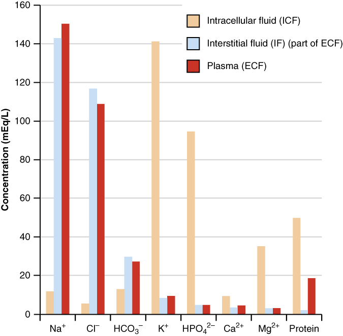
Figure \(\PageIndex{4}\): Concentrations of elements in key bodily fluids. The graph shows the composition of the ICF, IF, and plasma. The compositions of plasma and IF are similar to one another but are quite different from the composition of the ICF.
Most body fluids have a neutral charge, meaning they contain a balanced number of positive ions (cations) and negative ions (anions). Two of the most important ions are sodium (Na⁺) and chloride (Cl⁻), which are found mostly outside your cells (in the IF, the fluid between cells; and in your blood plasma). Another important ion, potassium (K⁺), is found mostly inside your cells (in the ICF).
Even though sodium and potassium can slowly leak in and out of cells through tiny pores, your body works hard to keep their levels in the right places. To do this, cells use sodium-potassium pumps — special proteins in the cell membrane that use energy from ATP to move sodium out of the cell and potassium in. This pump helps maintain the proper balance of ions, which is important for things like nerve signals and muscle contractions. (See image below).
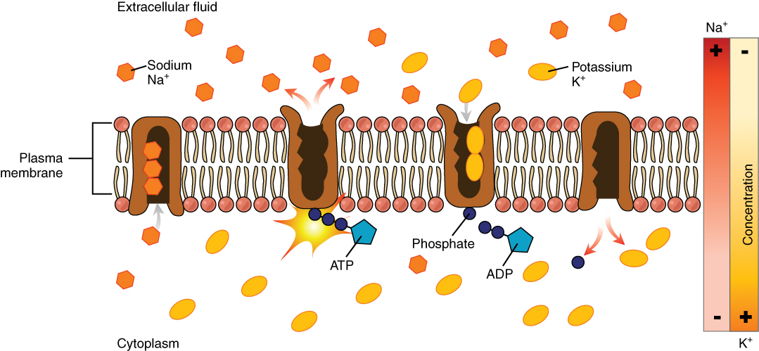
Figure \(\PageIndex{5}\): The sodium-potassium pump. The sodium-potassium pump is powered by ATP to transfer sodium out of the cytoplasm and into the ECF. The pump also transfers potassium out of the ECF and into the cytoplasm. (credit: modification of work by Mariana Ruiz Villarreal)