
2.11: Passive Movements across the Cell Membrane — Facilitated Diffusion and Osmosis
- Page ID
- 90504

\( \newcommand{\vecs}[1]{\overset { \scriptstyle \rightharpoonup} {\mathbf{#1}} } \)
\( \newcommand{\vecd}[1]{\overset{-\!-\!\rightharpoonup}{\vphantom{a}\smash {#1}}} \)
\( \newcommand{\dsum}{\displaystyle\sum\limits} \)
\( \newcommand{\dint}{\displaystyle\int\limits} \)
\( \newcommand{\dlim}{\displaystyle\lim\limits} \)
\( \newcommand{\id}{\mathrm{id}}\) \( \newcommand{\Span}{\mathrm{span}}\)
( \newcommand{\kernel}{\mathrm{null}\,}\) \( \newcommand{\range}{\mathrm{range}\,}\)
\( \newcommand{\RealPart}{\mathrm{Re}}\) \( \newcommand{\ImaginaryPart}{\mathrm{Im}}\)
\( \newcommand{\Argument}{\mathrm{Arg}}\) \( \newcommand{\norm}[1]{\| #1 \|}\)
\( \newcommand{\inner}[2]{\langle #1, #2 \rangle}\)
\( \newcommand{\Span}{\mathrm{span}}\)
\( \newcommand{\id}{\mathrm{id}}\)
\( \newcommand{\Span}{\mathrm{span}}\)
\( \newcommand{\kernel}{\mathrm{null}\,}\)
\( \newcommand{\range}{\mathrm{range}\,}\)
\( \newcommand{\RealPart}{\mathrm{Re}}\)
\( \newcommand{\ImaginaryPart}{\mathrm{Im}}\)
\( \newcommand{\Argument}{\mathrm{Arg}}\)
\( \newcommand{\norm}[1]{\| #1 \|}\)
\( \newcommand{\inner}[2]{\langle #1, #2 \rangle}\)
\( \newcommand{\Span}{\mathrm{span}}\) \( \newcommand{\AA}{\unicode[.8,0]{x212B}}\)
\( \newcommand{\vectorA}[1]{\vec{#1}} % arrow\)
\( \newcommand{\vectorAt}[1]{\vec{\text{#1}}} % arrow\)
\( \newcommand{\vectorB}[1]{\overset { \scriptstyle \rightharpoonup} {\mathbf{#1}} } \)
\( \newcommand{\vectorC}[1]{\textbf{#1}} \)
\( \newcommand{\vectorD}[1]{\overrightarrow{#1}} \)
\( \newcommand{\vectorDt}[1]{\overrightarrow{\text{#1}}} \)
\( \newcommand{\vectE}[1]{\overset{-\!-\!\rightharpoonup}{\vphantom{a}\smash{\mathbf {#1}}}} \)
\( \newcommand{\vecs}[1]{\overset { \scriptstyle \rightharpoonup} {\mathbf{#1}} } \)
\(\newcommand{\longvect}{\overrightarrow}\)
\( \newcommand{\vecd}[1]{\overset{-\!-\!\rightharpoonup}{\vphantom{a}\smash {#1}}} \)
\(\newcommand{\ket}[1]{\left| #1 \right>}\)
\(\newcommand{\bra}[1]{\left< #1 \right|}\)
\(\newcommand{\braket}[2]{\left< #1 \vphantom{#2} \right| \left. #2 \vphantom{#1} \right>}\)
\(\newcommand{\braopket}[3]{\left< #1 \vphantom{#2}\vphantom{#3} \right| #2 \vphantom{#1}\vphantom{#3} \left| #3 \vphantom{#1}\vphantom{#2} \right>}\)
\(\newcommand{\qmvec}[1]{\mathbf{\vec{#1}}}\)
\(\newcommand{\op}[1]{\hat{\mathbf{#1}}}\)
\(\newcommand{\expect}[1]{\langle #1 \rangle}\)
\(\newcommand{\dfn}[1]{\emph{\textbf{#1}}}\)
Facilitated diffusion is a process by which molecules are transported across the plasma membrane with the help of membrane proteins.
- Describe the differences between simple diffusion, facilitated diffusion, and osmosis.
- Explain the roles of channel proteins and carrier proteins in facilitated diffusion.
- Predict the direction of water movement during osmosis based on solute concentration.
Facilitated Diffusion: When Simple Diffusion Needs a Sidekick
In the last section, you learned about simple diffusion — the simplest form of passive transport, where small, nonpolar molecules like oxygen and carbon dioxide slip right through the lipid bilayer and move down their concentration gradient all by themselves.
But not all molecules are so lucky.
Some are simply too big — or not lipophilic enough (because they are polar or carry a charge) — to sneak through the lipophilic center of the phospholipid bilayer. These molecules cannot pass directly through the cell membrane, no matter how steep the concentration gradient is. They need a little help.
That is where facilitated diffusion comes in. Like simple diffusion, it is still passive (no ATP required) and still moves molecules down their concentration gradient: from high to low concentration. The key difference? These molecules need a transport protein to get across the membrane. The transport proteins are integral membrane proteins, meaning they span the whole cell membrane — they are transmembrane proteins — and they function as either channels for the material or carriers.
You can think of it like this:
-
Simple diffusion is like walking through an open doorway.
-
Facilitated diffusion is like being let in by a doorman — someone (a protein!) opens the way for you when you cannot do it alone.
This process allows essential molecules like glucose, amino acids, and ions to enter or leave cells safely and efficiently. By the way, for those of you who have not had any chemistry: Ions are atoms (or molecules) that carry an electric charge because they have either gained or lost one or more electrons.
As mentioned above, these "doorman" proteins come in two forms:
1) Channels
Channel proteins create water-filled (hydrophilic) passageways through which polar and charged substances can pass. Channel proteins can be open at all times, constantly allowing a particular substance into or out of the cell, depending on the concentration gradient; or they can be gated and can only be opened by a particular biological signal.

Channels are specific for the substance that is being transported. Channel proteins have hydrophilic domains exposed to the intracellular and extracellular fluids; they additionally have a hydrophilic channel through their core that provides a hydrated opening through the membrane layers. Passage through the channel allows polar compounds to avoid the nonpolar central layer of the plasma membrane that would otherwise slow or prevent their entry into the cell. Aquaporins are channel proteins that allow water molecules (which are polar) to pass through the membrane at a very high rate.
Channel proteins are either open at all times or they are “gated,” which controls the opening of the channel. The attachment of a particular ion to the channel protein may control the opening or other mechanisms or substances may be involved. In some tissues, sodium and chloride ions pass freely through open channels, whereas in other tissues, a gate must be opened to allow passage. An example of this occurs in the kidney, where both forms of channels are found in different parts of the renal tubules. Cells involved in the transmission of electrical impulses, such as nerve and muscle cells, have gated channels for sodium, potassium, and calcium in their membranes. Opening and closing of these channels changes the relative concentrations on opposing sides of the membrane of these ions, resulting in the facilitation of electrical transmission along membranes (in the case of nerve cells) or in muscle contraction (in the case of muscle cells).
2) Carriers
Carrier proteins bind to a particular substance, then alter their shape to bring that substance into or out of the cell.
Carrier proteins are typically specific for a single substance. This adds to the overall selectivity of the plasma membrane. The exact mechanism for the change of shape is poorly understood. Proteins can change shape when their hydrogen bonds are affected, but this may not fully explain this mechanism. Each carrier protein is specific to one substance, and there are a finite number of these proteins in any membrane. This can cause problems in transporting enough of the material for the cell to function properly.

A great example of facilitated diffusion happens in the kidneys.
When your blood is filtered in the kidneys, useful substances like glucose, water, salts, ions, and amino acids are removed temporarily from the blood into a fluid called filtrate. But your body does not want to lose all those valuable nutrients — so in another part of the kidney, these substances are reabsorbed back into the blood.
For glucose, this reabsorption depends on carrier proteins in the kidney tubule cells. These proteins help glucose move back into the bloodstream by facilitated diffusion. But here is the catch: there are only so many glucose carrier proteins available. If too much glucose is present—like in uncontrolled diabetes—the carrier proteins cannot keep up. Once they reach their maximum capacity, any extra glucose stays in the filtrate and is excreted in the urine.
This is why people with high blood sugar are sometimes said to be “spilling glucose into the urine.”
Elsewhere in the body, another set of specialized carrier proteins — called GLUTs (glucose transporters) — are responsible for moving glucose and similar sugars across the membranes of different cells. These transporters help regulate where glucose goes, depending on the needs of each tissue.
Channel and carrier proteins transport material at different rates. Channel proteins transport much more quickly than do carrier proteins. Channel proteins facilitate diffusion at a rate of tens of millions of molecules per second, whereas carrier proteins work at a rate of a thousand to a million molecules per second.
Osmosis: When Water Takes the Lead
By now, you have seen how molecules can move across membranes without using energy — as long as they go from an area of high concentration to an area of low concentration.
-
With simple diffusion, small nonpolar molecules pass right through the membrane.
-
With facilitated diffusion, larger or charged molecules get help from protein channels or carriers.
Now let us talk about a special case of passive transport that involves water — and only water. This process is called osmosis.
So What Is Osmosis?
Osmosis is the passive movement of water across a selectively permeable membrane according to the concentration gradient of water across the membrane. Osmosis occurs until the concentration gradient of water goes to zero or until the hydrostatic pressure of the water balances the osmotic pressure.
The amount of water present is inversely proportional to the concentration of solutes. Remember: solutes are the substances (like salts, sugars, or ions) that are dissolved in a liquid solvent, usually water, to create a solution
While diffusion transports materials across membranes and within cells, osmosis transports only water across a membrane.
Here is the rule of thumb:
👉 Water moves toward the side with more solutes — because that side has less “free” water.
In simpler terms: water follows the crowd (of solutes) in an effort to even things out.
Osmosis vs. Facilitated Diffusion
While facilitated diffusion helps molecules like glucose or ions move across the membrane using transport proteins, osmosis moves the water that surrounds those solutes. In most cases, water cannot diffuse directly through the membrane, but rather it needs to use special water channels called aquaporins.
Why Osmosis Matters in Real Life
-
In your red blood cells, osmosis helps keep the right amount of water inside. If too much water enters, the cell can swell and burst. If too much leaves, the cell shrivels up.
-
In your kidneys, osmosis helps control how much water is retained or lost in the urine, depending on how salty the surrounding environment is.
Mechanism of Osmosis
Imagine two full glasses of water. One has a single teaspoon of sugar in it, whereas the second one contains one-quarter cup of sugar. If the total volume of the solutions in both cups is the same, which cup contains more water?
Answer: Because the large amount of sugar in the second cup takes up much more space than the teaspoon of sugar in the first cup, the first cup has more water in it.
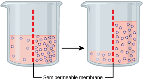
Now, imagine a beaker with a selectively permeable membrane separating the two sides or halves. On both sides of the membrane the water level is the same, but there are different concentrations of a dissolved substance, or solute, that cannot cross the membrane (otherwise the concentrations on each side would be balanced by the solute crossing the membrane).
If the volume of the solution on both sides of the membrane is the same but the concentrations of solute are different, then there are different amounts of water (the solvent) on either side of the membrane. If there is more solute in one area, then there is less water; if there is less solute in one area, then there must be more water.
A principle of diffusion is that the molecules move around due to their inherent kinetic energy. The molecules will therefore spread evenly throughout the medium if they can. However, only the material capable of passing through the membrane will diffuse through it. In this example, the solute cannot diffuse through the membrane, but the water can. Water has a concentration gradient in this system. Thus, water will diffuse down its concentration gradient, crossing the membrane to the side where it is less concentrated. This diffusion of water through the membrane — osmosis! — will continue until the concentration gradient of water goes to zero or until the hydrostatic pressure of the water balances the osmotic pressure. In the beaker example, this means that the level of fluid in the side with a higher solute concentration will go up. See the image below.