
9.4: Osteoporosis
- Page ID
- 21157
Learning Objectives
- Describe the test used to measure bone density.
- Identify test scores related to normal bone density, low bone density, and very low bone density.
- Describe osteoporosis, including its notable characteristics
- Discuss risk factors for osteoporosis.
- Describe prevention and treatment options for osteoporosis.
Osteoporosis is the excessive loss of bone over time. It leads to decreased bone strength and an increased risk for bone fractures. Osteoporosis affects more women than men, but men are also at risk for developing osteoporosis, especially after the age of seventy.
Measuring Bone Density
Bones grow and mineralize predominately during infancy, childhood, and puberty. During this time, bone growth exceeds bone loss. By age 20, bone growth is fairly complete and only a small amount (about 10%) of bone mass accumulates in the third decade of life. By age 30, bone mass is at its greatest in both men and women and then gradually declines after age 40. Bone mass refers to the total weight of bone tissue in the human body. The greatest quantity of bone tissue a person develops during his or her lifetime is called peak bone mass. The decline in bone mass after age 40 occurs because bone loss is greater than bone growth. On a cellular level, this means that the osteoclast-mediated bone degradation exceeds that of the bone building activity of osteoblasts. The increased bone degradation decreases the mineral content of bone tissue leading to a decrease in bone strength and increased fracture risk.
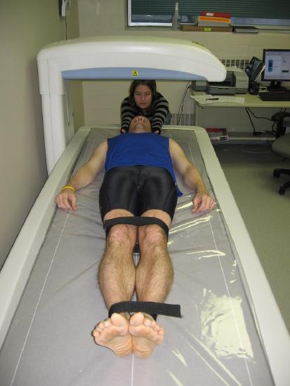
Detection and treatment of osteoporosis, before the occurrence of a fracture, can significantly improve the quality of life. To detect osteopenia (low bone density) or osteoporosis (very low bone density), bone density must be measured. Bone density is a measurement of the amount of calcified tissue in grams per centimeter squared of bone tissue. Bone density can be thought of as the total amount of bone mass in a defined area. When bone density is high, bone strength will be great. Similar to measuring blood pressure to predict the risk of stroke, a bone density measurement can help predict the risk of bone fracture. The most common tool used to measure bone density is called dual energy X-ray absorptiometry (DEXA). DEXA is the cheapest and most accurate way to measure bone density. During the procedure, a person lies on their back and a DEXA scanner passes two low-dose X-ray beams through their body (see Figure \(\PageIndex{1}\)). The amount of X-ray energy that passes through the bone is measured for both beams. The total amount of the X-ray energy that passes through a person varies depending on their bone thickness. Using this information and a defined area of bone, the amount of calcified tissue in grams per unit area (cm2) is calculated. Most often, the DEXA scan focuses on measuring bone density in the hip and the spine. These measurements are then used as indicators of overall bone strength and health.
The results of a DEXA scan are most often reported as T-scores. A T-score compares a person’s bone density to the average peak bone density of a healthy 30-year-old population of the same sex. The following bone density scores (T-scores) are used when diagnosing bone density:
- T-score between +1 and -1 indicates normal bone density
- T-score between -1 and -2.5 indicates low bone density (osteopenia) and increased risk for fractures
- T-score more negative than -2.5 indicates osteoporosis (very low bone density)
Osteoporosis
During the course of osteoporosis, bone density decreases and the bone tissue microarchitecture is compromised. Excessive bone resorption in the trabecular tissue increases the size of the holes in the lattice-like structure making it more porous and weaker. A disproportionate amount of resorption of the strong cortical bone causes it to become thinner. The deterioration of one or both types of bone tissue causes bones to weaken and, consequently, become more susceptible to fractures.
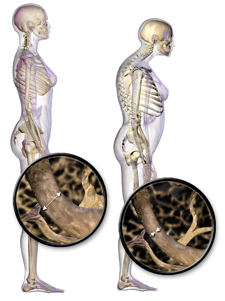
When the vertebral bone tissue is weakened, it can cause the spine to curve (Figure \(\PageIndex{2}\)). The increase in spine curvature not only causes pain, but also decreases a person’s height. Curvature of the upper spine produces what is called Dowager’s hump, also known as kyphosis.
Risk Factors for Osteoporosis
There are risk factors for osteoporosis that cannot be changed (unmodifiable), but there are also factors that can be modified to reduce risk for osteoporosis. Table \(\PageIndex{1}\) reviews the unmodifiable and modifiable risk factors for osteoporosis.
| Unmodifiable Risk Factors | Modifiable Risk Factors |
|---|---|
|
|
Prevention and Treatment for Osteoporosis
Preventing osteoporosis begins with building strong bones when you are growing. Maintaining healthy bones requires good nutrition, physical activity, and fall prevention.
Nutrition
Eating a balanced diet throughout life is helpful in preventing the onset of osteoporosis and deleterious fractures in old age. There is ample scientific evidence to suggest that low intakes of calcium and vitamin D in adulthood are linked to an increased risk for developing osteoporosis. Therefore, it is essential to make sure your diet contains adequate levels of these nutrients.
Physical Activity
Mechanical stress is one of the activation signals for bone remodeling and can increase bone strength. Exercises that apply forces to the bone increase bone density. The most helpful are weight-bearing exercises such as strength training with weights, and aerobic weight-bearing activities, such as walking, running, and stair climbing. Certain aerobic exercises such as biking and swimming do not build bones, although they are very good for cardiovascular fitness.
Fall Prevention
Reducing the number of falls a person has decreases the likelihood of sustaining a fracture. Fairly simple modifications to a person’s environment, such as installing night lights, railings on stairs, bars to hold onto in showers, and removing cords and throw rugs in walking paths can significantly reduce the likelihood of falling. Importantly, people at risk should have their vision and balance checked frequently.
Key Takeaways
- Bone density is an indicator of bone quality and correlates with bone strength.
- Bone density is a measurement of calcified bone tissue and positively correlates with overall bone health. DEXA is a clinical tool used to assess bone density.
- Excessive bone loss can lead to the development of osteopenia and eventually osteoporosis.
- Osteoporosis is influenced by unmodifiable and modifiable risk factors. Unmodifiable risk factors include age, sex, family history and race. Modifiable risk factors include physical inactivity, low calcium and vitamin D intake, cigarette smoking, low body weight and estrogen deficiency.
- Prevention of osteoporosis begins early in life with proper diet and exercise.