
2.2: Bones of the Skull
- Page ID
- 11430

The human skull is comprised of a total of 22 separate bones (excluding the ear ossicles and hyoid bone).
The cranial vault includes the following 8 bones:
- Frontal
- Parietals (2)
- Occipital
- Temporals (2)
- Ethmoid
- Sphenoid
.png?revision=1)
Skull. Anterior view
.png?revision=1)
Skull. Lateral view
.png?revision=1)
Skull. Inferior view
.png?revision=1)
Skull. Superior view
The face includes the following 14 bones:
- Lacrimals (2)
- Zygomatics (2)
- Maxillae (2)
- Mandible
- Palatines (2)
- Inferior nasal conchae (2)
- Nasals (2)
- Vomer
Flat Bones of the Skull: Frontal, Parietal, Occipital, and Temporal
The flat bones of the skull making up the neurocranium or braincase have three basic structural layers. These comprise the outer and inner layers of compact bone and an intervening layer of spongy, cancellous bone called diploe.
The inner and outer layers tend to run parallel to one another and the bones are somewhat rounded with the inner layer being concave. The areas of bone thickening or ridging generally reveal the points of muscle or ligamentous attachment. Each of the flat bones of the skull will now be reviewed individually. You should familiarize yourself with each of their distinguishing morphological features.
1. Frontal Bone
The frontal is a single bone which is comprised of two main parts, a squamous or flat portion which forms the forehead and articulates with the parietal bones and an orbital portion which provides a roof for the two orbits. The supraorbital or brow ridges are bony ridges just above the orbits. These bony ridges are quite well developed in the skulls of some forms of fossil man, but are less pronounced in modern man. The supraorbital notches or foramina are grooves or openings for the passage of neurovascular structures.
A trace of the metopic or frontal suture may be noted in the midsagittal region of this bone. The glabella is a roughened region or a bulging prominence on the frontal bone above the nasal root at about the level of the supraorbital ridges. The frontal eminences are paired prominences in the anterolateral regions of the squamous portion of the frontal bone. These prominences may vary in size and degree of development in individuals and are also a characteristic of sexual dimorphism. The median crest in the midline of the bone represents an area of muscle attachment and shows variability reflecting muscular robusticity of the individual. The paired temporal lines ascend superiorly and posteriorly from the zygomatic processes and constitute the superioranterior margin of the temporal fossae. Endocranially, note the frontal crest.
2. Parietal Bones
The parietals are paired bones which form the roof and sides of the calvaria. They articulate with one another medially at the sagittal suture and anteriorly with the frontal bone at the coronal suture. The coronal and sagittal suture intersect at a point called bregma. The bregmatic fontanelle or “anterior soft spot” exists here in infancy. Posteriorly the parietals articulate with the occipital bone at the lambdoidal suture and laterally at its squamous margin with the temporal bones. It should be noted that the lambdoidal suture has a beveled-concave surface on the parietal bones.
The temporal lines continue from the frontal bone onto the parietals, representing areas of muscle attachment. A slight elevation may be present along the sagittal suture, but tends to be poorly developed in modern man. The parietal foramina are present near the midline posteriorly and transmit veins to the sagittal sinus interiorly. Parietal foramina are a non-metric trait and may be present or absent on one or both sides. Additionally, size of the foramina should be noted as enlarged foramina may suggest heredity. Bilateral parietal eminences are prominences located postero-laterally on the parietal bones; they may or may not be present.
On the interior aspect of the parietal bones, depressions are present that are the result of the mid-meningeal arteries. To side the bone, hold the parietal in anatomical position and note that the arteries point superior and posterior; this will aid in identification of fragmentary finds. Also note the transverse sulcus or linear depression located at the inferior-posterior angle of the bone.
3. Temporal Bones
Each of these paired bones can be subdivided anatomically into a thin squamous portion which articulates with the parietal bone, a mastoid portion containing the mastoid sinuses and process, and a heavy-dense petrosal portion that contains the inner ear structure. The external auditory meatus or outer ear canal is readily apparent laterally. Projecting forward from each squama is the zygomatic process which articulates with the temporal process of the zygomatic bone. The zygomatic arch serves as an attachment for some of the muscles of mastication and is comprised of the zygomatic bone and the zygomatic process of the frontal, temporal and maxillary bones. In modern people this arch is delicate and relatively small in size and proportion.
The tympanic part and plate are located in the area surrounding the external auditory meatus. Note the mastoid crest located superior to the meatus.
Prominent, paired styloid processes may be seen projecting inferiorly and anteriorly directly below the mandibular fossae, which are also called the glenoid fossae. The condyles of the mandible articulate with the temporal bone at these fossaae. The posterior margin of the mandibular fossa is delimited by a small projection known as the postglenoid process. For siding, position the mastoid process to point inferior with the zygomatic arch pointing anterior. The external auditory meatus is lateral and the petrous process is medial.
4. Occipital Bone
A single occipital bone forms the posterior-inferior aspect of the neurocranium and is divided anatomically into a posterior-superior flat squamous part, and anteriorly projecting inferior basilar part and paired lateral parts or jugular processes. The occipital bone articulates with the two parietal bones at the lambdoid suture. Small islands of bone within this suture are called wormian bones. Lambda is a term used to designate the intersection of the lambdoid and sagittal sutures. Occassionally a transverse suture is found which separates the apex of the squamous portion from the rest of the bone. The separate apical portion is then called an Inca bone; a trait found at an especially high frequency in Peruvian peoples. The Inca bone may be singular, bipartite, or tripartite.
The large opening in the base of the occipital bone is the foramen magnum which permits the emergence of the spinal cord from the skull. The paired, kidney-shaped articular surfaces, called the occipital condyles, are situated anterior and lateral to the foramen magnum. These condyles articulate with the atlas or first cervical vertebra.
Superior to the foramen magnum and in the midsagittal plane is the external occipital protuberance. This process tends to be more prominent in males and reflects muscular robusticity. Extending downward from this projection is the external occipital crest, also called the median nuchal line. Projecting laterally from the external occipital protuberance are the supreme and superior nuchal lines. A pronounced ridge or torus defined by the superior nuchal lines is uncommon in moderns but may be quite pronounced in some forms of fossil hominids. Below the superior nuchal lines the inferior nuchal lines extend laterally. The nuchal musculature has a strong attachment to these ridges of bone and they are therefore usually more pronounced in males; in many modern skulls of bone sexes the lines are not sharply defined and may not be discernible.
The pharyngeal tubercle and fossa are two potential non-metric traits found on the basilar part of the occipital bone. Small foramina, the condylar and hypoglossal canals, allow the passage of neurovascular structures (hypoglossal nerve, etc) through the occipital bone. The condylar canal may be present, absent, or only a fossa on each side. The hypoglossal canal may be divided (internally or externally) or partially divided on each side.
Facial Bones, Sphenoid, Ethmoid, Ear Ossicles, and Hyoid
1. Zygomatic Bones
These paired quadrangularly shaped “cheek bones” are distinguished by their four separate processes. Three of the processes, the temporal, frontal, and maxillary are named according to their articulations. The fourth process which projects posteriorly from the frontal process is named the marginal process. The temporal process of the zygomatic bone and the zygomatic process of the temporal bone form the slender inferior-lateral portion of the zygomatic arches.
Occasionally, a suture separates the lower portion of the zygomatic. When present the inferior aspect of the bipartite zygomatic is termed the Os japonicum.
For siding, the concave surface is anterior, the masseter attachment is inferior, the orbital rim is smooth and rounded, and the sharp zygomatic process points posteriorly, and the long jagged articulation is medial.
2. Maxillary Bones
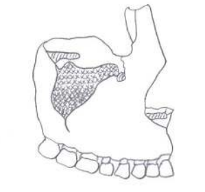
The paired maxillae contain the upper row of teeth, enclose the nasal cavity, form a portion of the orbital floors and form the anterior roof of the mouth. These bones form the major portion of the upper facial skeleton and, with the exception of the mandible, articulate with all of the other facial bones. The bodies of the maxillae contain the large maxillary sinuses which may be seen on a disarticulated skull specimen.
Four processes extend from the body of the maxillary bone. These comprise the zygomatic processes articulating with the zygomatic bone, the tooth bearing alveolar process (the alveolar arch is formed through the union of the two alveolar processes), the frontal process lateral to the nasal bone and superior to the nasal cavity, and the palatine processes which together form the greater portion of the hard palate. The infraorbital foramen located inferior to the orbit transmits cutaneous nerves to the face. Infraorbital sutures run between the infraorbital foramina and inferior orbital margins. Presence of these sutures is variable, present or absent on either side. The anterior nasal spine, subnasal groove, and nasal sill are all present at the anterior-inferior margin of the nasal cavity.
For siding, the dental arcade is inferior and sharp outline of the nasal aperture medial.
3. Nasal Bones
The thin paired nasal bones form the bridge of the nose and roof of the nasal cavity. They may vary considerably in size and configuration, shape of the suture should always be noted
4. Inferior Nasal Conchae
These paired structures comprise separate hook-like projections of bone which extend down from the lateral walls of the nasal cavity. The inferior nasal conchae articulate with the ethmoid, lacrimals, maxillae, and palatine bones throughout their extensive attachments.
5. Lacrimal Bones
The paired lacrimal bones are rectangular-shaped, small plates of bone located in the anterior medial aspect of the orbits. As the name implies these bones seat the lacrimal or tear ducts. A fine ridge of bone running superiorly-inferiorly through the central portion of this bone is called the posterior lacrimal crest. The anterior lacrimal crest is a portion of the maxillary bone.
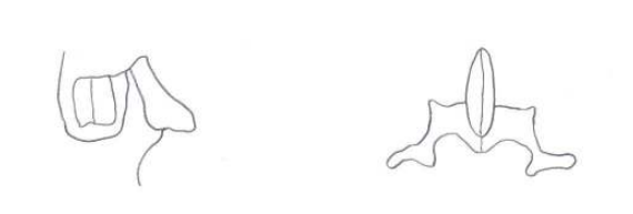
6. Vomer
This single bone is a thin plough-shaped structure which forms the inferior-posterior aspects of the nasal septum. The vomer divides the nasal cavity in the midsagittal plane. Frequently this bone is deviated to one side when viewed anteriorly. The small lateral projections superiorly and posteriorly are the alae or wings of the vomer.
7. Palatine Bones
The paired palatine bones form the posterior portion of the hard palate, a part of the floor and lateral walls of the nasal cavity and a portion of the orbital floor. Characteristic anatomic landmarks are the small, posteriorly projecting posterior nasal spine and the elevated anteriorly projecting bony ridge in the midline of the palate, called the palatine torus.
8. Mandible
The mandible or lower jaw is a large, strong, that articulates with the cranium and works in conjunction with the maxillae to masticate food. Anatomically it can be divided into a parabolically curved body containing the lower teeth and two vertically extending rami. The ramus on each side is composed of two distinct processes separated by the mandibular notch the most anterior is the coronoid process which gives attachment to the muscles of mastication. Posteriorly, the condyloid processes project superiorly to articulate with the mandibular fossae of the temporal bone. At the posterior and inferior of the mandible are located the gonial angles where the body and rami join. A mandibular symphysis is apparent during infancy, seen at midline on the anterior aspect of the mandibular body. The two halves of the mandible fuse by six months of age.
Projecting out below the symphysis is the mental tubercle or eminence which is an anatomic structure unique to man. the alveolar part contains the teeth and the paired mental foramina are found perforating the body of the mandible, located on the anterior aspect of the body and lateral to the mental eminence.
When viewing the posterior aspect of the mandible, several distinctive features can be seen. The mandibular foramen is located posterior and superior on the internal aspect of the mandible. Anterior to the mandibular foramen is the lingula, represented by a small tongue of bone. Rough, ridge-like attachments for the pterygoid muscles are found in the region of the inner aspect of the gonial angles. The mylohyoid ridge courses from posterior to anterior on the inner aspect of the mandibular body. Finally, the mental spine can be seen projecting posteriorly from the region of the symphysis where the two halves of the mandible articulate anteriorly.
9. Sphenoid
The single sphenoid bone is situated deep in the facial skeleton. This bone separates the face from the neurocranium and provides a seat called the sella turcica (or Turkish saddle) for the pituitary gland. the median or central part of this bone is called the body of the sphenoid. Extending laterally from the body are two sets of wings. The small wings form the posterior portions of the orbits while the large wings form the inferior and lateral portions of the orbits. Note that the lateral surface of the sphenoid also makes up part of the cranial vault, located just anterior to the temporal bones. on the inferior aspect of the sphenoid are projecting pointing downward called the pteryoid processes. These processes comprise lateral and medial plates; the most inferior "hook"
.png?revision=1)
Mandible, Top anterior view, middle posterior view, bottom lateral view.
of the medial plate is called the hamulus of the pterygoid. Additionally, the medial plate comprises part of the nasal walls.
Perforating the root of the large wing inferiorly are three foramina. These foramina are the foramen rotundum (for the maxillary nerve), the foramen ovale (for the mandibular nerve), and the foramen spinosum (for the middle meningeal artery). The foramen rotundum in the most medial and anterior of the three; ovale is the second most medial (and oval in shape), and the spinosum is the most lateral (and round in shape). The optic foramen (for the optic nerve) perforates the body of the sphenoid. Between the large and small wings the large superior orbital fissures are apparent.
10. Ethmoid
This single bone is essentially spongy in character and is located at the anterior aspect of the base of the neurocranium, between the two orbits. The ethmoid can be divided into four main parts: the perpendicular plate, horizontal plate, and two lateral masses. The perpendicular plate forms the upper portion of the nasal septum while the horizontal or cribriform plate is situated at a right angle to the perpendicular plate and the roof of the nasal cavity. Many small perforations pass through the cribriform plate which allows passage for branches of the olfactory (smell) nerve. An extension called the crista galli (cocks comb) is located centrally on the superior aspect of the cribriform plate. the paired lateral masses contain the conchae and the ethmoidal sinuses, as well as making up the postero-medial portion of the orbits.
11. Ear Ossicles
The three small bones of the middle ear are frequently lost during the excavation process of archaeological remains. These three bones are the incus (anvil in shape), malleus (hammer like in shape), and the stapes (stirrup shaped).
12. Hyoid
This single horseshoe-shpaed bone floats freely in the soft tissue of the nect just above the larynx. The hyoid bone has no bony articulations but gives support and attachment tot hte stylohyoid ligaments and pharyngeal musculature. This bone is divided anatomically into a central body and paired major and minor cornua or horns. If found broken in the area of the greater horns, strangulation or hanging are suggested.
.png?revision=1)
Lateral view of the skull.
.png?revision=1)
Frontal view of the skull.
.png?revision=1)
Inferior view of the skull.
.png?revision=1)
Top - Frontal Bone. Bottom - Parietal Bone.
.png?revision=1)
Top - Maxillary Bone.
.png?revision=1)
Bottom Left - Lacrimal Bone. Bottom Right - Palatine Bone.
.png?revision=1)
Top - Ethmoid. Middle - Zygomatic.
Bottom Left - Hyoid. Bottom Right - Vomer.
.png?revision=1)
Top - Temporal Bone. Bottom - Sphenoid Bone.
.png?revision=1)
Occipital Bone
.png?revision=1)
Mandible
.png?revision=1)
Top - Malleus. Middle - Incus. Bottom - Stapes.