
10.6: Appendicular Muscles of the Pectoral Girdle and Upper Limbs
- Page ID
- 22332

By the end of this section, you will be able to:
- Identify the muscles of the pectoral girdle and upper limbs
- Identify the movement and function of the pectoral girdle and upper limbs
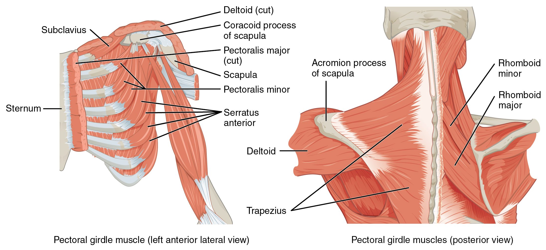
Muscles of the shoulder and upper limb can be divided into four groups: muscles that stabilize and position the pectoral girdle, muscles that move the arm, muscles that move the forearm, and muscles that move the wrists, hands, and fingers. The pectoral girdle, or shoulder girdle, consists of the lateral ends of the clavicle and scapula, along with the proximal end of the humerus, and the muscles covering these three bones to stabilize the shoulder joint. The girdle creates a base from which the head of the humerus, in its ball-and-socket joint with the glenoid fossa of the scapula, can move the arm in multiple directions.
Muscles That Position the Pectoral Girdle
Muscles that position the pectoral girdle are located either on the anterior thorax or on the posterior thorax (Figure \(\PageIndex{1}\) and Table \(\PageIndex{1}\)). The anterior muscles include the subclavius, pectoralis minor, and serratus anterior. The posterior muscles include the trapezius, rhomboid major, and rhomboid minor. When the rhomboids are contracted, your scapula moves medially, which can pull the shoulder and upper limb posteriorly.
| Prime Mover | Origin | Insertion | Movement | Target | Target motion direction | Position in the Thorax |
|---|---|---|---|---|---|---|
| Pectoralis Major | Anterior surfaces of certain ribs (2 - 4 or 3 - 5) | Corocoid process of scapula | Rotates shoulder anteriorly (throwing motion); assists with inhalation | Scapula; ribs | Scapula: depresses; ribs: elevates | Anterior thorax |
| Rhomboid major | Thoracic vertebrae (T2 - T5) | Medial border of scapula | Stabilizes scapula during pectoral girdle movement | Scapula | Retracts; rotates inferiorly | Posterior thorax |
| Rhomboid minor | Cervical and thoracic vertebrae (C7 and T1) | Medial border of scapula | Stabilizes scapula during pectoral girdle movement | Scapula | Retracts; rotates inferiorly | Posterior thorax |
| Serratus anterior | Muscle slips from certain ribs (1 - 8 or 1 - 9) | Anterior surface of vertebral border of scapula | Moves arm from side of body to front of body; assists with inhalation | Scapula; ribs | Scapula: protracts; ribs: elevates | Anterior thorax |
| Subclavius | First rib | Inferior surface of clavicle | Stabilizes clavicle during movement by depressing it | Clavicle | Depression | Anterior thorax |
| Trapezius | Skull; vertebral column | Acromion and spin of scapula; clavicle | Elevates shoulders (shrugging); pulls shoulder blades together; tilts head backwards | Scapula; cervical spine | Scapula: rotests inferiorly, retracts, elevates, and depresses; spine: extends | Posterior thorax |
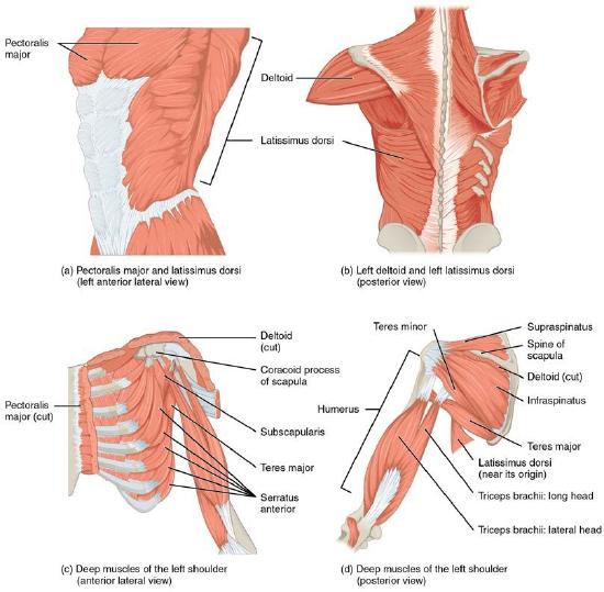
Muscles That Move the Humerus
Similar to the muscles that position the pectoral girdle, muscles that cross the shoulder joint and move the humerus bone of the arm include both axial and scapular muscles (Figure \(\PageIndex{2}\) and Table \(\PageIndex{2}\)). The two axial muscles are the pectoralis major and the latissimus dorsi. The pectoralis major is thick and fan-shaped, covering much of the superior portion of the anterior thorax. The broad, triangular latissimus dorsi is located on the inferior part of the back, where it inserts into a thick connective tissue shealth called an aponeurosis.
| Prime Mover | Origin | Insertion | Movement | Target | Target Motion Direction |
|---|---|---|---|---|---|
| Axial muscles | |||||
| Latissimus dorsi | Thoracic vertebrae (t7 - T12); lumbar vertebrae; lower ribs (9 - 12); iliac crest | Intertubercular sulcus of humerus | Moves elbow back (as in elbowing someone standing behind you); spreads elbows apart | Humerus; scapula |
Humerus: extension, adduction, and medial rotation Scapula: depression |
| Pectoralis major | Clavicle; sternum; cartilage of certain ribs (1 - 6 or 1 - 7); aponeurosis of external oblique muscle | Greater tubercle of humerus | Brings elbows together; moves elbow up (as during an uppercut punch) | Humerus | Flexion; adduction; medial rotation |
| Scapular Muscles | |||||
| Coracobra chialis | Coracoid process of scapula | Medial surface of humerus shaft | Moves elbow up and across body, as when putting hand on chest | Humerus | Flexion; adduction |
| Deltoid | Trapezius; clavicle; acromion and spine of scapula | Deltoid tuberosity of humerus | Lifts arms at shoulder | Humerus | Abduction; flexion; extension; medial and lateral rotation |
| Infraspinatus | Infraspinous fossa of scapula | Greater tubercle of humerus | Rotates elbow outwards, as during a tennis swing | Humerus | Extension; adduction |
| Subscapularis | Subscapular fossa of scapula | Lesser tubercle of humerus | Assists pectoralis major in bringing elbows together and stabilizes shoulder joint during movement of the pectoral girdle | Humerus | Medial rotation |
| Supraspinatus | Supraspinous fossa of scapula | Greater tubercle of humerus | Rotates elbow outwards, as during a tennis swing | Humerus | Abduction |
| Teres major | Posterior surface of scapula | Intertubercular sulcus of humerus | Assists infraspinatus in rotating elbow outwards | Humerus | Extension; adduction |
| Teres minor | Lateral border of dorsal scapular surface | Greater tubercle of humerus | Assists infraspinatus in rotating elbow outwards | Humerus | Extension; adduction |
The rest of the shoulder muscles originate on the scapula. The anatomical and ligamental structure of the shoulder joint and the arrangements of the muscles covering it, allows the arm to carry out different types of movements. The deltoid, the thick muscle that creates the rounded lines of the shoulder is the major abductor of the arm, but it also facilitates flexing and medial rotation, as well as extension and lateral rotation. The subscapularis originates on the anterior scapula and medially rotates the arm. Named for their locations, the supraspinatus (superior to the spine of the scapula) and the infraspinatus (inferior to the spine of the scapula) abduct the arm, and laterally rotate the arm, respectively. The thick and flat teres major is inferior to the teres minor and extends the arm, and assists in adduction and medial rotation of it. The long teres minor laterally rotates and extends the arm. Finally, the coracobrachialis flexes and adducts the arm.
The tendons of the deep subscapularis, supraspinatus, infraspinatus, and teres minor connect the scapula to the humerus, forming the rotator cuff (musculotendinous cuff), the circle of tendons around the shoulder joint. When baseball pitchers undergo shoulder surgery it is usually on the rotator cuff, which becomes pinched and inflamed, and may tear away from the bone due to the repetitive motion of bring the arm overhead to throw a fast pitch.
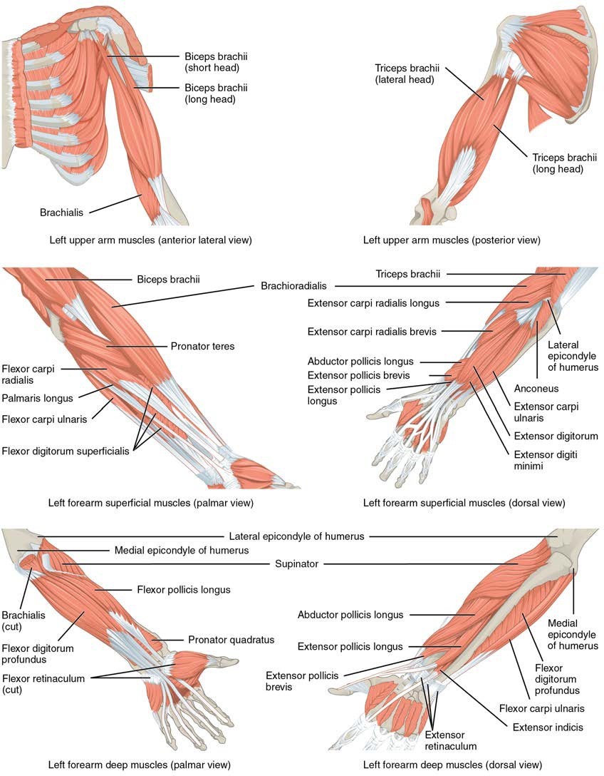
Muscles That Move the Forearm
The forearm, made of the radius and ulna bones, has four main types of action at the hinge of the elbow joint: flexion, extension, pronation, and supination. The forearm flexors include the biceps brachii, brachialis, and brachioradialis. The extensors are the triceps brachii and anconeus. The pronators are the pronator teres and the pronator quadratus, and the supinator is the only one that turns the forearm anteriorly. When the forearm faces anteriorly, it is supinated. When the forearm faces posteriorly, it is pronated.
The biceps brachii, brachialis, and brachioradialis flex the forearm. The two-headed biceps brachii crosses the shoulder and elbow joints to flex the forearm, also taking part in supinating the forearm at the radioulnar joints and flexing the arm at the shoulder joint. Deep to the biceps brachii, the brachialis provides additional power in flexing the forearm. Finally, the brachioradialis can flex the forearm quickly or help lift a load slowly. These muscles and their associated blood vessels and nerves form the anterior compartment of the arm (anterior flexor compartment of the arm) (Figure \(\PageIndex{3}\) and Table \(\PageIndex{3}\)).
| Prime Mover | Origin | Insertion | Movement | Target | Target Motion Direction |
|---|---|---|---|---|---|
| Anterior Muscles (Flexion) | |||||
| Biceps brachii | Coracoid process; tubercle above glenoid cavity | Radial tuberosity | Performs a bicep curl; also allows palm of hand to point toward body while flexing | Forearm | Flexion; supination |
| Brachialis | Front of distal humerus | Coronoid process of ulna | Performs a bicep curl | Forearm | Flexion |
| Brachioradialis | Lateral supracondylar ridge at distal end of humerus | Base of styloid process of radius | Assists and stabilizes elbow during bicep curl motion | Forearm | Flexion |
| Posterior Muscles (Extension) | |||||
| Anconeus | Lateral epicondyle of humerus | Lateral aspect of olecranon process of ulna | Assists in extending forearm; also allows forearm to extend away from the body | Forearm | Extension; abduction |
| Triceps brachii | Infraglenoid tubercle of scapula; posterior shaft of humerus; posterior humeral shaft distal to the radial groove | Olecranon process of ulna | Extends forearm, as during a punch | Forearm | Extension |
| Anterior Muscles (Pronation) | |||||
| Pronator quadratus | Distal portion of anterior ulnar shaft | Distal surface of anterior radius | Assists in turning hand palm down | Forearm | Pronation |
| Pronator teres | Medial epicondyle of humerus; coronoid process of ulna | Lateral radius | Turns hand palm down | Forearm | Pronation |
| Posterior Muscles (Supination) | |||||
| Supinator | Lateral epicondyle of humerus; proximal ulna | Proximal end of radius | Turns hand palm up | Forearm | Supination |
Muscles That Move the Wrist, Hand, and Fingers
Wrist, hand, and finger movements are facilitated by two groups of muscles. The forearm is the origin of the extrinsic muscles of the hand. The palm is the origin of the intrinsic muscles of the hand.
Muscles of the Arm That Move the Wrists, Hands, and Fingers
The muscles in the anterior compartment of the forearm (anterior flexor compartment of the forearm) originate on the humerus and insert onto different parts of the hand. These make up the bulk of the forearm. From lateral to medial, the superficial anterior compartment of the forearm includes the flexor carpi radialis, palmaris longus, flexor carpi ulnaris, and flexor digitorum superficialis. The flexor digitorum superficialis flexes the hand as well as the digits at the knuckles, which allows for rapid finger movements, as in typing or playing a musical instrument (Table \(\PageIndex{4}\) and Table \(\PageIndex{5}\)). However, poor ergonomics can irritate the tendons of these muscles as they slide back and forth with the carpal tunnel of the anterior wrist and pinch the median nerve, which also travels through the tunnel, causing Carpal Tunnel Syndrome. The deep anterior compartment produces flexion and bends fingers to make a fist. These are the flexor pollicis longus and the flexor digitorum profundus.
The muscles in the superficial posterior compartment of the forearm (superficial posterior extensor compartment of the forearm) originate on the humerus. These are the extensor radialis longus, extensor carpi radialis brevis, extensor digitorum, extensor digiti minimi, and the extensor carpi ulnaris.
The muscles of the deep posterior compartment of the forearm (deep posterior extensor compartment of the forearm) originate on the radius and ulna. These include the abductor pollicis longus, extensor pollicis brevis, extensor pollicis longus, and extensor indicis (Table \(\PageIndex{4}\)).
| Prime Mover | Origin | Insertion | Movement | Target | Target Motion Direction |
|---|---|---|---|---|---|
| Superficial Anterior Compartment of Forearm | |||||
| Flexor carpi ulnaris | Medial epicondyle of humerus; olecranon process; posterior surface of ulna | Pisiform bone; hamate bone; base of fifth metacarpal | Assists in bending hand up toward shoulder; tilts hand to side away from body; stabilizes wrist | Wrist; hand | Flexion; abduction |
| Flexor carpi radialis | Medial epicondyle of humerus | Base of second and third metacarpals | Bends wrist toward body; tilts hand to side away from body | Wrist; hand | Flexion; abduction |
| Flexor digitorum superficialis | Medial epicondyle of humerus; coronoid process of ulna; shaft of radius | Middle phalages of fingers 2 - 5 | Bends fingers to make fist | Wrist; fingers 2 - 5 | Flexion |
| Palmaris longus | Medical epicondyle of humerus | Palmar aponeurosis; skin and fascia of palm | Assists in bending hand up toward shoulder | Wrist | Flexion |
| Deep Anterior Compartment of Forearm | |||||
| Flexor digitorum profundus | Coronoid process; anteromedial surface of ulna; interosseous membrane | Distal phalanges of fingers 2 - 5 | Bends fingers to make a fist; also bends wrist toward body | Wrist; fingers | Flexion |
| Flexor pollicus longus | Anterior surface of radius; interosseous membrane | Distal phalanx of thumb | Bends tip of thumb | Thumb | Flexion |
| Superficial Posterior Compartment of Forearm | |||||
| Extensor carpi radialis brevis | Lateral epicondyle of humerus | Base of third metacarpal | Assists extensor radialis longus in extending and abducting wrist; also stabilizes hand during finger flexion | Wrist | Extension; abduction |
| Extensor carpi ulnaris | Lateral epicondyle of humerus; posterior border of ulna | Base of fifth metacarpal | Straightens wrist away from body; tilts hand to side toward body | Wrist | Extension; adduction |
| Extensor digiti minimi | Lateral epicondyle of humerus | Extensor expansion; distal phalanx of finger 5 (little finger) | Extends finger 5 (little finger) | Finger 5 (little finger) | Extension |
| Extensor digitorum | Lateral epicondyle of humerus | Extensor expansions; distal phalanges of fingers | Opens fingers and moves them sideways away from the body | Wrist; fingers | Extension; abduction |
| Extensor radialis longus | Lateral supracondylar ridge of humerus | Base of second metacarpal | Straightens wrist away from body | Wrist | Extension; abduction |
| Deep Posterior Compartment of Forearm | |||||
| Abductor pollicis longus | Posterior surface of radius and ulna; interosseous membrane | Base of first metacarpal; trapezium | Moves thumb sideways toward body; extends thumb; moves hand sideways toward body | Wrist; thumb |
Thumb: abduction; extension Wrist: abduction |
| Extensor indicis | Posterior surface of distal ulna; interosseous membrane | Tendon of extensor digitorum of finger 2 (index finger) | Extends finger 2 (index finger); straightens wrist away from body | Wrist; finger 2 (index finger) | Extension |
| Extensor pollicis brevis | Dorsal shaft of radius and ulna; interosseous membrane | Base of proximal phalanx of thumb | Extends thumb | Thumb | Extension |
| Extensor pollicis longus | Dorsal shaft of radius and ulna; interosseous membrane | Base of distal phalanx of thumb | Extends thumb | Thumb | Extension |
The tendons of the forearm muscles attach to the wrist and extend into the hand. Fibrous bands called retinacula sheath the tendons at the wrist. The flexor retinaculum extends over the palmar surface of the hand while the extensor retinaculum extends over the dorsal surface of the hand.
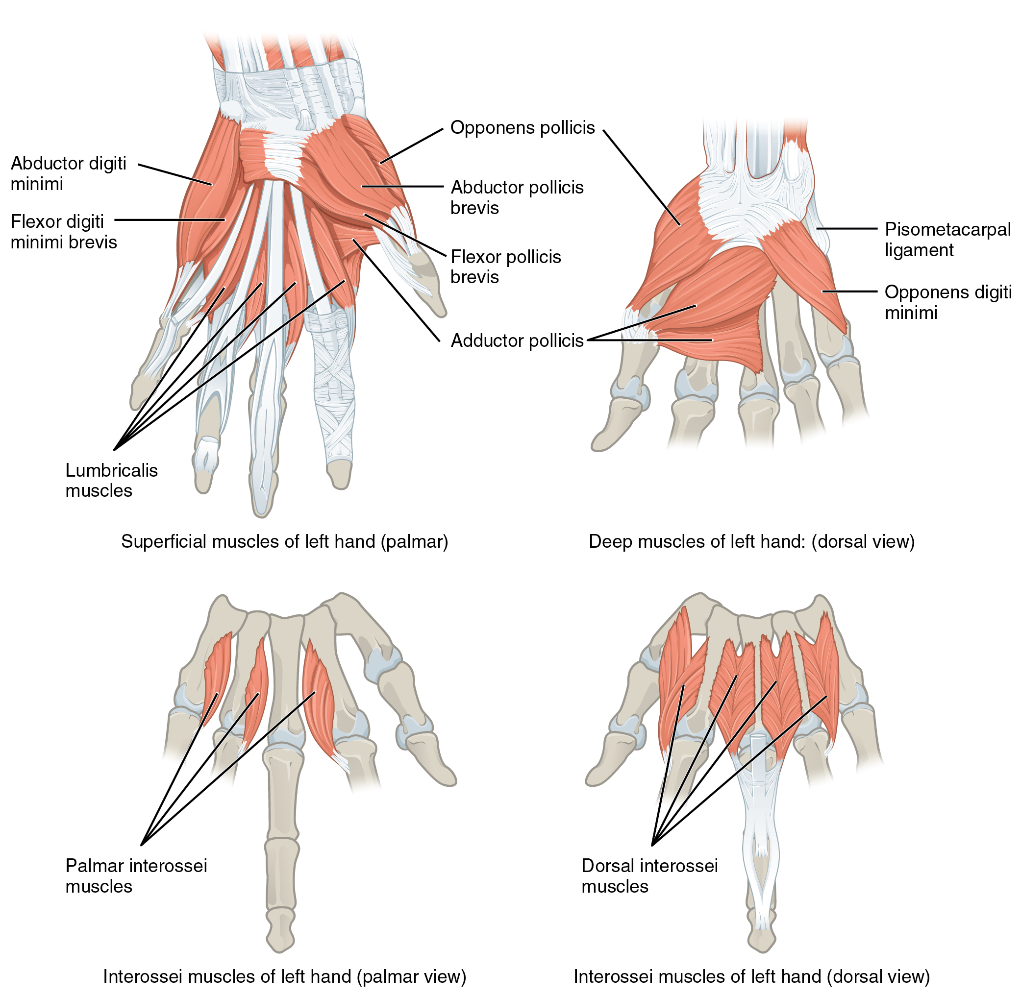
Intrinsic Muscles of the Hand
The intrinsic muscles of the hand both originate and insert within it (Figure \(\PageIndex{4}\)). These muscles allow your fingers to also make precise movements for actions, such as typing or writing. These muscles are divided into three groups. The thenar muscles are on the radial aspect of the palm. The hypothenar muscles are on the medial aspect of the palm, and the intermediate muscles are midpalmar.
The thenar muscles include the abductor pollicis brevis, opponens pollicis, flexor pollicis brevis, and the adductor pollicis. These muscles form the thenar eminence, the rounded contour of the base of the thumb, and all act on the thumb. The movements of the thumb play an integral role in most precise movements of the hand.
The hypothenar muscles include the abductor digiti minimi, flexor digiti minimi brevis, and the opponens digiti minimi. These muscles form the hypothenar eminence, the rounded contour of the little finger, and as such, they all act on the little finger. Finally, the intermediate muscles act on all the fingers and include the lumbrical, the palmar interossei, and the dorsal interossei.
| Prime mover | Origin | Insertion | Movement | Target | Target Motion Direction |
|---|---|---|---|---|---|
| Thenar Muscles | |||||
| Abductor pollicis brevis | Flexor retinaculum; and nearby carpals | Lateral base of proximal phalanx of thumb | Moves thumb toward body | Thumb | Abduction |
| Adductor pollicis | Capitate bone; bases of metacarpals 2 - 4; front of metacarpal 3 | Medial base of proximal phalanx of thumb | Moves thumb away from body | Thumb | Adduction |
| Flexor pollicis brevis | Flexor retinaculum; trapezium | Lateral base of proximal phalanx of thumb | Flexes thumb | Thumb | Flexion |
| Opponens pollicis | Flexor retinaculum; trapezium | Anterior of first metacarpal | Moves thumb across palm to touch other fingers | Thumb | Opposition |
| Hypothenar Muscles | |||||
| Abductor digiti minimi | Pisiform bone | Medial side of proximal phalanx of finger 5 (little finger) | Moves finger 5 (little finger) toward body | Finger 5 (little finger) | Abduction |
| Flexor digiti minimi brevis | Hamate bone; flexor retinaculum | Medial side of proximal phalanx of finger 5 (little finger) | Flexes finger 5 (little finger) | Finger 5 (little finger) | Flexor |
| Opponens digiti minimi | Hamate bone; flexor retinaculum | Medial side of fifth metacarpal | Moves finger 5 (little finger) across palm to touch thumb | Finger 5 (little finger) | Opposition |
| Intermediate Muscles | |||||
| Dorsal interossei | Sides of metacarpals | Both sides of finger 3; for each other finger, extensor expansion over first phalanx on side opposite finger 3 | Abducts and flexes the three middle fingers at metacarpo-phalangeal joints; extends the three middle fingers at interphalangeal joints | Fingers | Abduction; flexion; extension |
| Lumbricals | Palm (lateral sides of tendons in flexor digitorum profundus) | Fingers 2–5 (lateral edges of extensional expansions on first phalanges) | Flexes each finger at metacarpo-phalangeal joints; extends each finger at interphalangeal joints | Fingers | Flexion |
| Palmar interossei | Side of each metacarpal that faces metacarpal 3 (absent from metacarpal 3) | Extensor expansion on first phalanx of each finger (except finger 3) on side facing finger 3 | Adducts and flexes each finger at metacarpo-phalangeal joints; extends each finger at interphalangeal joints | Fingers | Adduction; flexion; extension |
Concept Review
The clavicle and scapula make up the pectoral girdle, which provides a stable origin for the muscles that move the humerus. The muscles that position and stabilize the pectoral girdle are located on the thorax. The anterior thoracic muscles are the subclavius, pectoralis minor, and the serratus anterior. The posterior thoracic muscles are the trapezius, levator scapulae, rhomboid major, and rhomboid minor. Nine muscles cross the shoulder joint to move the humerus. The ones that originate on the axial skeleton are the pectoralis major and the latissimus dorsi. The deltoid, subscapularis, supraspinatus, infraspinatus, teres major, teres minor, and coracobrachialis originate on the scapula.
The forearm flexors include the biceps brachii, brachialis, and brachioradialis. The extensors are the triceps brachii and anconeus. The pronators are the pronator teres and the pronator quadratus. The supinator is the only one that turns the forearm anteriorly.
The extrinsic muscles of the hands originate along the forearm and insert into the hand in order to facilitate crude movements of the wrists, hands, and fingers. The superficial anterior compartment of the forearm produces flexion. These muscles are the flexor carpi radialis, palmaris longus, flexor carpi ulnaris, and the flexor digitorum superficialis. The deep anterior compartment produces flexion as well. These are the flexor pollicis longus and the flexor digitorum profundus. The rest of the compartments produce extension. The extensor carpi radialis longus, extensor carpi radialis brevis, extensor digitorum, extensor digiti minimi, and extensor carpi ulnaris are the muscles found in the superficial posterior compartment. The deep posterior compartment includes the abductor longus, extensor pollicis brevis, extensor pollicis longus, and the extensor indicis.
Finally, the intrinsic muscles of the hands allow our fingers to make precise movements, such as typing and writing. They both originate and insert within the hand. The thenar muscles, which are located on the lateral part of the palm, are the abductor pollicis brevis, opponens pollicis, flexor pollicis brevis, and adductor pollicis. The hypothenar muscles, which are located on the medial part of the palm, are the abductor digiti minimi, flexor digiti minimi brevis, and opponens digiti minimi. The intermediate muscles, located in the middle of the palm, are the lumbricals, palmar interossei, and dorsal interossei.
Review Questions
Q. The rhomboid major and minor muscles are deep to the ________.
A. rectus abdominis
B. scalene muscles
C. trapezius
D. ligamentum nuchae
- Answer
-
Answer: C
Q. Which muscle extends the forearm?
A. biceps brachii
B. triceps brachii
C. brachialis
D. deltoid
- Answer
-
Answer: B
Q. What is the origin of the wrist flexors?
A. the lateral epicondyle of the humerus
B. the medial epicondyle of the humerus
C. the carpal bones of the wrist
D. the deltoid tuberosity of the humerus
- Answer
-
Answer: B
Q. Which muscles stabilize the pectoral girdle?
A. axial and scapular
B. axial
C. appendicular
D. axial and appendicular
- Answer
-
Answer: A
Critical Thinking Questions
Q. The tendons of which muscles form the rotator cuff? Why is the rotator cuff important?
- Answer
-
A. Tendons of the infraspinatus, supraspinatus, teres minor, and the subscapularis form the rotator cuff, which forms a foundation on which the arms and shoulders can be stabilized and move.
Q. List the general muscle groups of the shoulders and upper limbs as well as their subgroups.
- Answer
-
A. The muscles that make up the shoulders and upper limbs include the muscles that position the pelvic girdle, the muscles that move the humerus, the muscles that move the forearm, and the muscles that move the wrists, hands, and fingers.
Glossary
- abductor digiti minimi
- muscle that abducts the little finger
- adductor pollicis
- muscle that adducts the thumb
- abductor pollicis brevis
- muscle that abducts the thumb
- abductor pollicis longus
- muscle that inserts into the first metacarpal
- anconeus
- small muscle on the lateral posterior elbow that extends the forearm
- anterior compartment of the arm
- (anterior flexor compartment of the arm) the biceps brachii, brachialis, brachioradialis, and their associated blood vessels and nerves
- anterior compartment of the forearm
- (anterior flexor compartment of the forearm) deep and superficial muscles that originate on the humerus and insert into the hand
- biceps brachii
- two-headed muscle that crosses the shoulder and elbow joints to flex the forearm while assisting in supinating it and flexing the arm at the shoulder
- brachialis
- muscle deep to the biceps brachii that provides power in flexing the forearm.
- brachioradialis
- muscle that can flex the forearm quickly or help lift a load slowly
- coracobrachialis
- muscle that flexes and adducts the arm
- deep anterior compartment
- flexor pollicis longus, flexor digitorum profundus, and their associated blood vessels and nerves
- deep posterior compartment of the forearm
- (deep posterior extensor compartment of the forearm) the abductor pollicis longus, extensor pollicis brevis, extensor pollicis longus, extensor indicis, and their associated blood vessels and nerves
- deltoid
- shoulder muscle that abducts the arm as well as flexes and medially rotates it, and extends and laterally rotates it
- dorsal interossei
- muscles that abduct and flex the three middle fingers at the metacarpophalangeal joints and extend them at the interphalangeal joints
- extensor carpi radialis brevis
- muscle that extends and abducts the hand at the wrist
- extensor carpi ulnaris
- muscle that extends and adducts the hand
- extensor digiti minimi
- muscle that extends the little finger
- extensor digitorum
- muscle that extends the hand at the wrist and the phalanges
- extensor indicis
- muscle that inserts onto the tendon of the extensor digitorum of the index finger
- extensor pollicis brevis
- muscle that inserts onto the base of the proximal phalanx of the thumb
- extensor pollicis longus
- muscle that inserts onto the base of the distal phalanx of the thumb
- extensor radialis longus
- muscle that extends and abducts the hand at the wrist
- extensor retinaculum
- band of connective tissue that extends over the dorsal surface of the hand
- extrinsic muscles of the hand
- muscles that move the wrists, hands, and fingers and originate on the arm
- flexor carpi radialis
- muscle that flexes and abducts the hand at the wrist
- flexor carpi ulnaris
- muscle that flexes and adducts the hand at the wrist
- flexor digiti minimi brevis
- muscle that flexes the little finger
- flexor digitorum profundus
- muscle that flexes the phalanges of the fingers and the hand at the wrist
- flexor digitorum superficialis
- muscle that flexes the hand and the digits
- flexor pollicis brevis
- muscle that flexes the thumb
- flexor pollicis longus
- muscle that flexes the distal phalanx of the thumb
- flexor retinaculum
- band of connective tissue that extends over the palmar surface of the hand
- hypothenar
- group of muscles on the medial aspect of the palm
- hypothenar eminence
- rounded contour of muscle at the base of the little finger
- infraspinatus
- muscle that laterally rotates the arm
- intermediate
- group of midpalmar muscles
- intrinsic muscles of the hand
- muscles that move the wrists, hands, and fingers and originate in the palm
- latissimus dorsi
- broad, triangular axial muscle located on the inferior part of the back
- lumbrical
- muscle that flexes each finger at the metacarpophalangeal joints and extend each finger at the interphalangeal joints
- opponens digiti minimi
- muscle that brings the little finger across the palm to meet the thumb
- opponens pollicis
- muscle that moves the thumb across the palm to meet another finger
- palmar interossei
- muscles that abduct and flex each finger at the metacarpophalangeal joints and extend each finger at the interphalangeal joints
- palmaris longus
- muscle that provides weak flexion of the hand at the wrist
- pectoral girdle
- shoulder girdle, made up of the clavicle and scapula
- pectoralis major
- thick, fan-shaped axial muscle that covers much of the superior thorax
- pectoralis minor
- muscle that moves the scapula and assists in inhalation
- pronator quadratus
- pronator that originates on the ulna and inserts on the radius
- pronator teres
- pronator that originates on the humerus and inserts on the radius
- retinacula
- fibrous bands that sheath the tendons at the wrist
- rhomboid major
- muscle that attaches the vertebral border of the scapula to the spinous process of the thoracic vertebrae
- rhomboid minor
- muscle that attaches the vertebral border of the scapula to the spinous process of the thoracic vertebrae
- rotator cuff
- (also, musculotendinous cuff) the circle of tendons around the shoulder joint
- serratus anterior
- large and flat muscle that originates on the ribs and inserts onto the scapula
- subclavius
- muscle that stabilizes the clavicle during movement
- subscapularis
- muscle that originates on the anterior scapula and medially rotates the arm
- superficial anterior compartment of the forearm
- flexor carpi radialis, palmaris longus, flexor carpi ulnaris, flexor digitorum superficialis, and their associated blood vessels and nerves
- superficial posterior compartment of the forearm
- extensor radialis longus, extensor carpi radialis brevis, extensor digitorum, extensor digiti minimi, extensor carpi ulnaris, and their associated blood vessels and nerves
- supinator
- muscle that moves the palm and forearm anteriorly
- supraspinatus
- muscle that abducts the arm
- teres major
- muscle that extends the arm and assists in adduction and medial rotation of it
- teres minor
- muscle that laterally rotates and extends the arm
- thenar
- group of muscles on the lateral aspect of the palm
- thenar eminence
- rounded contour of muscle at the base of the thumb
- trapezius
- muscle that stabilizes the upper part of the back
- triceps brachii
- three-headed muscle that extends the forearm
Contributors and Attributions
OpenStax Anatomy & Physiology (CC BY 4.0). Access for free at https://openstax.org/books/anatomy-and-physiology