
12.5E: Trochlear (IV) Nerve
- Page ID
- 7670
The trochlear nerve (cranial nerve IV) is a motor nerve that innervates a single muscle: the superior oblique muscle of the eye.
- Describe the trochlear nerve (cranial nerve IV)
Key Points
- The trochlear nerve innervates the superior oblique muscle of the eye.
- The trochlear nerve contains the smallest number of axons of all the cranial nerves and has the greatest intracranial length.
- The two major clinical syndromes that can arise from damage to the trochlear nerve are vertical and torsional diplopia.
Key Terms
- contralateral: On the opposite side of the body.
- cerebral aqueduct: The channel in the brain that connects the third ventricle to the fourth ventricle. Also called the aqueduct of Sylvius, it is surrounded by periaqueductal gray matter.
The trochlear nerve (cranial nerve IV) is a motor nerve that innervates a single muscle: the superior oblique muscle of the eye.
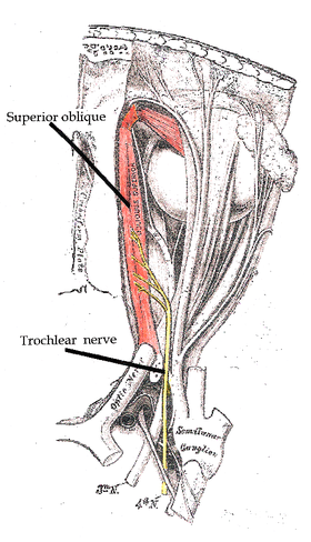
The trochlear nerve: The trocheal nerve and where it innervates.
The trochlear nerve is unique among the cranial nerves in several respects.
- It is the smallest nerve in terms of the number of axons it contains and it has the greatest intracranial length.
- Other than the optic nerve (cranial nerve II), it is the only cranial nerve that decussates (crosses to the other side) before innervating its target.
- It is the only cranial nerve that exits from the dorsal aspect of the brainstem.
The nucleus of the trochlear nerve is located in the caudal mesencephalon beneath the cerebral aqueduct. It is immediately below the nucleus of the oculomotor nerve (III) in the rostral mesencephalon.
The trochlear nucleus is unique in that its axons run dorsally and cross the midline before emerging from the brainstem—so a lesion of the trochlear nucleus affects the contralateral eye. Lesions of all other cranial nuclei affect the ipsilateral side (except of course the optic nerve, cranial nerve II, which innervates both eyes).
Homologous trochlear nerves are found in all jawed vertebrates. The unique features of the trochlear nerve, including its dorsal exit from the brainstem and its contralateral innervation, are seen in the primitive brains of sharks.
The human trochlear nerve is derived from the basal plate of the embryonic midbrain.
Clinical Syndromes
There are two major clinical syndromes that can manifest through damage to the trochlear nerve:
- Vertical diplopia: Injury to the trochlear nerve causes weakness of downward eye movement with consequent vertical diplopia (double vision).
- Torsional diplopia: Weakness of intorsion results in torsional diplopia, in which two different visual fields, tilted with respect to each other, are seen at the same time. To compensate for this, patients with trochlear nerve palsies tilt their heads to the opposite side, in order to fuse the two images into a single visual field.
The clinical syndromes can originate from both peripheral and central lesions. A peripheral lesion is damage to the bundle of nerves, in contrast to a central lesion, which is damage to the trochlear nucleus.