
14.5: Parts of the Nervous System
- Page ID
- 2266

When we describe the nervous system of vertebrates we usually divide it into two parts (see diagram 14.5).
- 1. The central nervous system (CNS) which consists of the brain and spinal cord.
- 2. The peripheral nervous system (PNS) which consists of the nerves that connect to the brain and spinal cord (cranial and spinal nerves) as well as the autonomic (or involuntary) nervous system.
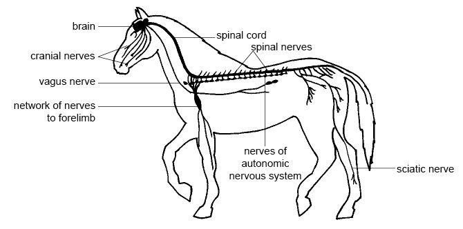
Diagram 14.5 - The nervous system of a horse
The Central Nervous System
The central nervous system consists of the brain and spinal cord. It acts as a kind of ‘telephone exchange’ where a vast number of cross connections are made.
When you look at the brain or spinal cord some regions appear creamy white (white matter) and others appear grey (grey matter). White matter consists of masses of nerve axons and the grey matter consists of the nerve cells. In the brain the grey matter is on the outside and in the spinal cord it is on the inside (see diagram 14.2).
The Brain
The major part of the brain lies protected within the sturdy “box” of skull called the cranium. Surrounding the fragile brain tissue (and spinal cord) are protective membranes called the meninges (see diagram 14.6), and a crystal-clear fluid called cerebrospinal fluid, which protects and nourishes the brain tissue. This fluid also fills four cavities or ventricles that lie within the brain.
Brain tissue is extremely active and, even when an animal is resting, it uses up to 20% of the oxygen taken into the body by the lungs. The carotid artery, a branch off the dorsal aorta, supplies it with the oxygen and nutrients it requires. Brain damage occurs if brain tissue is deprived of oxygen for only 4–8 minutes.
The brain consists of three major regions:
- 1. the fore brain which includes the cerebral hemispheres, hypothalamus and pituitary gland;
- 2. the hind brain or brain stem, contains the medulla oblongata and pons and
- 3. the cerebellum or “little brain” (see diagram 14.6).

Diagram 14.6 - Longitudinal section through the brain of a dog
Mapping the brain
In humans and some animals the functions of the different regions of the cerebral cortex have been mapped (see diagram 14.7).
Diagram 14.7 - The functions of the regions of the human cerebral cortex
The Forebrain
The cerebral hemispheres are the masses of brain tissue that sit on the top of the brain. The surface is folded into ridges and furrows called sulci (singular sulcus). They make this part of the brain look rather like a very large walnut kernel. The two hemispheres are separated by a deep groove although they are connected internally by a thick bundle of nerve fibres. The outer layer of each hemisphere is called the cerebral cortex and this is where the main functions of the cerebral hemispheres are carried out.
The cerebral cortex is large and convoluted in mammals compared to other vertebrates and largest of all in humans because this is where the so-called “higher centres” concerned with memory, learning, reasoning and intelligence are situated.
Nerves from the eyes, ears, nose and skin bring sensory impulses to the cortex where they are interpreted. Appropriate voluntary movements are initiated here in the light of the memories of past events.
Different regions of the cortex are responsible for particular sensory and motor functions, e.g. vision, hearing, taste, smell, or moving the fore-limbs, hind-limbs or tail. For example, when a dog sniffs a scent, sensory impulses from the organ of smell in the nose pass via the olfactory (smelling) nerve to the olfactory centres of the cerebral hemispheres where the impulses are interpreted and co-ordinated.
In humans and some animals the functions of the different regions of the cerebral cortex have been mapped (see diagram 14.8).

Diagram 14.8 - The functions of the regions of the cerebral cortex
The hypothalamus is situated at the base of the brain and is connected by a “stalk” to the pituitary gland, the “master” hormone-producing gland (see chapter 16). The hypothalamus can be thought of as the bridge between the nervous and endocrine (hormone producing) systems. It produces some of the hormones that are released from the pituitary gland and controls the release of others from it.
It is also an important centre for controlling the internal environment of the animal and therefore maintaining homeostasis. For example, it helps regulate the movement of food through the gut and the temperature, blood pressure and concentration of the blood. It is also responsible for the feeling of being hungry or thirsty and it controls sleep patterns and sex drive.
The Hindbrain
The medulla oblongata is at the base of the brain and is a continuation of the spinal cord. It carries all signals between the spinal cord and the brain and contains centres that control vital body functions like the basic rhythm of breathing, the rate of the heartbeat and the activities of the gut. The medulla oblongata also co-ordinates swallowing, vomiting, coughing and sneezing.
The Cerebellum
The cerebellum (little brain) looks rather like a smaller version of the cerebral hemispheres attached to the back of the brain. It receives impulses from the organ of balance (vestibular organ) in the inner ear and from stretch receptors in the muscles and tendons. By co-ordinating these it regulates muscle contraction during walking and running and helps maintain the posture and balance of the animal. When the cerebellum malfunctions it causes a tremor and uncoordinated movement.
The Spinal Cord
The spinal cord is a cable of nerve tissue that passes down the channel in the vertebrae from the hindbrain to the end of the tail. It becomes progressively smaller as paired spinal nerves pass out of the cord to parts of the body. Protective membranes or meninges cover the cord and these enclose cerebral spinal fluid (see diagram 14.9).

Diagram 14.9 - The spinal cord
If you cut across the spinal cord you can see that it consists of white matter on the outside and grey matter in the shape of an H or butterfly on the inside.
The Peripheral Nervous System
The peripheral nervous system consists of nerves that are connected to the brain (cranial nerves), and nerves that are connected to the spinal cord (spinal nerves). The autonomic nervous system is also part of the peripheral nervous system.
Cranial Nerves
There are twelve pairs of cranial nerves that come from the brain. Each passes through a hole in the cranium (brain case). The most important of these are the olfactory, optic, acoustic and vagus nerves.
- The olfactory nerves - (smell) carry impulses from the olfactory organ of the nose to the brain.
- The optic nerves - (sight) carry impulses from the retina of the eye to the brain.
- The auditory (acoustic) nerves - (hearing) carry impulses from the cochlear of the inner ear to the brain.
- The vagus nerve - controls the muscles that bring about swallowing. It also controls the muscles of the heart, airways, lungs, stomach and intestines (see diagram 14.5).
Spinal Nerves
Spinal nerves connect the spinal cord to sense organs, muscles and glands in the body. Pairs of spinal nerves leave the spinal cord and emerge between each pair of adjacent vertebrae (see diagram 14.9).
The sciatic nerve is the largest spinal nerve in the body (see diagram 14.5). It leaves the spinal cord as several nerves that join to form a flat band of nervous tissue. It passes down the thigh towards the hind leg where it gives off branches to the various muscles of this limb.
The Autonomic Nervous System
The autonomic nervous system controls internal body functions that are not under conscious control. For example when a prey animal is chased by a predator the autonomic nervous system automatically increases the rate of breathing and the heartbeat. It dilates the blood vessels that carry blood to the muscles, releases glucose from the liver, and makes other adjustments to provide for the sudden increase in activity. When the animal has escaped and is safe once again the nervous system slows down all these processes and resumes all the normal body activities like the digestion of food.
The nerves of the autonomic nervous system originate in the spinal cord and pass out between the vertebrae to serve the various organs (see diagram 14.10).There are two main parts to the autonomic nervous system—the sympathetic system and the parasympathetic system.
The sympathetic system stimulates the “flight, fright, fight” response that allows an animal to face up to an attacker or make a rapid departure. It increases the heart and respiratory rates, as well as the amount of blood flowing to the skeletal muscles while blood flow to less critical regions like the gut and skin is reduced. It also causes the pupils of the eyes to dilate. Note that the effects of the sympathetic system are similar to the effects of the hormone adrenaline (see Chapter 16).
The parasympathetic system does the opposite to the sympathetic system. It maintains the normal functions of the relaxed body. These are sometimes known as the “housekeeping” functions. It promotes effective digestion, stimulates defaecation and urination and maintains a regular heartbeat and rate of breathing.

Diagram 14.10 - The function of the sympathetic and parasympathetic nervous systems