
27.3A: Trophoblast Development
- Page ID
- 8271
Trophoblasts are the outer layer of cells that provide nutrients to the embryo and form part of the placenta.
- Describe the trophoblast
Key Points
- The trophoblast is subdivided into two layers, the inner cytotrophoblast, which proliferates upon implantation, and the outer syncytiotrophoblast, which supports cytotrophoblast proliferation through contact with the maternal blood.
- The cytotrophoblast, at the edges of the villi in the placenta, further differentiates into extravillous trophoblasts, which penetrate into the uterus and attach to the placenta and the mother to promote placental vasculature.
- Some trophoblasts replace endothelial cells in the uterine spiral arteries and remodel them into wide-bore conduits that are independent of maternal vasoconstriction. This ensures the fetus receives a steady supply of blood, and that the placenta is not sensitive to damaging fluctuations in oxygen.
Key Terms
- decidualization: The changes in the endometrial lining after ovulation, characterized by its transformation into a secretory lining in preparation to accept an embryo.
- extravillous trophoblast: These grow out from the placenta and penetrate into the decidualized uterus.
- trophoblast: The membrane of cells that forms the wall of a blastocyst during early pregnancy, and also provides nutrients to the embryo and later develops into part of the placenta.
Trophoblasts
Trophoblasts (from the Greek words trephein, to feed, and blastos, germinator) are cells that form the outer layer of a blastocyst. These cells provide nutrients to the embryo and develop into a large part of the placenta.
They are formed during the first stage of pregnancy and are the first cells to differentiate from the fertilized egg. This layer of trophoblasts is also collectively referred to as the trophoblast, or, after gastrulation, the trophectoderm, as it is then contiguous with the ectoderm of the embryo.
Trophoblast Function
Trophoblasts play an important role in embryo implantation and interaction with the endometrium of the maternal uterus following decidualization. The trophoblast is composed of two layers: an inner cytotrophoblast and an outer syncytiotrophoblast.
The syncytiotrophoblast is non-proliferative and thus relies on fusion of the underlying cytotrophoblast cells to expand. The syncytiotrophoblast are the cells in direct contact with the maternal blood that reaches the placental surface, and thus facilitates the exchange of nutrients, wastes, and gases between the maternal and fetal systems.
Cytotrophoblast in the tips of villi can differentiate into another type of trophoblast called the extravillous trophoblast. Extravillous trophoblasts grow out from the placenta and penetrate into the decidualized uterus.
This process is essential not only for physically attaching the placenta to the mother, but also for altering the vasculature in the uterus to allow it to provide an adequate blood supply to the growing fetus as pregnancy progresses.
Some of the trophoblast even replaces the endothelial cells in the uterine spiral arteries as they remodel these vessels into wide bore conduits that are independent of maternal vasoconstriction. This ensures the fetus receives a steady supply of blood, and the placenta is not sensitive to fluctuations in oxygen that could cause it damage.
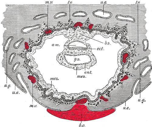
Extraembryonic coelom or chorionic cavitiy: A blastocyst embedded in the uterine decidua. (am. Amniotic cavity, b.c. Blood clot., b.s. Body stalk., etc Embryonic ectoderm, ent. Entoderm., mes. Mesoderm, m.v. Maternal vessels., tr. Trophoblast., u.e. Uterine epithelium, u.g. Uterine glands., y.s. Yolk-sac.)
Clinical Example
Intrauterine growth restriction (IUGR) can result in an undersized fetus with poor organ development or even death. The primary factor in IUGR is placental dysfunction caused by a failure of the extravillous trophoblasts to penetrate and modify the uterine spiral arteries. The result is a poorly-oxygenated environment and reduced fetal growth.