
26.5D: Female Duct System
- Page ID
- 8253
The Fallopian tubes, or oviducts, connect the ovaries to the uterus.
- Describe the structures of the female duct system
Key Points
- The Fallopian tube allows passage of the egg from the ovary to the uterus.
- The lining of the Fallopian tubes are ciliated and have several segments, including the infundibulum, ampullary, isthmus, and interstitial regions.
- Interspersed between the ciliated cells are peg cells, which contain apical granules and produce the tubular fluid that contains nutrients for spermatozoa, oocytes, and zygotes.
- Occasionally, the embryo implants into the Fallopian tube instead of the uterus, creating an ectopic pregnancy.
Key Terms
- oviduct: A duct through which an ovum passes from an ovary to the uterus or to the exterior.
- fallopian tubes: Also known as oviducts, uterine tubes, and salpinges (singular salpinx), two very fine tubes lined with ciliated epithelia, leading from the ovaries of female mammals into the uterus via the uterotubal junction.
- ovarian follicle: The basic units of female reproductive biology, each composed of roughly spherical aggregations of cells found in the ovary.
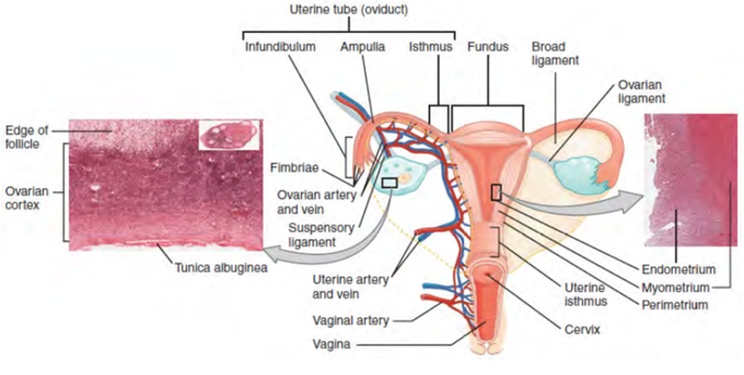
The Fallopian tubes, also known as oviducts, uterine tubes, and salpinges (singular salpinx), are two very fine tubes lined with ciliated epithelia, leading from the ovaries of female mammals into the uterus via the uterotubal junction. In non-mammalian vertebrates, the equivalent structures are the oviducts. These tubes allows passage of the egg from the ovary to the uterus.
The different segments of the fallopian tube are ( lateral to medial):
- The infundibulum with associated fimbriae near the ovary
- The ampullary region that represents the major portion of the lateral tube
- The isthmus, which is the narrower part of the tube that links to the uterus
- The interstitial (intramural) part that transverses the uterine musculature
The tubal ostium is the point at which the tubal canal meets the peritoneal cavity, while the uterine opening of the Fallopian tube is the entrance into the uterine cavity, the uterotubal junction.
Uterine Segments: Illustrative drawing of the anterior view of the uterus showing the uterine segments
There are two types of cells within the simple columnar epithelium of the Fallopian tube. Ciliated cells predominate throughout the tube, but are most numerous in the infundibulum and ampulla. Estrogen increases the production of cilia on these cells.
Interspersed between the ciliated cells are peg cells, which contain apical granules and produce the tubular fluid. This fluid contains nutrients for spermatozoa, oocytes, and zygotes. The secretions also promote capacitation of the sperm by removing glycoproteins and other molecules from the plasma membrane of the sperm. Progesterone increases the number of peg cells, while estrogen increases their height and secretory activity. Tubal fluid flows against the action of the ciliae, toward the fimbrated end.
When an ovum is developing in an ovary, it is encapsulated in a sac known as an ovarian follicle. On maturation, the follicle and the ovary’s wall rupture, allowing the ovum to escape. The egg is caught by the fimbriated end and travels to the ampulla where typically the sperm are met and fertilization occurs. The fertilized ovum, now a zygote, travels towards the uterus aided by the tubal cilia and tubal muscle. After about five days, the new embryo enters the uterine cavity and implants about a day later. Occasionally, the embryo implants into the Fallopian tube instead of the uterus, creating an ectopic pregnancy.