
3.3: Open Cervical Surgery for Congenital H-type Tracheoesophageal Fistulae
- Page ID
- 15442

OPEN ACCESS ATLAS OF OTOLARYNGOLOGY, HEAD & NECK OPERATIVE SURGERY
OPEN CERVICAL SURGERY FOR CONGENITAL H-TYPE TRACHEOESOPHAGEAL FISTULAE (TOF)
Mark Quick, Shyan Vijayasekaran
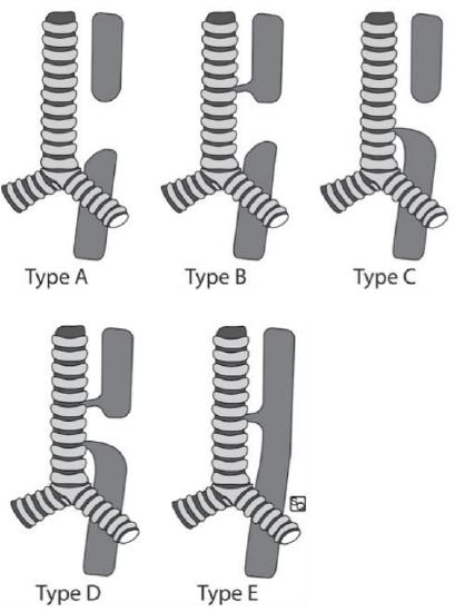
Figure 1: Gross TOF classification4
Esophageal atresia (OA) with or without a tracheoesophageal fistula (TOF) has an incidence of 1 in 2500 – 4500 live births.1 Two distinct TOF classification systems are used, with the initial system by Vogt published in 1929, that was later modified by Gross in 1953 into five types of TOF (Figure 1). 2,3
The most common TOF is Type C (86%) comprising of a proximal blind esophageal pouch and distal TOF into the distal esophagus (Figure 1). Type E, better known as an H-type (Figure 1) fistula without OA, has an incidence of 4-7% of all TOFs.4 H-type fistulae have occurs in 1 in 50,000 to 80,000 live births.5
Endoscopic procedures have been employed to obliterate TOFs and include using fibrin glue, electrocautery, or laser coagulation of the fistula tract.6,7 Surgery remains the standard treatment with low recurrence rates.4,8-10 An open cervical approach is recommended for TOFs located above the level of T211 which accounts for majority of H-type TOFs.
Etiology
The etiology of TOF/OA is poorly understood. Although a TOF may occur in isolation, 50% are associated with congenital anomalies with cardiac anomalies being the most common. These associated anomalies are summarized in the genetic syndrome VACTERL (vertebral, anorectal, cardiac, tracheoesophageal, renal and limb abnormalities) (Table 1). Other rare associated syndromes include Pierre-Robin, CHARGE or Di George with its chromosomal abnormality on 22q11 associated with esophageal atresia.
| Type | Incidence | Anomalies |
| Vertebral | 17% | Scoliosis |
| Anal | 12% | Imperforate anus, Duodenal atresia |
| Cardiac | 20% | VSD, PDA, Tetralogy of Fallot, ASD |
| Renal | 16% | Renal agenesis / dysplasia, hypospadias, polycystic |
| Limb | 10% | Radial anomalies, polydactyly |
Table 1: Congenital anomalies associated with TOF/OA14
Anatomy and Embryology
The trachea and esophagus develop from a ventral diverticulum of the foregut. Proliferation of endodermal cells around week 3 of intrauterine life occurs over the diverticulum. The division of this cell mass forms the trachea and esophagus. A TOF occurs during this period because of disruption to normal development. The primary mechanism of this disruption is unknown. However several key mutations including that of the Sonic hedgehog (Shh) gene identified in animal models result in failure of tracheoesophageal separation and fistula formation.12
Diagnosis
Presentation and examination
TOFs are not easily diagnosed antenatally. Polyhydramnios identified on prenatal ultrasound may be secondary to esophageal obstruction but is nonspecific and should not be used solely as a diagnostic marker.

Figures 2 a, b: Chest X-ray showing curled nasogastric tube at blind-ending esophageal lumen and gastric bubble suspicion for TOF
The diagnosis of OA is made early following delivery and is confirmed by inability to pass a nasogastric tube into the stomach (Figure 2).

Figure 3: MLB with posterior tracheal wall TOF identified
The type of TOF is determined at microlaryngoscopy and bronchoscopy (MLB); however, subtle deformities and the level of a TOF can be missed (Figure 3).
H-type TOFs has more variable presentations. A delay or even misdiagnosis can occur as the esophagus remains intact without disruption. Common clinical symptoms include choking or cyanotic spells during feeding, abdominal distension leading to splinting during inhalation, or recurrent chest infections. These symptoms can be misdiagnosed as gastroesophageal reflux. A high index of suspicion for H-type TOF is important particularly with patients presenting with recurrent chest infections.
Investigations
Endoscopic evaluation including MLB and esophagoscopy can identify the location, size, and number of TOFs, which are all important for surgical planning. Other associated laryngeal or tracheal abnormalities can be identified at the time of MLB.

Figure 4: Contrast radiographic study showing communication between esophagus and trachea
Contrast radiographic studies (tube esophagograms) are useful in premature and very small neonates where MLB is technically difficult. It avoids a general anesthetic in neonates with complex underlying medical conditions. The neonate is placed supine or in a lateral position and contrast is injected into a nasogastric tube while slowly withdrawing it within the esophagus. If there is a fistula, contrast will spill into the trachea. This is not routinely employed by the senior author (Figure 4).
Management
Immediate

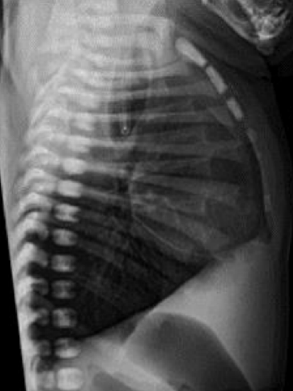
Figure 5: Chest X-ray showing the NGT in the stomach with significant gastric inflation from a suspected TOF
A neonate presenting with difficulties with swallowing or respiratory distress with abdominal splinting following delivery as outlined above, should be transferred to a neonatal intensive care unit. A nasogastric tube (NGT) should be inserted, and its position confirmed on x-ray. Increasing abdominal distension with inhalation and a confirmed gastric position of a NGT should raise the suspicion of an H-type TOF (Figure 5).
Aspiration of abdominal gas to alleviate abdominal splinting can be performed to improve breathing. Neonatologists, pediatric ENTs, pediatric general surgeons, and pediatricians should be consulted.
Tracheoesophageal fistula identification

Figure 6: Esophagoscopy identifying Foley catheter cannulated through the TOF from the trachea
Initial management is directed at identifying the TOF, including the level, size, and number of fistulae. Using MLB, the TOF can be cannulated with a size 5/6 FG Foley catheter and then confirmed with esophagoscopy (Figure 6). TOFs located above T2 are amenable to open cervical surgery.
Surgical techniques to close H-type TOF: open cervical approaches
Two open cervical surgical options can be used for the H-type TOF i.e., extraluminal or transtracheal ‘keyhole’ techniques. The open cervical approach is selected at the time of the MLB. Patients are intubated with an age-appropriate endotracheal tube (ETT) to confirm that the cuff is located distal to the TOF.
Extraluminal technique
- Following MLB, place the neonate or child in a supine position on a shoulder roll +/- head ring
- Administer a single dose of prophylactic antibiotics
- Identify anterior neck landmarks including thyroid cartilage, trachea, and sternal notch
- Mark a standard horizontal tracheostomy neck incision
- Infiltrate the subcutaneous tissues with local anesthetic (1% lignocaine) and 1:100,000 adrenaline, not exceeding the maximum safe dose
- Prep the skin, and drape to protect the eyes, but to cover the mouth with the anterior neck and upper chest exposed
- Incise skin and subcutaneous tissue
- Raise subplatysmal flaps and retract the flaps with silk stay sutures
- Separate the strap muscles along the median raphe and retract them laterally
- Identify the trachea
- Divide the thyroid isthmus
- Create extraluminal dissection planes along the trachea up to the tracheoesophageal groove, staying on the tracheal adventitia
- Staying close to tracheal adventitia reduces the risk of injury to the recurrent laryngeal nerves
- Palpating a Foley catheter placed through the TOF at the time of the MLB can assist with identification of the TOF

Figure 7: Extraluminal approach with vessel loop isolating the TOF and stay sutures securing the subplatysmal flaps4
- Having identified and circumferentially isolated the TOF, place a vessel loop around the TOF and withdraw the Foley catheter if previously placed through the TOF (Figure 7)
- Sharply divide the TOF
- Individually close the tracheal and esophageal sides of the TOF
- Close the tracheal side with long lasting absorbable suture 4/0 PDS (Ethicon, Inc., Johnson and Johnson, New Jersey, US)
- Close the esophageal defect using an inverting Connell suture technique with braided absorbable suture 4/0 Vicryl (Ethicon, Inc., Johnson and Johnson, New Jersey, US)
- Reinforce the closure by interposing a local rotation muscle flap using one of the strap muscles, or a free graft of perichondrium from the manubrium sterni, secured with either 4/0 Vicryl and Tisseel glue (Baxter International Inc., Illinois, US)
- Do a layered closure of the neck with absorbable sutures over a Yates drain and apply a skin dressing
Transtracheal ‘Keyhole’ technique
The transtracheal technique reduces the risk of injuring the recurrent laryngeal nerves as it does not require circumferential extraluminal (tracheal) dissection. It permits a pouch-free closure of the trachea but requires an airtight seal and repair of the extended tracheotomy incision.
- Following MLB, place the neonate or child in a supine position on a shoulder roll +/- head ring
- Administer a single dose of prophylactic antibiotics
- Identify the anterior neck landmarks including thyroid cartilage, trachea, and sternal notch
- Mark a standard horizontal tracheostomy neck incision
- Infiltrate the subcutaneous tissues with local anesthetic (1% lignocaine) and 1:100,000 adrenaline, not exceeding the maximum safe dose
- Prep the skin, and drape to protect the eyes, but to cover the mouth with the anterior neck and upper chest exposed
- Incise skin and subcutaneous tissue
- Raise subplatysmal flaps and retract the flaps with silk stay sutures
- Separate the strap muscles along the median raphe and retract them laterally
- Identify and expose the anterior trachea
- Divide the thyroid isthmus
- The number of tracheal rings exposed is dependent on TOF position and the tracheostomy site
- The level of the TOF has already been identified at MLB or is reconfirmed by flexible bronchoscopy via the ETT and withdrawing the ETT proximal to the TOF
- Place tracheal stay sutures at each side of the trachea
- Make a vertical extended transtracheal incision over the TOF position extending several rings inferiorly to incorporate a tracheostomy
- A temporary tracheostomy tube (reinforced / armored ETT) is placed and secured to the anterior chest wall and the transoral ETT is removed
- The vertical tracheostomy incision is usually incorporated into the extended transtracheal incision, or can be a separate incision
- Identify the TOF on the posterior tracheal wall

Figure 8: Transtracheal technique with extended vertical tracheostomy incision, temporary tracheostomy (reinforced ETT below TOF) and confirmation of TOF with Foley catheter cannulation
- Cannulate the TOF with a Foley catheter transtracheally as a guide (Figure 8)
- Make an elliptical incision around the TOF in a vertical plane to isolate the TOF

Figure 9: TOF isolated freely with elliptical incision from the posterior tracheal wall4
- Dissect the TOF free of the trachealis muscle and retract it anteriorly with a stay suture (Figure 9)
- Once isolated (remove Foley catheter if used as a guide), close the TOF with a Connell suture technique as for the extraluminal technique
- The repair can be reinforced through the posterior tracheal defect with periosteum harvested from the manubrium sterni

Figure 10: Intraluminal closure of the trachealis muscle4
- Close the trachealis muscle with 5/0 Vicryl to create well-opposed muscle edges to reduce formation of a tracheal pouch (Figure 10)
- Intubate the child transnasally such that the cuff of the ETT is inflated below the level of the transtracheal incision, and mark the position of the ETT at the nasal aperture
- Remove the temporary tracheostomy tube
- Close the anterior tracheal incision with interrupted 5/0 PDS
- Tisseel glue can be applied over the tracheal repair
- Perform a Valsalva maneuver to confirm an airtight seal
- Do a layered closure of the neck with absorbable sutures over a Yates drain and apply a skin dressing
Postoperative care
- Transfer the intubated patient to the neonatal or pediatric intensive care unit (NICU/ PICU)
- Extubate in NICU/PICU at 48-72 hours depending on clinical parameters
- Remove the Yates drain at 72 hours
- Continue IV antibiotics until the Yates drain is removed
- Request a contrast swallow study 1 week after surgery
Complications and long-term follow up
- Recurrent laryngeal nerve injury: Staying close to the tracheal adventitia with the extraluminal approach reduces the risk of injury
- Persistent TOF or recurrence: Reinforcing the closure with a rotation muscle flap and/or perichondrium reduces the small risk of failure and recurrence
- Surgical emphysema: Ensure an airtight tracheal closure
- Persistent, large, or recurrent TOF: These are difficult to manage surgically. Slide tracheoplasty has been used to treat these complex fistulae13
References
- Goyal A, Jones MO, Couriel JM, Losty PD. Esophageal atresia and tracheooesophageal fistula. Arch Dis Child Fetal Neonatal Ed. 2006;91(5): F381-F384
- Gross RE. The Surgery of Infancy and Childhood: Its Principles and Techniques. WB Saunders; 1953
- Vogt E. Congenital esophageal atresia. Am J Roentgenol. 1929;22:463-5
- Quick ME, Giblett N, Uwiera TC, Herbert H, Vijayasekaran S. A novel approach in managing challenging tracheoesophageal fistulae. Int J Pediatr Otorhinolaryngol. 2020;138:110261
- Zani A, Jamal L, Cobellis G, et al. Long-term outcomes following H-type tracheoesophageal fistula repair in infants. Pediatr Surg Int. 2017;33(2): 187-90
- Bhatnagar V, Lal R, Sriniwas M, Agarwala S, Mitra DK. Endoscopic treatment of tracheoesophageal fistula using electrocautery and the Nd:YAG laser. J Pediatr Surg. 1999;34(3):464- 7
- Tzifa KT, Maxwell EL, Chait P, et al. Endoscopic treatment of congenital HType and recurrent tracheoesophageal fistula with electrocautery and histoacryl glue. Int J Pediatr Otorhinolaryngol. 2006;70(5):925-30
- Coran AG. Redo esophageal surgery: The diagnosis and management of recurrent tracheoesophageal fistula. Pediatr Surg Int. 2013;29(10):995-9
- Kovesi T, Rubin S. Long-term complications of congenital esophageal atresia and / or tracheoesophageal fistula. Chest. 2004;126:915-25
- Bruch SW, Hirschl RB, Coran AG. The diagnosis and management of recurrent tracheoesophageal fistulas. J Pediatr Surg. 2010;45(2):337-40
- Al-Salem AH, Mohaidly M Al, AlBuainain HMH, Aljadaan S, Raboei E. Congenital H-type tracheoesopha-geal fistula: a national multicenter study. Pediatr Surg Int. 2016;32(5): 487-91
- Ioannides AS, Henderson DJ, Spitz L, Copp AJ. Role of Sonic hedgehog in the development of the trachea and oesophagus. J Pediatr Surg. 2003;38 (1):29-36
- Provenzano MJ, Rutter MJ, Von Allmen D, et al. Slide tracheoplasty for the treatment of tracheoesophageal fistulas. J Pediatr Surg. 2014;49(6): 910-4
- Gayle JA, Gómez SL, Baluch A, Fox C, Lock S, Kaye AD. Anesthetic considerations for the neonate with tracheoesophageal fistula. Middle East J Anaesthesiol. 2008;19(6):1241-54
Authors
Mark Quick BSc, MBBS, MEng
Department of Otolaryngology
Perth Children’s Hospital
Nedlands, WA, Australia
quickymark@gmail.com
Shyan Vijayasekaran MBBS, FRACS
Department of Otolaryngology
Perth Children’s Hospital
Nedlands, WA, Australia
Faculty of Health and Medical Sciences, University of Western Australia, WA, Australia
shyan_vijayasekaran@me.com
Pediatric Section Editor
Nico Jonas MBChB, FCORL, MMed
Pediatric Otolaryngologist
Addenbrooke’s Hospital
Cambridge, United Kingdom
nicojonas@gmail.com
Editor
Johan Fagan MBChB, FCS (ORL), MMed
Professor and Chairman
Division of Otolaryngology
University of Cape Town
Cape Town, South Africa
johannes.fagan@uct.ac.za