
4.1: Bones in the Hand
- Page ID
- 11443

Eight carpal bones, in two rows, make up the bones of each wrist. The articular surface formed by the proximal row is convex and articulates with the distal end of the radius. The ulna takes no direct part in the wrist joint. Observe that the palmar side of the carpals are arched so as to form a tunnel through which pass the tendons and muscles of the forearm responsible for flexion of the fingers. The extensor tendons similarly lie on the back of the wrist, less protected. The majority of the movements of the hand, with the major exception of the opposition of the thumb, are brought about by muscles which lie in the forearm; some of these muscles originate as high as the epicondyles of the humerus.
The bones of the palm are known as the metacarpals, four of which are bound together by ligaments while the thumb is freely moveable. Those bones making up the fingers are the phalanges. The phalanges closest to the write are the proximal, further out medial, and making up the finger tips the distal phalanges.
Notes on Siding*
Navicular/Scaphoid – Concave surface toward you with the tubercle pointing superiorly; the bone belongs to the side to which the tubercle points.
Lunate – Place the flat surface down, concave surface facing you, the remaining facet will rise upward toward the side from which it comes.
Triquetral– Between your fingers, place the two facets that come together. The bone should be vertical with the largest facet toward you. The remaining facet will point toward the side from which it comes.
Pisiform – With the non-articular surface pointing superiorly and the facet facing you, a groove will be located on the side from which the bone comes.
Greater Multangular – Lay the bone on the table with the tubercle pointing superiorly and away from you, with the concave surfaces lateral and there will be a groove next to the tubercle on the side from which the bone comes.
Lesser Multangular – This bone has a boot shape in appearance. Take the boot and put the sole on the table, with the v-shaped toe pointing toward you. The toe of the boot points toward the side from which it comes.
Capitate – The head should be placed superiorly and the long narrow articulation toward you, the bone belongs to the side on which the long narrow articulation is observed.
Hamate – The hook and facets should be positioned away from you, with the flat surface down and the hook will lean toward the side from which it comes. *Siding of the hands after White 2000.
.png?revision=1)
Navicular. Right side. Left view from capitate and Right radial articulation.
.png?revision=1)
Lunate. Right side. Left view from the capitate and non-articular view.
.png?revision=1)
Pisiform. Right side. Left view from triquetral and Left palmar view.
.png?revision=1)
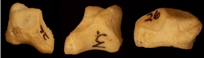
Greater Multangular or Trapezium. Right side.
.png?revision=1)
Capitate. Left side. Left view from the hamate and right view from the scaphoid and trapezoid.
.png?revision=1)
Hamate. Right side. Left view from triquetral, Middle left view from the fourth and fifth metacarpal bases, Middle right view from the capitate.
.png?revision=1)
Triquetral. Left side. Middle view from the triquetral.
.png?revision=1)
Metacarpal 1. Right side. Left palmar and Right dorsal.
.png?revision=1)
Metacarpal 2. Left side. Left medial, Middle left lateral, Middle right dorsal, Right proximal base.
.png?revision=1)
Metacarpal 3. Right side. Left medial, Middle left palmar, Middle right lateral, Right proximal base.
.png?revision=1)
Metacarpal 4. Right side. Left medial, Middle left palmar, Middle right lateral, Right proximal base.
.png?revision=1)
Metacarpal 5. Right side. Left medial, Middle left palmar, Middle right lateral, Right proximal base.
.png?revision=1)
First Proximal Phalange of the Hand. Left dorsal, Middle palmar, Right proximal base.
.png?revision=1)
Proximal Non-First Phalanges of the Hand. Left dorsal, Middle palmar, Right proximal base.
.png?revision=1)
Medial Phalanges of the Hand. Left dorsal and Right palmar.
.png?revision=1)
First Distal Phalange of the Hand. Left dorsal and Right palmar.
.png?revision=1)
Hand