
1.5: Body Cavities
- Page ID
- 90475

\( \newcommand{\vecs}[1]{\overset { \scriptstyle \rightharpoonup} {\mathbf{#1}} } \)
\( \newcommand{\vecd}[1]{\overset{-\!-\!\rightharpoonup}{\vphantom{a}\smash {#1}}} \)
\( \newcommand{\dsum}{\displaystyle\sum\limits} \)
\( \newcommand{\dint}{\displaystyle\int\limits} \)
\( \newcommand{\dlim}{\displaystyle\lim\limits} \)
\( \newcommand{\id}{\mathrm{id}}\) \( \newcommand{\Span}{\mathrm{span}}\)
( \newcommand{\kernel}{\mathrm{null}\,}\) \( \newcommand{\range}{\mathrm{range}\,}\)
\( \newcommand{\RealPart}{\mathrm{Re}}\) \( \newcommand{\ImaginaryPart}{\mathrm{Im}}\)
\( \newcommand{\Argument}{\mathrm{Arg}}\) \( \newcommand{\norm}[1]{\| #1 \|}\)
\( \newcommand{\inner}[2]{\langle #1, #2 \rangle}\)
\( \newcommand{\Span}{\mathrm{span}}\)
\( \newcommand{\id}{\mathrm{id}}\)
\( \newcommand{\Span}{\mathrm{span}}\)
\( \newcommand{\kernel}{\mathrm{null}\,}\)
\( \newcommand{\range}{\mathrm{range}\,}\)
\( \newcommand{\RealPart}{\mathrm{Re}}\)
\( \newcommand{\ImaginaryPart}{\mathrm{Im}}\)
\( \newcommand{\Argument}{\mathrm{Arg}}\)
\( \newcommand{\norm}[1]{\| #1 \|}\)
\( \newcommand{\inner}[2]{\langle #1, #2 \rangle}\)
\( \newcommand{\Span}{\mathrm{span}}\) \( \newcommand{\AA}{\unicode[.8,0]{x212B}}\)
\( \newcommand{\vectorA}[1]{\vec{#1}} % arrow\)
\( \newcommand{\vectorAt}[1]{\vec{\text{#1}}} % arrow\)
\( \newcommand{\vectorB}[1]{\overset { \scriptstyle \rightharpoonup} {\mathbf{#1}} } \)
\( \newcommand{\vectorC}[1]{\textbf{#1}} \)
\( \newcommand{\vectorD}[1]{\overrightarrow{#1}} \)
\( \newcommand{\vectorDt}[1]{\overrightarrow{\text{#1}}} \)
\( \newcommand{\vectE}[1]{\overset{-\!-\!\rightharpoonup}{\vphantom{a}\smash{\mathbf {#1}}}} \)
\( \newcommand{\vecs}[1]{\overset { \scriptstyle \rightharpoonup} {\mathbf{#1}} } \)
\(\newcommand{\longvect}{\overrightarrow}\)
\( \newcommand{\vecd}[1]{\overset{-\!-\!\rightharpoonup}{\vphantom{a}\smash {#1}}} \)
\(\newcommand{\avec}{\mathbf a}\) \(\newcommand{\bvec}{\mathbf b}\) \(\newcommand{\cvec}{\mathbf c}\) \(\newcommand{\dvec}{\mathbf d}\) \(\newcommand{\dtil}{\widetilde{\mathbf d}}\) \(\newcommand{\evec}{\mathbf e}\) \(\newcommand{\fvec}{\mathbf f}\) \(\newcommand{\nvec}{\mathbf n}\) \(\newcommand{\pvec}{\mathbf p}\) \(\newcommand{\qvec}{\mathbf q}\) \(\newcommand{\svec}{\mathbf s}\) \(\newcommand{\tvec}{\mathbf t}\) \(\newcommand{\uvec}{\mathbf u}\) \(\newcommand{\vvec}{\mathbf v}\) \(\newcommand{\wvec}{\mathbf w}\) \(\newcommand{\xvec}{\mathbf x}\) \(\newcommand{\yvec}{\mathbf y}\) \(\newcommand{\zvec}{\mathbf z}\) \(\newcommand{\rvec}{\mathbf r}\) \(\newcommand{\mvec}{\mathbf m}\) \(\newcommand{\zerovec}{\mathbf 0}\) \(\newcommand{\onevec}{\mathbf 1}\) \(\newcommand{\real}{\mathbb R}\) \(\newcommand{\twovec}[2]{\left[\begin{array}{r}#1 \\ #2 \end{array}\right]}\) \(\newcommand{\ctwovec}[2]{\left[\begin{array}{c}#1 \\ #2 \end{array}\right]}\) \(\newcommand{\threevec}[3]{\left[\begin{array}{r}#1 \\ #2 \\ #3 \end{array}\right]}\) \(\newcommand{\cthreevec}[3]{\left[\begin{array}{c}#1 \\ #2 \\ #3 \end{array}\right]}\) \(\newcommand{\fourvec}[4]{\left[\begin{array}{r}#1 \\ #2 \\ #3 \\ #4 \end{array}\right]}\) \(\newcommand{\cfourvec}[4]{\left[\begin{array}{c}#1 \\ #2 \\ #3 \\ #4 \end{array}\right]}\) \(\newcommand{\fivevec}[5]{\left[\begin{array}{r}#1 \\ #2 \\ #3 \\ #4 \\ #5 \\ \end{array}\right]}\) \(\newcommand{\cfivevec}[5]{\left[\begin{array}{c}#1 \\ #2 \\ #3 \\ #4 \\ #5 \\ \end{array}\right]}\) \(\newcommand{\mattwo}[4]{\left[\begin{array}{rr}#1 \amp #2 \\ #3 \amp #4 \\ \end{array}\right]}\) \(\newcommand{\laspan}[1]{\text{Span}\{#1\}}\) \(\newcommand{\bcal}{\cal B}\) \(\newcommand{\ccal}{\cal C}\) \(\newcommand{\scal}{\cal S}\) \(\newcommand{\wcal}{\cal W}\) \(\newcommand{\ecal}{\cal E}\) \(\newcommand{\coords}[2]{\left\{#1\right\}_{#2}}\) \(\newcommand{\gray}[1]{\color{gray}{#1}}\) \(\newcommand{\lgray}[1]{\color{lightgray}{#1}}\) \(\newcommand{\rank}{\operatorname{rank}}\) \(\newcommand{\row}{\text{Row}}\) \(\newcommand{\col}{\text{Col}}\) \(\renewcommand{\row}{\text{Row}}\) \(\newcommand{\nul}{\text{Nul}}\) \(\newcommand{\var}{\text{Var}}\) \(\newcommand{\corr}{\text{corr}}\) \(\newcommand{\len}[1]{\left|#1\right|}\) \(\newcommand{\bbar}{\overline{\bvec}}\) \(\newcommand{\bhat}{\widehat{\bvec}}\) \(\newcommand{\bperp}{\bvec^\perp}\) \(\newcommand{\xhat}{\widehat{\xvec}}\) \(\newcommand{\vhat}{\widehat{\vvec}}\) \(\newcommand{\uhat}{\widehat{\uvec}}\) \(\newcommand{\what}{\widehat{\wvec}}\) \(\newcommand{\Sighat}{\widehat{\Sigma}}\) \(\newcommand{\lt}{<}\) \(\newcommand{\gt}{>}\) \(\newcommand{\amp}{&}\) \(\definecolor{fillinmathshade}{gray}{0.9}\)Vertebrates have fluid-filled spaces called body cavities that contain the organs.
- Distinguish between the dorsal and the ventral body cavities, identifying their subdivisions and representative organs found in each.
By the broadest definition, a body cavity is any fluid-filled space in a multicellular organism. Blood vessels are not considered cavities but may be held within the body cavities. Most cavities provide room for the organs to adjust to changes in the organism’s position. They usually contains protective membranes and sometimes bones that protect the organs.
Overview of Body Cavities
 The human body has several major cavities that protect internal organs and allow them to move and change shape. The two primary body cavities — the dorsal and ventral cavities — remain when internal organs are removed (see details below).
In addition to these, there are smaller cavities formed by serous membranes.
A serous membrane (or serosa) is a thin, slippery tissue that lines certain internal body cavities and covers the organs within them. Each serous membrane has two layers:
-
Parietal layer: lines the walls of the cavity
-
Visceral layer: covers the surface of the organs
Between these two layers is a small amount of serous fluid, which reduces friction so the organs can move smoothly against each other — like when your lungs expand or your heart beats.
Examples are the pleural cavity (around the lungs; see image on the right), the pericardial cavity (around the heart), and the peritoneal cavity (around abdominal organs).
As mentioned above, the body has two major cavities, each of which is further divided into smaller compartments. These cavities house and protect delicate internal organs. The ventral cavity, in particular, is designed to accommodate significant changes in the size and shape of organs as they function. For example, the lungs, heart, stomach, and intestines can expand and contract as needed, without interfering with surrounding tissues or disrupting the activity of nearby organs
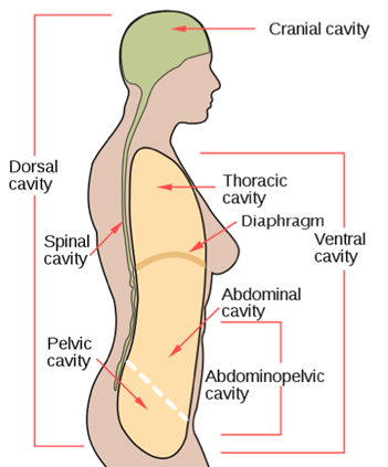
🟦 Dorsal Body Cavity (toward the back)
The dorsal cavity, in the back of the body, contains the primary organs of the nervous system, specifically the brain and the spinal cord. Just as the brain and spinal cord make up a continuous, uninterrupted structure, the cranial and spinal cavities that house them are also continuous. This cavity is surrounded by bone for protection (skull bones and the vertebral column), and by cerebrospinal fluid, a colorless fluid produced by the brain, which cushions the brain and spinal cord.
-
Cranial Cavity
The cranial cavity is the portion of the dorsal cavity consisting of the space inside the skull. This cavity contains the brain, the meninges of the brain, and cerebrospinal fluid
-
Spinal (Vertebral) Cavity
The The spinal or vertebral cavity is the posterior portion of the dorsal cavity and contains the structures within the vertebral column. These include the spinal cord, the meninges of the spinal cord, and the fluid-filled spaces between them. This is the most narrow of all body cavities, sometimes described as threadlike.
🟦 Ventral Body Cavity
The ventral cavity is the large internal space located at the front of the body. It contains many of the body’s major organ systems, and the organs housed within it are collectively referred to as the viscera.
This cavity is divided into two main subdivisions: the thoracic cavity and the abdominopelvic cavity. Each of these is further subdivided, as described below.
Separating the thoracic and abdominal cavities is the diaphragm, a sheet of muscle that plays a vital role in breathing by helping move air in and out of the lungs.
-
Thoracic Cavity
The thoracic cavity is found within the rib cage in the torso. It houses the primary organs of the cardiovascular and respiratory systems, such as the heart and lungs, but also includes organs from other systems, such as the esophagus and the thymus gland. The thoracic cavity is lined by two types of serous membranes, the pleura lining the pleural cavity and the lungs, and the pericardium lining the pericardial cavity and the heart. (See above).
-
Pleural Cavities (2)
Each houses a lung, and is lined with the pleura, both visceral and parietal.
-
Pericardial Cavity –
Contains the heart, and is lined with the pericardium, both visceral and parietal.
-
Abdominopelvic Cavity
Located below the diaphragm, it is the largest cavity in the body. Although there is no physical membrane separating the abdominal and pelvic regions, the space is commonly divided into two parts for anatomical and clinical purposes.
-
Abdominal Cavity
The abdominal cavity is not contained within bone and houses many organs of the digestive (like the stomach, liver, intestines) and renal systems, as well as some organs of the endocrine system, such as the adrenal glands.
-
Pelvic cavity
The pelvic cavity is contained within the pelvis and houses the bladder, the reproductive system, and part of the large intestine.