
12: Cranial and Spinal Nerves
- Page ID
- 12532
The Spinal Cord
The description of the CNS is concentrated on the structures of the brain, but the spinal cord is another major organ of the system. Whereas the brain develops out of expansions of the neural tube into primary and then secondary vesicles, the spinal cord maintains the tube structure and is only specialized into certain regions. As the spinal cord continues to develop in the newborn, anatomical features mark its surface. The anterior midline is marked by the anterior median fissure, and the posterior midline is marked by the posterior median sulcus. Axons enter the posterior side through the dorsal (posterior) nerve root, which marks the posterolateral sulcus on either side. The axons emerging from the anterior side do so through the ventral (anterior) nerve root. Note that it is common to see the terms dorsal (dorsal = “back”) and ventral (ventral = “belly”) used interchangeably with posterior and anterior, particularly in reference to nerves and the structures of the spinal cord. You should learn to be comfortable with both.
Gray Horns
In cross-section, the gray matter of the spinal cord has the appearance of an ink-blot test, with the spread of the gray matter on one side replicated on the other—a shape reminiscent of a bulbous capital “H.” As shown in Figure 12.1, the gray matter is subdivided into regions that are referred to as horns.
The posterior horn is responsible for sensory processing. The anterior horn sends out motor signals to the skeletal muscles. The lateral horn, which is only found in the thoracic, upper lumbar, and sacral regions, is the central component of the sympathetic division of the autonomic nervous system.
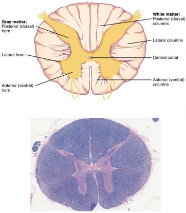
Figure 12.1 Cross-section of Spinal Cord The cross-section of a thoracic spinal cord segment shows the posterior, anterior, and lateral horns of gray matter, as well as the posterior, anterior, and lateral columns of white matter. LM × 40. (Micrograph provided by the Regents of University of Michigan Medical School © 2012)
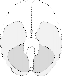
Cranial Nerves
The nerves attached to the brain are the cranial nerves, which are primarily responsible for the sensory and motor functions of the head and neck (one of these nerves targets organs in the thoracic and abdominal cavities as part of the parasympathetic nervous system). There are twelve cranial nerves, which are designated CNI through CNXII for “Cranial Nerve,” using Roman numerals for 1 through 12. They can be classified as sensory nerves, motor nerves, or a combination of both, meaning that the axons in these nerves originate out of sensory ganglia external to the cranium or motor nuclei within the brain stem. Sensory axons enter the brain to synapse in a nucleus. Motor axons connect to skeletal muscles of the head or neck. Three of the nerves are solely composed of sensory fibers; five are strictly motor; and the remaining four are mixed nerves.
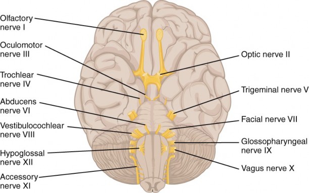
Figure 12.2 The Cranial Nerves The anatomical arrangement of the roots of the cranial nerves observed from an inferior view of the brain.
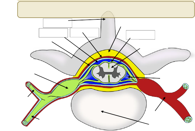
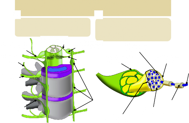
Label the following: Dura mater, Arachnoid mater, Pia mater, Epidural space, Subarachnoid space, Spinal cord, Denticulate ligament, Dorsal Root Ganglion, Body (of vertebrae), Spinous process (of vertebrae) , Spinal nerve, Dorsal Ramus, Ventral ramus
10
9
5
8
3
4
7
1
11
2
12
4
13
6
LAB 12 EXERCISES 12-2
LABEL THE FOLLOWING: DORSAL HORN, LATERAL HORN, VENTRAL HORN, DORSAL COLUMN, LATERAL COLUMN, VENTRAL COLUMN, CENTRAL CANAL, GREY COMMISURE
1
5
2
3
6
7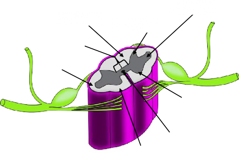
8
LAB 12 EXERCISES 12-3
SPINAL CORD AND NERVE ANATOMY
A
B
3
Label the following: Dorsal root, Ventral root, Dorsal root ganglion, Sympathetic chain ganglion, Spinal nerve, Rami communicantes, Dorsal ramus, Ventral ramus
Label the parts of a nerve: Blood vessels, Schwann cell, Fascicle, Epineurium, Perineurium, Endoneurium, Axon
1
5
6
3
2
7
1
4
5
4
8
6

2
7
LAB 12 EXERCISES 12-4
VERTEBRAL COLUMN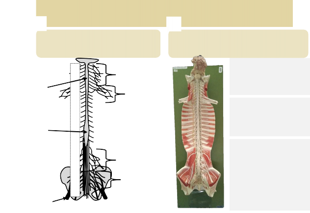
A
B
Identify the following: Cervical plexus, Brachial plexus, Lumbar plexus, Sacral plexus, Sciatic nerve, Cervical enlargement, Lumbar enlargement.
Match these items to their relative position: Cervical enlargement, Lumbar enlargement*, Conus medullaris, Cauda equina, Filum terminale
(* trickier than you might expect)
4
|
Cervical region: |
|
Thoracic region: |
|
Lumbo-sacral region: |
C1 2
3
Cervical 4
nerves 5
1
6
5
7
8
T1 2
3
4
5
Thoracic 6
nerves 7
2
8
9
10
11
12
6
L1 2
Lumbar 3
nerves 4
5
7
S1
Sacral 2
nerves 3
4
5
3
LAB 12 EXERCISES 12-5

PERIPHERAL NERVES AND PLEXUSES
A
B


2
1
3
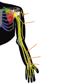
Label the nerves Label the following: Axillary nerve, Median nerve, Radial nerve, Ulnar nerve, Musculocutaneous nerve
1
2
5
4
LAB 12 EXERCISES 12-6
DRAW YOUR OWN CRANIAL NERVE STUDY SHEET:
|
Mnemonic |
Cranial nerve name |
|
|---|---|---|
|
I |
||
|
II |
||
|
III |
||
|
IV |
||
|
V |
||
|
VI |
||
|
VII |
||
|
VIII |
||
|
IX |
||
|
X |
||
|
XI |
||
|
XII |
MODELS
Spinal Cord, Spinal Columns (Flat), Spinal Cord in Vertebral Column, Blank Brain Picture (For CN)
Spinal cord anatomy
Meningeal layers
- Dura mater
Epidural space
- Arachnoid mater
Subarachnoid space
- Pia mater
Denticulate ligaments
Conus medullaris
Cauda equina
Filum terminale
Cervical enlargement
Lumbar enlargement
Spinal nerve:
- Ventral root
- Dorsal root
- Dorsal root ganglion
- Ventral Ramus
- Dorsal Ramus
Spinal cord cross sectional anatomy:
- Dorsal horn
- Lateral horn
- Ventral horn
- Dorsal column
- Lateral column
- Ventral column
- Gray commissure
- Central canal
- Dorsal median sulcus
- Ventral median fissure
Cranial nerves
- I Olfactory
- II Optic
- III Occulomotor
- IV Trochlear
- V Trigeminal
- VI Abducens
- VII Facial
- VIII Acoustic / Vestibulocochlear
- IX Glossopharyngeal
- X Vagus
- XI (spinal) Accessory
- XII Hypoglossal
Peripheral Nerves
Nerve anatomy:
- Epineurium
- Perineurium
- Endoneurium
- Fascicle
Cervical Plexus
- Phrenic n.
Brachial Plexus
- Axillary n.
- Radial.
- Ulnar n.
- Median n.
- Musculocutaneous n.
Intercostal nerves
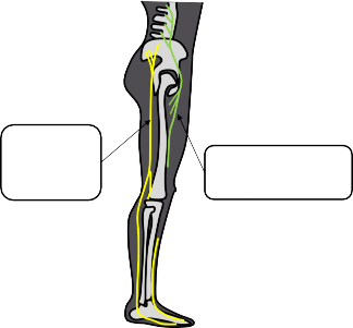
Lumbar plexus
- Femoral n.
Sacral plexus
- Sciatic n.
- Pudendal n.
Autonomic NS
- Sympathetic chain ganglion
- Sympathetic chain
- Rami communicantes
- Lateral horn
Lab list continued on ne
- Note: remember that dorsal/posterior, and ventral/anterior are interchangeable.

Histology
- Spinal cord & dorsal root ganglia
See cross sectional anatomy
- Nerve cross section
- Spinal cord & dorsal root ganglia