
8.4A: Structure of Synovial Joints
- Page ID
- 7516
")
\( \newcommand{\vecs}[1]{\overset { \scriptstyle \rightharpoonup} {\mathbf{#1}} } \)
\( \newcommand{\vecd}[1]{\overset{-\!-\!\rightharpoonup}{\vphantom{a}\smash {#1}}} \)
\( \newcommand{\dsum}{\displaystyle\sum\limits} \)
\( \newcommand{\dint}{\displaystyle\int\limits} \)
\( \newcommand{\dlim}{\displaystyle\lim\limits} \)
\( \newcommand{\id}{\mathrm{id}}\) \( \newcommand{\Span}{\mathrm{span}}\)
( \newcommand{\kernel}{\mathrm{null}\,}\) \( \newcommand{\range}{\mathrm{range}\,}\)
\( \newcommand{\RealPart}{\mathrm{Re}}\) \( \newcommand{\ImaginaryPart}{\mathrm{Im}}\)
\( \newcommand{\Argument}{\mathrm{Arg}}\) \( \newcommand{\norm}[1]{\| #1 \|}\)
\( \newcommand{\inner}[2]{\langle #1, #2 \rangle}\)
\( \newcommand{\Span}{\mathrm{span}}\)
\( \newcommand{\id}{\mathrm{id}}\)
\( \newcommand{\Span}{\mathrm{span}}\)
\( \newcommand{\kernel}{\mathrm{null}\,}\)
\( \newcommand{\range}{\mathrm{range}\,}\)
\( \newcommand{\RealPart}{\mathrm{Re}}\)
\( \newcommand{\ImaginaryPart}{\mathrm{Im}}\)
\( \newcommand{\Argument}{\mathrm{Arg}}\)
\( \newcommand{\norm}[1]{\| #1 \|}\)
\( \newcommand{\inner}[2]{\langle #1, #2 \rangle}\)
\( \newcommand{\Span}{\mathrm{span}}\) \( \newcommand{\AA}{\unicode[.8,0]{x212B}}\)
\( \newcommand{\vectorA}[1]{\vec{#1}} % arrow\)
\( \newcommand{\vectorAt}[1]{\vec{\text{#1}}} % arrow\)
\( \newcommand{\vectorB}[1]{\overset { \scriptstyle \rightharpoonup} {\mathbf{#1}} } \)
\( \newcommand{\vectorC}[1]{\textbf{#1}} \)
\( \newcommand{\vectorD}[1]{\overrightarrow{#1}} \)
\( \newcommand{\vectorDt}[1]{\overrightarrow{\text{#1}}} \)
\( \newcommand{\vectE}[1]{\overset{-\!-\!\rightharpoonup}{\vphantom{a}\smash{\mathbf {#1}}}} \)
\( \newcommand{\vecs}[1]{\overset { \scriptstyle \rightharpoonup} {\mathbf{#1}} } \)
\(\newcommand{\longvect}{\overrightarrow}\)
\( \newcommand{\vecd}[1]{\overset{-\!-\!\rightharpoonup}{\vphantom{a}\smash {#1}}} \)
\(\newcommand{\avec}{\mathbf a}\) \(\newcommand{\bvec}{\mathbf b}\) \(\newcommand{\cvec}{\mathbf c}\) \(\newcommand{\dvec}{\mathbf d}\) \(\newcommand{\dtil}{\widetilde{\mathbf d}}\) \(\newcommand{\evec}{\mathbf e}\) \(\newcommand{\fvec}{\mathbf f}\) \(\newcommand{\nvec}{\mathbf n}\) \(\newcommand{\pvec}{\mathbf p}\) \(\newcommand{\qvec}{\mathbf q}\) \(\newcommand{\svec}{\mathbf s}\) \(\newcommand{\tvec}{\mathbf t}\) \(\newcommand{\uvec}{\mathbf u}\) \(\newcommand{\vvec}{\mathbf v}\) \(\newcommand{\wvec}{\mathbf w}\) \(\newcommand{\xvec}{\mathbf x}\) \(\newcommand{\yvec}{\mathbf y}\) \(\newcommand{\zvec}{\mathbf z}\) \(\newcommand{\rvec}{\mathbf r}\) \(\newcommand{\mvec}{\mathbf m}\) \(\newcommand{\zerovec}{\mathbf 0}\) \(\newcommand{\onevec}{\mathbf 1}\) \(\newcommand{\real}{\mathbb R}\) \(\newcommand{\twovec}[2]{\left[\begin{array}{r}#1 \\ #2 \end{array}\right]}\) \(\newcommand{\ctwovec}[2]{\left[\begin{array}{c}#1 \\ #2 \end{array}\right]}\) \(\newcommand{\threevec}[3]{\left[\begin{array}{r}#1 \\ #2 \\ #3 \end{array}\right]}\) \(\newcommand{\cthreevec}[3]{\left[\begin{array}{c}#1 \\ #2 \\ #3 \end{array}\right]}\) \(\newcommand{\fourvec}[4]{\left[\begin{array}{r}#1 \\ #2 \\ #3 \\ #4 \end{array}\right]}\) \(\newcommand{\cfourvec}[4]{\left[\begin{array}{c}#1 \\ #2 \\ #3 \\ #4 \end{array}\right]}\) \(\newcommand{\fivevec}[5]{\left[\begin{array}{r}#1 \\ #2 \\ #3 \\ #4 \\ #5 \\ \end{array}\right]}\) \(\newcommand{\cfivevec}[5]{\left[\begin{array}{c}#1 \\ #2 \\ #3 \\ #4 \\ #5 \\ \end{array}\right]}\) \(\newcommand{\mattwo}[4]{\left[\begin{array}{rr}#1 \amp #2 \\ #3 \amp #4 \\ \end{array}\right]}\) \(\newcommand{\laspan}[1]{\text{Span}\{#1\}}\) \(\newcommand{\bcal}{\cal B}\) \(\newcommand{\ccal}{\cal C}\) \(\newcommand{\scal}{\cal S}\) \(\newcommand{\wcal}{\cal W}\) \(\newcommand{\ecal}{\cal E}\) \(\newcommand{\coords}[2]{\left\{#1\right\}_{#2}}\) \(\newcommand{\gray}[1]{\color{gray}{#1}}\) \(\newcommand{\lgray}[1]{\color{lightgray}{#1}}\) \(\newcommand{\rank}{\operatorname{rank}}\) \(\newcommand{\row}{\text{Row}}\) \(\newcommand{\col}{\text{Col}}\) \(\renewcommand{\row}{\text{Row}}\) \(\newcommand{\nul}{\text{Nul}}\) \(\newcommand{\var}{\text{Var}}\) \(\newcommand{\corr}{\text{corr}}\) \(\newcommand{\len}[1]{\left|#1\right|}\) \(\newcommand{\bbar}{\overline{\bvec}}\) \(\newcommand{\bhat}{\widehat{\bvec}}\) \(\newcommand{\bperp}{\bvec^\perp}\) \(\newcommand{\xhat}{\widehat{\xvec}}\) \(\newcommand{\vhat}{\widehat{\vvec}}\) \(\newcommand{\uhat}{\widehat{\uvec}}\) \(\newcommand{\what}{\widehat{\wvec}}\) \(\newcommand{\Sighat}{\widehat{\Sigma}}\) \(\newcommand{\lt}{<}\) \(\newcommand{\gt}{>}\) \(\newcommand{\amp}{&}\) \(\definecolor{fillinmathshade}{gray}{0.9}\)A synovial joint or diarthrosis occurs at articulating bones to allow movement. It is distinguished by a surrounding synovial capsule.
- Identify the structures of the synovial joint that allow it to move freely
Key Points
- The bones of a synovial joint are surrounded by a synovial capsule, which secretes synovial fluid to lubricate and nourish the joint while acting as a shock absorber.
- The ends of the joint bones are covered with smooth, glass-like hyaline cartilage which reduces friction during movement.
- A synovial joint contains a synovial cavity and dense, irregular connective tissue that forms the articular capsule normally associated with accessory ligaments.
Key Terms
- articulation: A joint or the collection of joints at which something is articulated, or hinged, for bending.
- synovial membrane: A thin membrane of joints comprised of smooth connective tissue and that secretes synovial fluid.
- synovial fluid: A viscous, non-Newtonian fluid found in the cavities of synovial joints. With its yolk-like consistency, its principal role is to reduce friction between the articular cartilage of synovial joints during movement.
- articular cartilage: A tough, elastic, fibrous connective tissue found in various parts of the body such as the joints, outer ear, and larynx. A major constituent of the embryonic and young vertebrate skeleton, converted largely to bone with maturation.
- diarthrosis: A joint that can move freely in various planes.
A synovial joint, also known as a diarthrosis, is the most common and most movable type of joint in a mammal’s body. Diarthroses are freely movable articulations. In these joints, the contiguous bony surfaces are covered with articular cartilage and connected by ligaments lined by synovial membrane. The joint may be divided, completely or incompletely, by an articular disk or meniscus, the periphery of which is continuous with the fibrous capsule while its free surfaces are covered by synovial membrane.
The articular capsule is fibrous and continuous with the periosteum of articulating bones, surrounding the diarthrosis and uniting the articulating bones. The articular capsule also consists of two layers: (1) the outer fibrous membrane that may contain ligaments and (2) the inner synovial membrane that secretes the lubricating, shock-absorbing, and joint-nourishing synovial fluid. The bones of a synovial joint are covered by a layer of hyaline cartilage that lines the epiphyses of joint ends of bone with a smooth, slippery surface that does not bind them together. This articular cartilage functions to absorb shock and reduce friction during movement.
Synovial Membrane and Components
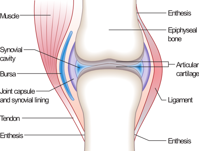
Synovial Joint: An illustration of the structure of a synovial joint.
A synovial membrane (or synovium) is the soft tissue found between the articular capsule (joint capsule) and the joint cavity of synovial joints. Synovial fluid is the clear, viscid, lubricating fluid secreted by synovial membranes. The morphology of synovial membranes may vary, but it often consists of two layers. The outer layer, or subintima, can be fibrous, fatty, or loosely areolar. The inner layer, or intima, consists of a sheet of cells thinner than a piece of paper.
Where the underlying subintima is loose, the intima sits on a pliable membrane called the synovial membrane. This membrane, together with the cells of the intima, acts like an inner tube, sealing the synovial fluid from the surrounding tissue and effectively stopping the joints from being squeezed dry when subjected to impact (such as when running). As with most other joints, synovial joints achieve movement at the point of contact of the articulating bones. The main structural differences between synovial and fibrous joints are the existence of capsules surrounding the articulating surfaces of a synovial joint and the presence of lubricating synovial fluid within those capsules (synovial cavities).
Synoviocytes
The intimal cells are termed synoviocytes and can be either fibroblastic (type B synoviocytes) and macrophagic (type A synoviocytes). Both types have differences from similar cells in other tissues. The type B synoviocytes manufacture a long-chain sugar polymer called hyaluronan, which combines with a molecule called lubricin to give the synovial fluid a stringy, egg-white consistency. The water component of synovial fluid is effectively trapped in the joint space by the hyaluronan due to its large, highly negatively charged moieties. The macrophages are responsible for the removal of undesirable substances from the synovial fluid.
Structure of Synovium
The surface of a synovium may be flat or covered with finger-like projections (villi) to allow the soft tissue to change shape as the joint surfaces move on one another. Just beneath the intima, most synovia have a dense net of small blood vessels that provide nutrients for the synovia and the avascular cartilage.
In any one position, much of the cartilage is close enough to get nutrition directly from the synovium. Some areas of cartilage have to obtain nutrients indirectly and may do so either from diffusion through cartilage or by the stirring of synovial fluid.
Synovial Bursa
The synovial bursa is a small, fluid-filled sac lined by synovial membrane containing synovial fluid. It provides a cushion between bones and tendons and/or muscles around a joint.