
6.3: Endocrine System
- Page ID
- 150552

\( \newcommand{\vecs}[1]{\overset { \scriptstyle \rightharpoonup} {\mathbf{#1}} } \)
\( \newcommand{\vecd}[1]{\overset{-\!-\!\rightharpoonup}{\vphantom{a}\smash {#1}}} \)
\( \newcommand{\dsum}{\displaystyle\sum\limits} \)
\( \newcommand{\dint}{\displaystyle\int\limits} \)
\( \newcommand{\dlim}{\displaystyle\lim\limits} \)
\( \newcommand{\id}{\mathrm{id}}\) \( \newcommand{\Span}{\mathrm{span}}\)
( \newcommand{\kernel}{\mathrm{null}\,}\) \( \newcommand{\range}{\mathrm{range}\,}\)
\( \newcommand{\RealPart}{\mathrm{Re}}\) \( \newcommand{\ImaginaryPart}{\mathrm{Im}}\)
\( \newcommand{\Argument}{\mathrm{Arg}}\) \( \newcommand{\norm}[1]{\| #1 \|}\)
\( \newcommand{\inner}[2]{\langle #1, #2 \rangle}\)
\( \newcommand{\Span}{\mathrm{span}}\)
\( \newcommand{\id}{\mathrm{id}}\)
\( \newcommand{\Span}{\mathrm{span}}\)
\( \newcommand{\kernel}{\mathrm{null}\,}\)
\( \newcommand{\range}{\mathrm{range}\,}\)
\( \newcommand{\RealPart}{\mathrm{Re}}\)
\( \newcommand{\ImaginaryPart}{\mathrm{Im}}\)
\( \newcommand{\Argument}{\mathrm{Arg}}\)
\( \newcommand{\norm}[1]{\| #1 \|}\)
\( \newcommand{\inner}[2]{\langle #1, #2 \rangle}\)
\( \newcommand{\Span}{\mathrm{span}}\) \( \newcommand{\AA}{\unicode[.8,0]{x212B}}\)
\( \newcommand{\vectorA}[1]{\vec{#1}} % arrow\)
\( \newcommand{\vectorAt}[1]{\vec{\text{#1}}} % arrow\)
\( \newcommand{\vectorB}[1]{\overset { \scriptstyle \rightharpoonup} {\mathbf{#1}} } \)
\( \newcommand{\vectorC}[1]{\textbf{#1}} \)
\( \newcommand{\vectorD}[1]{\overrightarrow{#1}} \)
\( \newcommand{\vectorDt}[1]{\overrightarrow{\text{#1}}} \)
\( \newcommand{\vectE}[1]{\overset{-\!-\!\rightharpoonup}{\vphantom{a}\smash{\mathbf {#1}}}} \)
\( \newcommand{\vecs}[1]{\overset { \scriptstyle \rightharpoonup} {\mathbf{#1}} } \)
\(\newcommand{\longvect}{\overrightarrow}\)
\( \newcommand{\vecd}[1]{\overset{-\!-\!\rightharpoonup}{\vphantom{a}\smash {#1}}} \)
\(\newcommand{\avec}{\mathbf a}\) \(\newcommand{\bvec}{\mathbf b}\) \(\newcommand{\cvec}{\mathbf c}\) \(\newcommand{\dvec}{\mathbf d}\) \(\newcommand{\dtil}{\widetilde{\mathbf d}}\) \(\newcommand{\evec}{\mathbf e}\) \(\newcommand{\fvec}{\mathbf f}\) \(\newcommand{\nvec}{\mathbf n}\) \(\newcommand{\pvec}{\mathbf p}\) \(\newcommand{\qvec}{\mathbf q}\) \(\newcommand{\svec}{\mathbf s}\) \(\newcommand{\tvec}{\mathbf t}\) \(\newcommand{\uvec}{\mathbf u}\) \(\newcommand{\vvec}{\mathbf v}\) \(\newcommand{\wvec}{\mathbf w}\) \(\newcommand{\xvec}{\mathbf x}\) \(\newcommand{\yvec}{\mathbf y}\) \(\newcommand{\zvec}{\mathbf z}\) \(\newcommand{\rvec}{\mathbf r}\) \(\newcommand{\mvec}{\mathbf m}\) \(\newcommand{\zerovec}{\mathbf 0}\) \(\newcommand{\onevec}{\mathbf 1}\) \(\newcommand{\real}{\mathbb R}\) \(\newcommand{\twovec}[2]{\left[\begin{array}{r}#1 \\ #2 \end{array}\right]}\) \(\newcommand{\ctwovec}[2]{\left[\begin{array}{c}#1 \\ #2 \end{array}\right]}\) \(\newcommand{\threevec}[3]{\left[\begin{array}{r}#1 \\ #2 \\ #3 \end{array}\right]}\) \(\newcommand{\cthreevec}[3]{\left[\begin{array}{c}#1 \\ #2 \\ #3 \end{array}\right]}\) \(\newcommand{\fourvec}[4]{\left[\begin{array}{r}#1 \\ #2 \\ #3 \\ #4 \end{array}\right]}\) \(\newcommand{\cfourvec}[4]{\left[\begin{array}{c}#1 \\ #2 \\ #3 \\ #4 \end{array}\right]}\) \(\newcommand{\fivevec}[5]{\left[\begin{array}{r}#1 \\ #2 \\ #3 \\ #4 \\ #5 \\ \end{array}\right]}\) \(\newcommand{\cfivevec}[5]{\left[\begin{array}{c}#1 \\ #2 \\ #3 \\ #4 \\ #5 \\ \end{array}\right]}\) \(\newcommand{\mattwo}[4]{\left[\begin{array}{rr}#1 \amp #2 \\ #3 \amp #4 \\ \end{array}\right]}\) \(\newcommand{\laspan}[1]{\text{Span}\{#1\}}\) \(\newcommand{\bcal}{\cal B}\) \(\newcommand{\ccal}{\cal C}\) \(\newcommand{\scal}{\cal S}\) \(\newcommand{\wcal}{\cal W}\) \(\newcommand{\ecal}{\cal E}\) \(\newcommand{\coords}[2]{\left\{#1\right\}_{#2}}\) \(\newcommand{\gray}[1]{\color{gray}{#1}}\) \(\newcommand{\lgray}[1]{\color{lightgray}{#1}}\) \(\newcommand{\rank}{\operatorname{rank}}\) \(\newcommand{\row}{\text{Row}}\) \(\newcommand{\col}{\text{Col}}\) \(\renewcommand{\row}{\text{Row}}\) \(\newcommand{\nul}{\text{Nul}}\) \(\newcommand{\var}{\text{Var}}\) \(\newcommand{\corr}{\text{corr}}\) \(\newcommand{\len}[1]{\left|#1\right|}\) \(\newcommand{\bbar}{\overline{\bvec}}\) \(\newcommand{\bhat}{\widehat{\bvec}}\) \(\newcommand{\bperp}{\bvec^\perp}\) \(\newcommand{\xhat}{\widehat{\xvec}}\) \(\newcommand{\vhat}{\widehat{\vvec}}\) \(\newcommand{\uhat}{\widehat{\uvec}}\) \(\newcommand{\what}{\widehat{\wvec}}\) \(\newcommand{\Sighat}{\widehat{\Sigma}}\) \(\newcommand{\lt}{<}\) \(\newcommand{\gt}{>}\) \(\newcommand{\amp}{&}\) \(\definecolor{fillinmathshade}{gray}{0.9}\)Overview and Functions
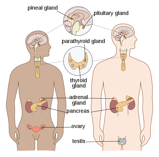
The purpose of the endocrine system (Figure 6.10) is to regulate various organs by releasing hormones. It is a key player in maintaining homeostasis within the body, which it does by sending chemical signals to one or more glands in order to control and coordinate hormones. Sometimes these chemical signals have immediate effect, whereas others take time for changes to occur. For example, target cells may take up to 48 hours to respond to reproductive hormones, but adrenal hormones, such as epinephrine and norepinephrine, are released within seconds when you are confronted with a dangerous or frightening situation.
(CrashCourse, 2015)
Components of the Endocrine System
Hypothalamus:
The hypothalamus is a structure in the brain. It is located in front of and below the thalamus and both produces and secretes many hormones.
Pineal gland: This gland is positioned below and slightly behind the thalamus. It is a very small gland whose functions are not entirely understood. Some of the specialized cells in the pineal gland are known to produce and secrete the hormone melatonin.
Pituitary gland: This small, bean-sized organ is divided into two parts. The first part is the posterior pituitary, which does not produce hormones but stores and secretes hormones produced by the hypothalamus. The other part is the anterior pituitary, which does produce hormones.
Thyroid gland: This butterfly-shaped organ is located in front of the trachea, just below the larynx. It produces hormones that regulate the body’s metabolic rate and control digestive, muscle, and heart functions, as well as brain development and bone maintenance.
Parathyroid glands: These glands are tiny, round structures found on the rear surface of the thyroid gland. They produce and secrete parathyroid hormone, the major hormone involved in the regulation of blood calcium levels.
Adrenal glands: The triangular-shaped adrenal glands are found on the top of the kidneys. They have a rich blood supply and actually possess one of the highest rates of blood flow in the whole body. They produce hormones that help regulate metabolism, the immune system, blood pressure, the body’s response to stress, and other essential functions.
Pancreas: This long, slender organ is located posterior to the bottom half of the stomach. It is primarily an exocrine gland, secreting a variety of digestive enzymes, but it also has an endocrine function. Its pancreatic islets secrete the hormones glucagon, insulin, somatostatin, and pancreatic polypeptide.
Ovaries: The ovaries are female reproductive organs. The primary hormones produced by the ovaries are estrogens, which include estradiol, estriol, and estrone. Estrogens play an important role in many physiological processes, including the development of the female reproductive system, regulation of the menstrual cycle, the development of female secondary sexual characteristics, the development of breast tissue, and the maintenance of pregnancy. Another important ovarian hormone is progesterone, which contributes to regulation of the menstrual cycle and is necessary to prepare the body for pregnancy and maintain pregnancy.
Testes: The testes are male reproductive organs. The primary hormone produced by the testes is testosterone, which is a steroid hormone important in the development of the male reproductive system, the maturation of sperm cells, and the development of male secondary sex characteristics, which include a deepened voice, body hair, and increased muscle mass.
Combining Forms
| COMBINING FORM | MEANING | EXAMPLE OF USE IN MEDICAL TERMS |
|---|---|---|
| adenoid/o | adenoid gland | adenoidectomy |
| adren/o | adrenal gland | adrenopathy |
| adrenal/o | adrenal gland | adrenalectomy |
| hypophys/o | pituitary gland | hypophysectomy |
| oophor/o | ovary | oophorectomy |
| ovari/o | ovary | ovarian |
| orch/o | testis | orchitis |
| orchi/o | testis | orchioplasty |
| orchid/o | testis | orchidotomy |
| pancreat/o | pancreas | pancreatitis |
| parathyroid/o | parathyroid gland | parathyroidectomy |
| pituitary/o | pituitary gland | hyperpituitarism |
| thyroaden/o | thyroid gland | thyroadenitis |
| thyroid/o | thyroid gland | thyroidotomy |
Common Pathologies
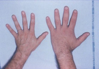
Acromegaly:
This disorder affects adults and is caused by abnormally high levels of growth hormone that results in increased growth of the bones in the face, hands, and feet. Figure 6.11 shows a typical presentation of the hand of a person with this pathology (right) compared to the hand of someone who does not have the condition (left).
Addison’s disease: This condition is caused by the hyposecretion of corticosteroids, which also results in low blood glucose levels and low blood sodium levels. An Addisonian crisis is a life-threatening condition that presents with severely low blood pressure as a result of low corticosteroid levels (Ernstmeyer & Christman, 2020).
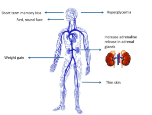
Cushing’s syndrome: Excessive production of the hormone cortisol results in this pathology. The signs and symptoms are rapid weight gain, depression, anxiety, high blood sugar, moon-shaped face, and fatigue (Figure 6.12). Individuals also experience weak muscles and bone pain.
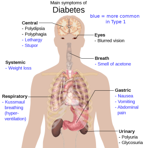
Diabetes mellitus: This condition results from dysfunction in the production and secretion of insulin or target cell responsiveness to insulin. There are two types of diabetes:
- Type 1: No insulin is produced, and individuals must take insulin injections to maintain blood sugar levels.
- Type 2: Usually develops later in life and generally develops because of lifestyle factors such as diet and exercise. This type of diabetes can usually be controlled through diet, exercise, and oral medications. Some individuals do end up needing insulin injections, though that is rare.
Gigantism: A disorder in children, gigantism is caused by the secretion of abnormally high levels of growth hormone, resulting in excessive growth.
Goiter: This condition is an increase in the size of the thyroid gland and appears as a lump in the neck. It can result from hypothyroidism.
Graves’ disease: This is a type of hyperthyroidism. The causative factor is an autoimmune reaction in which antibodies overstimulate the thyroid gland.
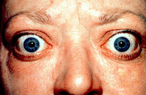
Hyperthyroidism: This is an elevated blood level of thyroid hormones that is often caused by a pituitary or thyroid tumour. Individuals can present with an increased metabolic rate, diarrhea, weight loss, increased heart rate, sweating, and tremors. They may also present with bulging eyes, which is known as exophthalmic goiter (Figure 6.14), and a goiter in the region of the thyroid.
Hypothyroidism: This condition is characterized by low levels of thyroid hormone in the blood. Inflammation of the thyroid gland can cause hypothyroidism, but it can also result from dietary deficiency of iodine (usually found in iodized salt). Individuals experience cold extremities, reduced libido, weight gain, reduced mental activity, and possibly menstrual irregularities.
Exercise
Attribution
Unless otherwise indicated, material on this page has been adapted from the following resource:
Betts, J. G., Young, K. A., Wise, J. A., Johnson, E., Poe, B., Kruse, D. H., Korol, O., Johnson, J. E., Womble, M., & DeSaix, P. (2013). Anatomy and physiology. OpenStax. https://openstax.org/details/books/anatomy-and-physiology licensed under CC BY 4.0
References
CrashCourse. (2015, June 22). Endocrine system, part 1 – Glands & hormones: Crash Course A&P #23 [Video]. YouTube. https://www.youtube.com/watch?v=eWHH9je2zG4&list=PL8dPuuaLjXtOAKed_MxxWBNaPno5h3Zs8&index=24
Ernstmeyer, K., & Christman, E. (Eds.). (2020). Nursing pharmacology. Chippewa Valley Technical College. https://wtcs.pressbooks.pub/pharmacology/ licensed under CC BY 4.0
Image Credits (images are listed in order of appearance)
by OpenStax, Tomáš Kebert, and umimeto, CC BY-SA 4.0
Acromegaly hands by Philippe Chanson and Sylvie Salenave, CC BY 2.0
The Cushing’s syndrome by Shiva.D, CC BY-SA 4.0
Main symptoms of diabetes by Mikael Häggström, Public domain
Proptosis and lid retraction from Graves’ Disease by Jonathan Trobe, CC BY 3.0
removal of the adenoid gland
Disease of the adrenal glands
Removal of one or both adrenal glands
Surgical removal of the pituitary gland
Removal of the ovaries
Pertaining to the ovaries
Inflammation of the testis
Surgical repair of the testes
An incision into the testis
Inflammation of the pancreas
Removal of the parathyroid gland
An overactive pituitary gland
Inflammation of the thyroid gland
An incision into the thyroid gland