
14.3: Autonomic Reflexes and Homeostasis
- Page ID
- 61592

- Compare the structure of somatic and autonomic reflex arcs
- Explain the differences in sympathetic and parasympathetic reflexes
- Differentiate between short and long reflexes
- Determine the effect of the autonomic nervous system on the regulation of the various organ systems on the basis of the signaling molecules involved
- Describe the effects of drugs that affect autonomic function
The autonomic nervous system regulates organ systems through circuits that resemble the reflexes described in the somatic nervous system. The main difference between the somatic and autonomic systems is in what target tissues are effectors. Somatic responses are solely based on skeletal muscle contraction. The autonomic system, however, targets cardiac and smooth muscle, as well as glandular tissue. Whereas the basic circuit is a reflex arc, there are differences in the structure of those reflexes for the somatic and autonomic systems.
The Structure of Reflexes
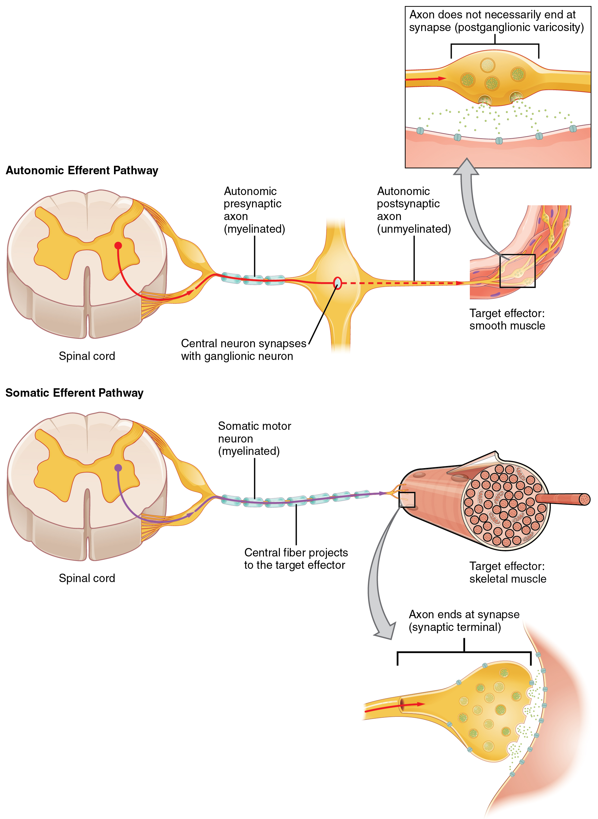
One difference between a somatic reflex, such as the withdrawal reflex, and a visceral reflex, which is an autonomic reflex, is in the efferent branch. The output of a somatic reflex is the lower motor neuron in the ventral horn of the spinal cord that projects directly to a skeletal muscle to cause its contraction. The output of a visceral reflex is a two-step pathway starting with the preganglionic fiber emerging from a lateral horn neuron in the spinal cord, or a cranial nucleus neuron in the brain stem, to a ganglion—followed by the postganglionic fiber projecting to a target effector. The other part of a reflex, the afferent branch, is often the same between the two systems. Sensory neurons receiving input from the periphery—with cell bodies in the sensory ganglia, either of a cranial nerve or a dorsal root ganglion adjacent to the spinal cord—project into the CNS to initiate the reflex (Figure \(\PageIndex{1}\)). The Latin root “effere” means “to carry.” Adding the prefix “ef-” suggests the meaning “to carry away,” whereas adding the prefix “af-” suggests “to carry toward or inward.”
Afferent Branch
The afferent branch of a reflex arc does differ between somatic and visceral reflexes in some instances. Many of the inputs to visceral reflexes are from special or somatic senses, but particular senses are associated with the viscera that are not part of the conscious perception of the environment through the somatic nervous system. For example, there is a specific type of mechanoreceptor, called a baroreceptor, in the walls of the aorta and carotid sinuses that senses the stretch of those organs when blood volume or pressure increases. You do not have a conscious perception of having high blood pressure, but that is an important afferent branch of the cardiovascular and, particularly, vasomotor reflexes. The sensory neuron is essentially the same as any other general sensory neuron. The baroreceptor apparatus is part of the ending of a unipolar neuron that has a cell body in a sensory ganglion. The baroreceptors from the carotid arteries have axons in the glossopharyngeal nerve, and those from the aorta have axons in the vagus nerve.
Though visceral senses are not primarily a part of conscious perception, those sensations sometimes make it to conscious awareness. If a visceral sense is strong enough, it will be perceived. The sensory homunculus—the representation of the body in the primary somatosensory cortex—only has a small region allotted for the perception of internal stimuli. If you swallow a large bolus of food, for instance, you will probably feel the lump of that food as it pushes through your esophagus, or even if your stomach is distended after a large meal. If you inhale especially cold air, you can feel it as it enters your larynx and trachea. These sensations are not the same as feeling high blood pressure or blood sugar levels.
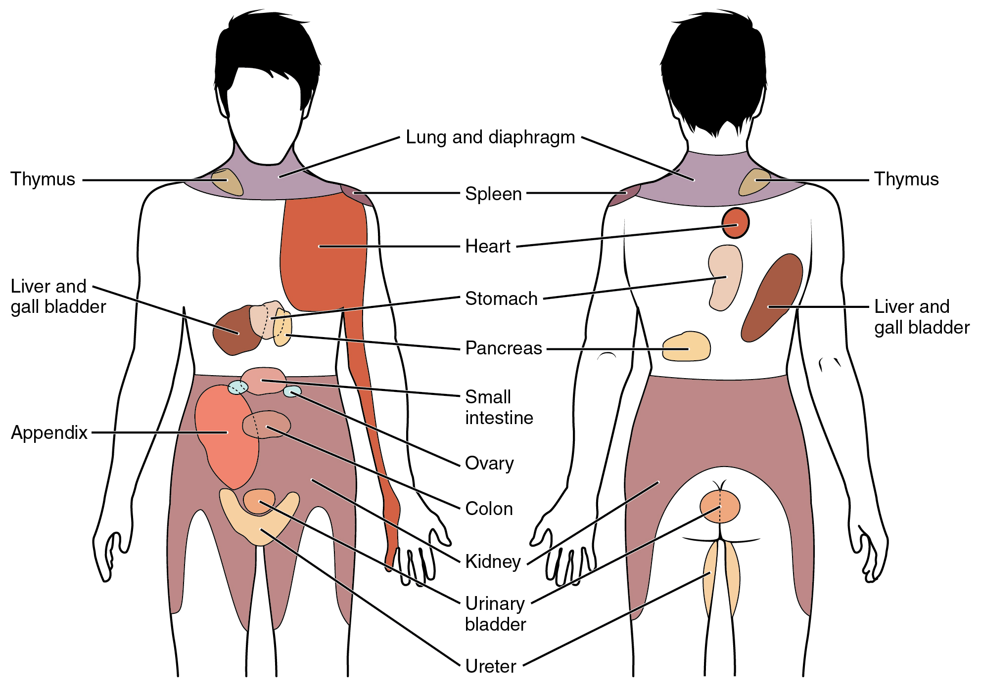
When particularly strong visceral sensations rise to the level of conscious perception, the sensations are often felt in unexpected places. For example, strong visceral sensations of the heart will be felt as pain in the left shoulder and left arm. This irregular pattern of projection of conscious perception of visceral sensations is called referred pain. Depending on the organ system affected, the referred pain will project to different areas of the body (Figure \(\PageIndex{2}\)). The location of referred pain is not random, but a definitive explanation of the mechanism has not been established. The most broadly accepted theory for this phenomenon is that the visceral sensory fibers enter into the same level of the spinal cord as the somatosensory fibers of the referred pain location. By this explanation, the visceral sensory fibers from the mediastinal region, where the heart is located, would enter the spinal cord at the same level as the spinal nerves from the shoulder and arm, so the brain misinterprets the sensations from the mediastinal region as being from the axillary and brachial regions. Projections from the medial and inferior divisions of the cervical ganglia do enter the spinal cord at the middle to lower cervical levels, which is where the somatosensory fibers enter.
DISORDERS OF THE...
Nervous System: Kehr’s Sign
Kehr’s sign is the presentation of pain in the left shoulder, chest, and neck regions following rupture of the spleen. The spleen is in the upper-left abdominopelvic quadrant, but the pain is more in the shoulder and neck. How can this be? The sympathetic fibers connected to the spleen are from the celiac ganglion, which would be from the mid-thoracic to lower thoracic region whereas parasympathetic fibers are found in the vagus nerve, which connects in the medulla of the brain stem. However, the neck and shoulder would connect to the spinal cord at the mid-cervical level of the spinal cord. These connections do not fit with the expected correspondence of visceral and somatosensory fibers entering at the same level of the spinal cord.
The incorrect assumption would be that the visceral sensations are coming from the spleen directly. In fact, the visceral fibers are coming from the diaphragm. The nerve connecting to the diaphragm takes a special route. The phrenic nerve is connected to the spinal cord at cervical levels 3 to 5. The motor fibers that make up this nerve are responsible for the muscle contractions that drive ventilation. These fibers have left the spinal cord to enter the phrenic nerve, meaning that spinal cord damage below the mid-cervical level is not fatal by making ventilation impossible. Therefore, the visceral fibers from the diaphragm enter the spinal cord at the same level as the somatosensory fibers from the neck and shoulder.
The diaphragm plays a role in Kehr’s sign because the spleen is just inferior to the diaphragm in the upper-left quadrant of the abdominopelvic cavity. When the spleen ruptures, blood spills into this region. The accumulating hemorrhage then puts pressure on the diaphragm. The visceral sensation is actually in the diaphragm, so the referred pain is in a region of the body that corresponds to the diaphragm, not the spleen.
Efferent Branch
The efferent branch of the visceral reflex arc begins with the projection from the central neuron along the preganglionic fiber. This fiber then makes a synapse on the ganglionic neuron that projects to the target effector.
The effector organs that are the targets of the autonomic system range from the iris and ciliary body of the eye to the urinary bladder and reproductive organs. The thoracolumbar output, through the various sympathetic ganglia, reaches all of these organs. The cranial component of the parasympathetic system projects from the eye to part of the intestines. The sacral component picks up with the majority of the large intestine and the pelvic organs of the urinary and reproductive systems.
Chapter Review
Autonomic nervous system function is based on the visceral reflex. This reflex is similar to the somatic reflex, but the efferent branch is composed of two neurons. The central neuron projects from the spinal cord or brain stem to synapse on the ganglionic neuron that projects to the effector. The afferent branch of the somatic and visceral reflexes is very similar, as many somatic and special senses activate autonomic responses. However, there are visceral senses that do not form part of conscious perception. If a visceral sensation, such as cardiac pain, is strong enough, it will rise to the level of consciousness. However, the sensory homunculus does not provide a representation of the internal structures to the same degree as the surface of the body, so visceral sensations are often experienced as referred pain, such as feelings of pain in the left shoulder and arm in connection with a heart attack.
The role of visceral reflexes is to maintain a balance of function in the organ systems of the body. The two divisions of the autonomic system each play a role in effecting change, usually in competing directions. The sympathetic system increases heart rate, whereas the parasympathetic system decreases heart rate. The sympathetic system dilates the pupil of the eye, whereas the parasympathetic system constricts the pupil. The competing inputs can contribute to the resting tone of the organ system. Heart rate is normally under parasympathetic tone, whereas blood pressure is normally under sympathetic tone. The heart rate is slowed by the autonomic system at rest, whereas blood vessels retain a slight constriction at rest.
In a few systems of the body, the competing input from the two divisions is not the norm. The sympathetic tone of blood vessels is caused by the lack of parasympathetic input to the systemic circulatory system. Only certain regions receive parasympathetic input that relaxes the smooth muscle wall of the blood vessels. Sweat glands are another example, which only receive input from the sympathetic system.
Interactive Link Questions
Read this article to learn about a teenager who experiences a series of spells that suggest a stroke. He undergoes endless tests and seeks input from multiple doctors. In the end, one expert, one question, and a simple blood pressure cuff answers the question. Why would the heart have to beat faster when the teenager changes his body position from lying down to sitting, and then to standing?
Answer: The effect of gravity on circulation means that it is harder to get blood up from the legs as the body takes on a vertical orientation.
Watch this video to learn about the pupillary reflexes. The pupillary light reflex involves sensory input through the optic nerve and motor response through the oculomotor nerve to the ciliary ganglion, which projects to the circular fibers of the iris. As shown in this short animation, pupils will constrict to limit the amount of light falling on the retina under bright lighting conditions. What constitutes the afferent and efferent branches of the competing reflex (dilation)?
Answer: The optic nerve still carries the afferent input, but the output is from the thoracic spinal cord, through the superior cervical ganglion, to the radial fibers of the iris.
Review Questions
Q. Which of the following represents a sensory input that is not part of both the somatic and autonomic systems?
A. vision
B. taste
C. baroreception
D. proprioception
Answer: C
Q. What is the term for a reflex that does not include a CNS component?
A. long reflex
B. visceral reflex
C. somatic reflex
D. short reflex
Answer: D
Q. What neurotransmitter will result in constriction of the pupil?
A. norepinephrine
B. acetylcholine
C. epinephrine
D. serotonin
Answer: B
Q. What gland produces a secretion that causes fight-or-flight responses in effectors?
A. adrenal medulla
B. salivatory gland
C. reproductive gland
D. thymus
Answer: A
Q. Which of the following is an incorrect pairing?
A. norepinephrine dilates the pupil
B. epinephrine increases blood pressure
C. acetylcholine decreases digestion
D. norepinephrine increases heart rate
Answer: C
Critical Thinking Questions
Q. Damage to internal organs will present as pain associated with a particular surface area of the body. Why would something like irritation to the diaphragm, which is between the thoracic and abdominal cavities, feel like pain in the shoulder or neck?
A. The nerves that carry sensory information from the diaphragm enter the spinal cord in the cervical region where somatic sensory fibers from the shoulder and neck would enter. The brain superimposes this experience onto the sensory homunculus where the somatic nerves are connected.
Q. Medical practice is paying more attention to the autonomic system in considering disease states. Why would autonomic tone be important in considering cardiovascular disease?
A. Within the cardiovascular system, different aspects demonstrate variation in autonomic tone. Heart rate is under parasympathetic tone, and blood pressure is under sympathetic tone. Pharmaceuticals that treat cardiovascular disorders may be more effective if they work with the normal state of the autonomic system. Alternatively, some disorders may be exacerbated by autonomic deficits and common therapies might not be as effective.
Glossary
- autonomic tone
- tendency of an organ system to be governed by one division of the autonomic nervous system over the other, such as heart rate being lowered by parasympathetic input at rest
- afferent branch
- component of a reflex arc that represents the input from a sensory neuron, for either a special or general sense
- baroreceptor
- mechanoreceptor that senses the stretch of blood vessels to indicate changes in blood pressure
- efferent branch
- component of a reflex arc that represents the output, with the target being an effector, such as muscle or glandular tissue
- long reflex
- reflex arc that includes the central nervous system
- referred pain
- the conscious perception of visceral sensation projected to a different region of the body, such as the left shoulder and arm pain as a sign for a heart attack
- reflex arc
- circuit of a reflex that involves a sensory input and motor output, or an afferent branch and an efferent branch, and an integrating center to connect the two branches
- short reflex
- reflex arc that does not include any components of the central nervous system
- somatic reflex
- reflex involving skeletal muscle as the effector, under the control of the somatic nervous system
- visceral reflex
- reflex involving an internal organ as the effector, under the control of the autonomic nervous system
Contributors and Attributions
OpenStax Anatomy & Physiology (CC BY 4.0). Access for free at https://openstax.org/books/anatomy-and-physiology