
2.11: Lab Exercise 13- The Brain
- Page ID
- 72641
\( \newcommand{\vecs}[1]{\overset { \scriptstyle \rightharpoonup} {\mathbf{#1}} } \)
\( \newcommand{\vecd}[1]{\overset{-\!-\!\rightharpoonup}{\vphantom{a}\smash {#1}}} \)
\( \newcommand{\dsum}{\displaystyle\sum\limits} \)
\( \newcommand{\dint}{\displaystyle\int\limits} \)
\( \newcommand{\dlim}{\displaystyle\lim\limits} \)
\( \newcommand{\id}{\mathrm{id}}\) \( \newcommand{\Span}{\mathrm{span}}\)
( \newcommand{\kernel}{\mathrm{null}\,}\) \( \newcommand{\range}{\mathrm{range}\,}\)
\( \newcommand{\RealPart}{\mathrm{Re}}\) \( \newcommand{\ImaginaryPart}{\mathrm{Im}}\)
\( \newcommand{\Argument}{\mathrm{Arg}}\) \( \newcommand{\norm}[1]{\| #1 \|}\)
\( \newcommand{\inner}[2]{\langle #1, #2 \rangle}\)
\( \newcommand{\Span}{\mathrm{span}}\)
\( \newcommand{\id}{\mathrm{id}}\)
\( \newcommand{\Span}{\mathrm{span}}\)
\( \newcommand{\kernel}{\mathrm{null}\,}\)
\( \newcommand{\range}{\mathrm{range}\,}\)
\( \newcommand{\RealPart}{\mathrm{Re}}\)
\( \newcommand{\ImaginaryPart}{\mathrm{Im}}\)
\( \newcommand{\Argument}{\mathrm{Arg}}\)
\( \newcommand{\norm}[1]{\| #1 \|}\)
\( \newcommand{\inner}[2]{\langle #1, #2 \rangle}\)
\( \newcommand{\Span}{\mathrm{span}}\) \( \newcommand{\AA}{\unicode[.8,0]{x212B}}\)
\( \newcommand{\vectorA}[1]{\vec{#1}} % arrow\)
\( \newcommand{\vectorAt}[1]{\vec{\text{#1}}} % arrow\)
\( \newcommand{\vectorB}[1]{\overset { \scriptstyle \rightharpoonup} {\mathbf{#1}} } \)
\( \newcommand{\vectorC}[1]{\textbf{#1}} \)
\( \newcommand{\vectorD}[1]{\overrightarrow{#1}} \)
\( \newcommand{\vectorDt}[1]{\overrightarrow{\text{#1}}} \)
\( \newcommand{\vectE}[1]{\overset{-\!-\!\rightharpoonup}{\vphantom{a}\smash{\mathbf {#1}}}} \)
\( \newcommand{\vecs}[1]{\overset { \scriptstyle \rightharpoonup} {\mathbf{#1}} } \)
\(\newcommand{\longvect}{\overrightarrow}\)
\( \newcommand{\vecd}[1]{\overset{-\!-\!\rightharpoonup}{\vphantom{a}\smash {#1}}} \)
\(\newcommand{\avec}{\mathbf a}\) \(\newcommand{\bvec}{\mathbf b}\) \(\newcommand{\cvec}{\mathbf c}\) \(\newcommand{\dvec}{\mathbf d}\) \(\newcommand{\dtil}{\widetilde{\mathbf d}}\) \(\newcommand{\evec}{\mathbf e}\) \(\newcommand{\fvec}{\mathbf f}\) \(\newcommand{\nvec}{\mathbf n}\) \(\newcommand{\pvec}{\mathbf p}\) \(\newcommand{\qvec}{\mathbf q}\) \(\newcommand{\svec}{\mathbf s}\) \(\newcommand{\tvec}{\mathbf t}\) \(\newcommand{\uvec}{\mathbf u}\) \(\newcommand{\vvec}{\mathbf v}\) \(\newcommand{\wvec}{\mathbf w}\) \(\newcommand{\xvec}{\mathbf x}\) \(\newcommand{\yvec}{\mathbf y}\) \(\newcommand{\zvec}{\mathbf z}\) \(\newcommand{\rvec}{\mathbf r}\) \(\newcommand{\mvec}{\mathbf m}\) \(\newcommand{\zerovec}{\mathbf 0}\) \(\newcommand{\onevec}{\mathbf 1}\) \(\newcommand{\real}{\mathbb R}\) \(\newcommand{\twovec}[2]{\left[\begin{array}{r}#1 \\ #2 \end{array}\right]}\) \(\newcommand{\ctwovec}[2]{\left[\begin{array}{c}#1 \\ #2 \end{array}\right]}\) \(\newcommand{\threevec}[3]{\left[\begin{array}{r}#1 \\ #2 \\ #3 \end{array}\right]}\) \(\newcommand{\cthreevec}[3]{\left[\begin{array}{c}#1 \\ #2 \\ #3 \end{array}\right]}\) \(\newcommand{\fourvec}[4]{\left[\begin{array}{r}#1 \\ #2 \\ #3 \\ #4 \end{array}\right]}\) \(\newcommand{\cfourvec}[4]{\left[\begin{array}{c}#1 \\ #2 \\ #3 \\ #4 \end{array}\right]}\) \(\newcommand{\fivevec}[5]{\left[\begin{array}{r}#1 \\ #2 \\ #3 \\ #4 \\ #5 \\ \end{array}\right]}\) \(\newcommand{\cfivevec}[5]{\left[\begin{array}{c}#1 \\ #2 \\ #3 \\ #4 \\ #5 \\ \end{array}\right]}\) \(\newcommand{\mattwo}[4]{\left[\begin{array}{rr}#1 \amp #2 \\ #3 \amp #4 \\ \end{array}\right]}\) \(\newcommand{\laspan}[1]{\text{Span}\{#1\}}\) \(\newcommand{\bcal}{\cal B}\) \(\newcommand{\ccal}{\cal C}\) \(\newcommand{\scal}{\cal S}\) \(\newcommand{\wcal}{\cal W}\) \(\newcommand{\ecal}{\cal E}\) \(\newcommand{\coords}[2]{\left\{#1\right\}_{#2}}\) \(\newcommand{\gray}[1]{\color{gray}{#1}}\) \(\newcommand{\lgray}[1]{\color{lightgray}{#1}}\) \(\newcommand{\rank}{\operatorname{rank}}\) \(\newcommand{\row}{\text{Row}}\) \(\newcommand{\col}{\text{Col}}\) \(\renewcommand{\row}{\text{Row}}\) \(\newcommand{\nul}{\text{Nul}}\) \(\newcommand{\var}{\text{Var}}\) \(\newcommand{\corr}{\text{corr}}\) \(\newcommand{\len}[1]{\left|#1\right|}\) \(\newcommand{\bbar}{\overline{\bvec}}\) \(\newcommand{\bhat}{\widehat{\bvec}}\) \(\newcommand{\bperp}{\bvec^\perp}\) \(\newcommand{\xhat}{\widehat{\xvec}}\) \(\newcommand{\vhat}{\widehat{\vvec}}\) \(\newcommand{\uhat}{\widehat{\uvec}}\) \(\newcommand{\what}{\widehat{\wvec}}\) \(\newcommand{\Sighat}{\widehat{\Sigma}}\) \(\newcommand{\lt}{<}\) \(\newcommand{\gt}{>}\) \(\newcommand{\amp}{&}\) \(\definecolor{fillinmathshade}{gray}{0.9}\)Lab Summary: This lab will give you an opportunity explore the anatomy and physiology of the brain. To enhance your learning, you will also perform a dissection and pinning exercise with a sheep or pig brain.
Your objectives for this lab are:
- In human brain models, you should be able to identify and list the function(s) of
- Ventricles: Lateral ventricles, Third ventricles, Fourth ventricle
- Cerebral hemispheres, right and left
- Convolutions – Sulcus and gyrus
- Corpus callosum
- Lobes: Frontal lobe, Parietal lobe, Temporal lobe, Occipital lobe, Insula
- Diencephalon: Pineal gland, Thalamus, Hypothalamus
- Choroid plexus
- Pituitary gland and infundibulum
- The pituitary gland is NOT part of the brain but is intimately connected physically and functionally to the hypothalamus
- Explain how the pituitary is connected to the brain physically
- Explain how the pituitary is connected to the hypothalamus functionally
- Brainstem: Pons, Medulla
- Cerebellum
- Meninges: Dura mater, Pia mater
- In a pig or sheep brain, you should be able to identify and list the function(s) of
- Ventricles: Lateral ventricles, Third ventricles, Fourth ventricle
- Cerebral hemispheres, right and left
- Convolutions – Sulcus and gyrus
- Corpus callosum
- Lobes: Frontal lobe, Parietal lobe, Temporal lobe, Occipital lobe, Insula
- Diencephalon: Pineal gland, Thalamus, Hypothalamus
- Pituitary gland
- Brainstem: Pons, Medulla
- Cerebellum
- Meninges: Dura mater, Pia mater
- Optic nerve and optic chiasma
- Olfactory bulb and olfactory nerve
- Explain three differences or similarities between human and sheep brains. “The sheep brain (or a particular lobe) is bigger” would not be an acceptable answer. Think about how humans and sheep (prey animals) use brain structures differently
- For cerebrospinal fluid,
- Explain its function
- Explain its relationship to the choroid plexus
- List the cells responsible for its synthesis
Background Information
As a quick reminder, the nervous system can be divided into two major regions: the central and peripheral nervous systems. The central nervous system (CNS) consists of the brain and spinal cord; the peripheral nervous system (PNS) consists of all other nervous system structures, such as cranial nerves, spinal nerves, and receptors (Figure \(\PageIndex{1}\)). The brain is contained within the cranial cavity of the skull, and the spinal cord is contained within the vertebral cavity of the vertebral column. It is a bit of an oversimplification to say that the CNS is what is inside these two cavities and the peripheral nervous system is outside of them, but that is one way to start to think about it. There are some elements of the peripheral nervous system that are within the cranial or vertebral cavities. The peripheral nervous system is so named because it is on the periphery—meaning beyond the brain and spinal cord. Depending on different aspects of the nervous system, the dividing line between central and peripheral is not necessarily universal.

Nervous Tissue Structures
Nervous tissue, present in both the CNS and PNS, contains two basic types of cells: neurons and glial cells. A glial cell is one of a variety of cells that provide a framework of tissue that supports the neurons and their activities, which were studied in a previous lab. The neuron is the more functionally important of the two, in terms of the communicative function of the nervous system.
Neurons are cells and therefore have a soma, or cell body, but they also have notable extensions of the cell; each extension is generally referred to as a process. There is one important process that nearly all neurons have called an axon, which is the fiber that connects a neuron with its target. Another type of process that branches off from the soma is the dendrite. Dendrites are responsible for receiving most of the input from other neurons. Looking at nervous tissue, there are regions that predominantly contain cell bodies and regions that are largely composed of axons. These two regions within nervous system structures are referred to as gray matter (the regions with many cell bodies and dendrites) or white matter (the regions with many highly myelinated axons). The colors ascribed to these regions are what would be seen in unstained, nervous tissue (Figure \(\PageIndex{2}\)). Gray matter is not necessarily gray. It can be pinkish because of blood content, or even slightly tan, depending on how long the tissue has been preserved. White matter is white because axons are insulated by a lipid-rich substance called myelin. Lipids can appear as white material, much like the fat on a raw piece of meat. Gray matter may have that color ascribed to it because next to the white matter, it is just darker— hence, gray.

The cell bodies of neurons or axons are often located in discrete anatomical structures that are named, depending on whether the structure is central or peripheral. A localized collection of neuron cell bodies in the CNS is referred to as a nucleus. In the PNS, a cluster of neuron cell bodies is referred to as a ganglion. A notable exception to this naming convention is a group of nuclei in the central nervous system that were once called the basal ganglia before “ganglion” became accepted as a description for a peripheral structure. Some sources refer to this group of nuclei as the “basal nuclei” which helps avoid confusion.
Terminology applied to bundles of axons also differs depending on location. A bundle of axons, or fibers, found in the CNS is called a tract whereas the same thing in the PNS would be called a nerve. Please note that both can be used to refer to the same bundle of axons. When those axons are in the PNS, the term is nerve, but if they are in the CNS, the term is tract. One example of this is the axons that project from the nervous tissue in the retina into the brain. Axons leaving the eye are called the optic nerve but as soon as they enter the cranium they are referred to as the optic tract.
Activity 13.1: Identifying Selected Structures in Brain Models
Procedure for Activity 13.1: For this activity, you will use human brain models. Use the figures and information below to locate the required anatomical features of the brain and its associated structure. These are listed in the objectives at the beginning of this lab. As you work, you should review and make notes about the functions of each part you study.
The Central Nervous System
The brain and the spinal cord make up the central nervous system. Although the spinal cord is a single structure, the adult brain is described in terms of four major regions: the cerebrum, the diencephalon, the brain stem, and the cerebellum. You will learn more about each of these regions and their specific parts as you work through this lab.
Cerebrum
The iconic gray mantle of the human brain, which appears to make up most of the mass of the brain, is the cerebrum (Figure \(\PageIndex{3}\)). The wrinkled outer portion is the cerebral cortex, and the rest of the structure is beneath that outer covering. The left and right hemispheres are separated by the longitudinal fissure. Deep within the cerebrum, the white matter of the corpus callosum provides the major pathway for communication between the two hemispheres of the cerebral cortex. Many of the higher neurological functions, such as memory, emotion, and consciousness, are the result of cerebral function.

Cerebral Cortex & Lobes
The cerebrum is covered by a continuous layer of gray matter that wraps around either side of the cerebral cortices. This thin, extensive region of wrinkled gray matter is responsible for the higher functions of the nervous system. A gyrus (plural = gyri) is a raised ridge of one of those wrinkles, and a sulcus (plural = sulci) is a depression between two gyri. The folding of the cortex maximizes the amount of gray matter in the cranial cavity. During embryonic development, the developing brain expands within the skull and goes through a regular course of growth that results in everyone’s brain having a similar pattern of folds. The surface of the cortex can be separated into four major regions, or lobes (Figure \(\PageIndex{4}\)). The fact that there is no obvious anatomical border between these lobes is consistent with the functions of these regions being interrelated.
The frontal lobe is responsible for many complex functions. Some of these functions include executing motor functions (planning and performing movements via commands sent to the spinal cord and nerves), mediating aspects of personality, moderating emotional decisions, task management, cognition and learning, making rational decisions based on previous experience, and voluntary eye movements. A specialized area within the frontal lobe called Broca’s area plays a significant role in the spoken language and the mechanics of speech (motor speech).
The parietal lobe receives and processes information from skin and proprioceptors (in joints, muscles) in the somatosensory cortices. Some of this information allows us to understand spatial discrimination. It also contains part of Wernicke’s area, which helps us to understand language (the meaning of words).
The occipital lobe is where visual processing begins, although the other parts of the brain can contribute to visual function. The occipital lobe receives visual information from receptors in the retina of the eye called rods (for shades of black and white) and cones (for blue, red, and green) via the optic nerve. Neurons here use past visual experiences to interpret visual stimuli, such as recognizing a red, round form as an apple or cherry.
The temporal lobe contains the cortical area for auditory processing and has regions crucial for memory formation related to sound (sound memory). Like the parietal lobe, the temporal lobe also contains part of Wernicke’s area. Conscious awareness of odors (olfaction) is also performed here. However, interpretations of smells and tastes must also involve opinions formed in the frontal lobe.
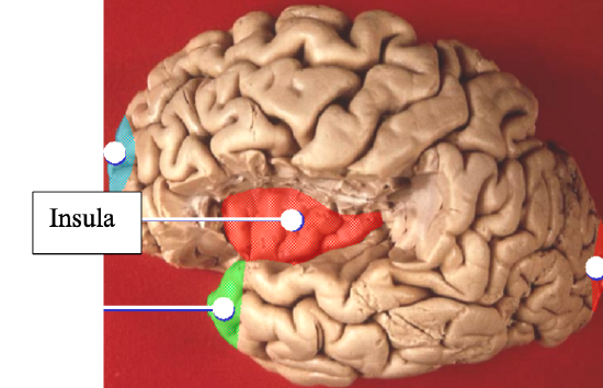
The insula (or insular lobe) located deep within the lateral sulcus is the fifth lobe of the brain (Figure \(\PageIndex{5}\)). It assists with conscious understanding of balance/vestibulation, interpretation of taste sensations (such as sweet or umami). Damage or atrophy (loss of nervous tissue) to the insula has also been linked to addiction and a variety of neuropsychiatric disorders, such as schizophrenia, hallucinations, poor judgment, impulsivity/disinhibition, and lack of empathy (Shura et al., 2014).

The Diencephalon
The thalamus (Figure \(\PageIndex{6}\)) is a collection of nuclei that relay information between the cerebral cortex and the periphery, spinal cord, or brain stem. All sensory information, except for olfaction, passes through the thalamus before processing by the cortex. The thalamus does not just pass the information on, it also processes that information. The cerebrum and basal nuclei also send motor information to the thalamus which usually involves interactions between the cerebellum and other nuclei in the brain stem as well.
The epithalamus contains the pineal gland (Figure \(\PageIndex{6}\)), which secretes the hormone melatonin. This hormone helps regulate Circadian rhythms and appears to play a role in the development of sexual maturity. The secretion of melatonin varies according to the level of light received from the environment. Additionally, children have higher melatonin levels than adults, which may prevent the release of gonadotropins from the anterior pituitary, thereby inhibiting the onset of puberty.
The hypothalamus (Figure \(\PageIndex{6}\)) is a collection of nuclei that are largely involved in regulating homeostasis:
- Regulates autonomic nervous system functions
- Regulates the endocrine system via hormones sent to the anterior pituitary and action potentials sent to the posterior pituitary
- Involved in memory and emotion as part of the limbic system
- Synthesizes hormones, including ADH (antidiuretic hormone), oxytocin, releasing hormones, inhibiting hormones
- Regulates thirst and water balance via osmoreceptors that detect water levels in blood
- Regulates hunger
- Assists in regulation of Circadian rhythms (sleep-wake cycles)

Brain stem
The midbrain, pons, and medulla oblongota are collectively referred to as the brain stem (Figure \(\PageIndex{6}\) and Figure \(\PageIndex{7}\)). The midbrain, a small region between the thalamus and pons. coordinates sensory representations of the visual, auditory, and somatosensory perceptual information. The pons plays a key role in regulating the depth and rate of breathing. The medulla regulates several crucial functions, including the heart rate (pulse), blood pressure, and respiratory rate; it also contains reflexive centers, such as those that control vomiting, coughing, sneezing, and swallowing. The major ascending and descending pathways between the spinal cord and brain, specifically the cerebrum, pass through the brain stem.

Cerebellum
The cerebellum, as the name suggests, is the “little brain” and accounts for approximately 10% of the brain’s mass. It is covered in gyri (humps) and sulci (depressions) like the cerebrum and looks like a miniature version of that part of the brain (Figure \(\PageIndex{8}\)). The cerebellum is largely responsible for coordinating vestibulation (sense of balance) by comparing information from the cerebrum with sensory feedback from the PNS via the spinal cord, including visual information, hearing information, and proprioceptive information. For example, if the cerebrum sends a command down to the spinal cord to initiate walking, a copy of that motor command is sent to the cerebellum. Sensory feedback from the muscles and joints (proprioceptive information) about the movements of walking, and sensations of balance are sent to the cerebellum, which then integrates all of that information. If walking is not coordinated, perhaps because the ground is uneven or a strong wind is blowing, then the cerebellum sends out a corrective command to compensate for the difference between the original command from the cerebrum and the sensory feedback from the periphery. The output of the cerebellum is into the midbrain, which then sends a descending input to the spinal cord to correct motor information going to skeletal muscles. Some research also suggests that the cerebellum may play a role in cognition and how we condition ourselves to respond to fear (Timmann, D et al., 2010)

Cerebrospinal Fluid
Cerebrospinal fluid (CSF) circulates throughout and around the CNS. In other tissues, water and small molecules are filtered through capillaries as the major contributor to the interstitial fluid. In the brain, CSF is produced by filtration of blood through the choroid plexus, which is found in some of the ventricles. The ventricles are the open spaces within the brain where CSF circulates. The CSF cycles through all of the ventricles to eventually emerge into the subarachnoid space where it will be reabsorbed into the blood. CSF circulates to remove metabolic wastes from the interstitial fluids of nervous tissues and return them to the blood stream. It also serves to cushion the brain and spinal cord from physical trauma. Along with the periosteal layer of the dura mater, CSF helps to buoy the weight of the brain, so that it does not become compressed under its own weight within the cranium.
The Meninges
The outer surface of the brain is covered by a series of membranes composed of connective tissue called the meninges, which protect the brain (Figure \(\PageIndex{9}\)). There are three major meningeal layers: the dura mater (having a periosteal layer and a meningeal layer), the arachnoid mater and the pia mater. Generally, you will not be able to see these layers in a brain model.
The dura mater is the thick, fibrous, outermost meninx, which provides a strong protective sheath over the entire brain and spinal cord and encloses the major blood vessels, as well. It is anchored to the inner surface of the cranium and to the very end of the vertebral cavity. The name comes from the Latin for “tough mother” to represent its physically protective role. In the cranial cavity, the dura mater has two layers: the periosteal layer, which knits its collagen fibers into those of the periosteum to anchor the brain and buoy its weight; and the meningeal layer, which sits adjacent to the arachnoid mater. In some places, these two layers of dura form sinuses, large, highly permeable vein-like structures. These sinuses help to recycle CSF.
The middle layer of the meninges is the arachnoid, named for the spider-web–like extensions between it and the pia mater. The arachnoid defines a sac-like enclosure around the CNS. The branching extensions are found in the subarachnoid space, which is filled with circulating CSF (cerebrospinal fluid). The arachnoid emerges into the dural sinuses as the arachnoid granulations, where the CSF is filtered back into the blood for drainage from the nervous system. The subarachnoid space is filled with circulating CSF, which also provides a liquid cushion to the brain and spinal cord. Like clinical blood work, a sample of CSF can be withdrawn to find chemical evidence of neuropathology or metabolic traces of the biochemical functions of nervous tissue.
The innermost layer of meninges is pia mater, a thin fibrous membrane that extends into every convolution of gyri and sulci in the cerebral cortex (contours of the brain) and other grooves and indentations. Its continuous layer of cells provides a fluid-impermeable membrane. The name pia mater comes from the Latin for “tender mother,” suggesting the thin membrane is a gentle covering for the brain.

Activity 13.2: Dissection of the Sheep or Pig Brains
Procedure for Activity 13.2: For this activity, you will dissect a pig or sheep brain and locate many of the same human brain structures you studied in the previous activity. You will need 2-3 partners for this activity. Before you begin, go to the supply area to obtain a brain, dissection pan, forceps, a scalpel, scissors, a probe, and a set of labeled dissection pins.
Each student must don goggles and gloves for this activity. Put these on before you open the brain’s packaging.
Read the following steps carefully before you begin.
- Place the sheep brain specimen in the tray, dorsal side up (Figure \(\PageIndex{10A}\)). Identify the dura mater (the thick, silvery gray membrane on the outside of the brain), left and right cerebral hemispheres, cerebrum, cerebellum, and the longitudinal fissure.
- Place the brain in the tray ventral side up (Figure \(\PageIndex{10B}\)). Identify the cerebellum, optic chiasma, optic nerves, and the pituitary gland.
- Turn the brain back over so its dorsal side up. Locate the longitudinal fissure. Now, you a knife or scalpel to cut the brain into two left and right halves (Figure \(\PageIndex{10D}\)). Be sure to cut all the way through the pituitary gland.
- Carefully remove the dura mater from one half, being careful not to remove the cerebellum, pituitary gland, or optic nerve.
- Use Figure \(\PageIndex{10}\), Figure \(\PageIndex{11}\), and Figure \(\PageIndex{12}\) to locate the following structures on either of the halves: pia mater, examples of gyri and sulci, the different lobes of the cerebrum, thalamus, hypothalamus, pineal gland, pons, medulla, corpus collosum, lateral ventricles, third ventricle, cerebellum, lobes (frontal, temporal, parietal, and occipital), pituitary gland, olfactory bulb, and olfactory nerve.
- Compare the structures that you see in your dissection specimen to those in the human brain models.
- Perform the pinning exercise as directed by your instructor.
- When you are done observing the pig/sheep brain specimen and have completed the pinning exercise, dispose of it as directed by your instructor. Wash and dry your dissection instruments, pins, and tray. Return these to the supply area.


Additional Learning Resources (see electronic document in Canvas to access weblinks):
- Watch these two videos to locate brain structures listed above for the human brain models:
- Watch these two videos to locate brain structures listed above for the dissected sheep brains:
- Watch this video to see human brain dissection and an explanation of functions:
- Practice identifying parts of the brain here:
- Practice identifying A&P of the brain in models with this document: Practice for Brain Model Labeling.pdf (found in the Canvas Module containing the electronic copy of the SLM)