
2.13: Lab Exercise 15- Reflexes and The Neurological Exam
- Page ID
- 72643
\( \newcommand{\vecs}[1]{\overset { \scriptstyle \rightharpoonup} {\mathbf{#1}} } \)
\( \newcommand{\vecd}[1]{\overset{-\!-\!\rightharpoonup}{\vphantom{a}\smash {#1}}} \)
\( \newcommand{\dsum}{\displaystyle\sum\limits} \)
\( \newcommand{\dint}{\displaystyle\int\limits} \)
\( \newcommand{\dlim}{\displaystyle\lim\limits} \)
\( \newcommand{\id}{\mathrm{id}}\) \( \newcommand{\Span}{\mathrm{span}}\)
( \newcommand{\kernel}{\mathrm{null}\,}\) \( \newcommand{\range}{\mathrm{range}\,}\)
\( \newcommand{\RealPart}{\mathrm{Re}}\) \( \newcommand{\ImaginaryPart}{\mathrm{Im}}\)
\( \newcommand{\Argument}{\mathrm{Arg}}\) \( \newcommand{\norm}[1]{\| #1 \|}\)
\( \newcommand{\inner}[2]{\langle #1, #2 \rangle}\)
\( \newcommand{\Span}{\mathrm{span}}\)
\( \newcommand{\id}{\mathrm{id}}\)
\( \newcommand{\Span}{\mathrm{span}}\)
\( \newcommand{\kernel}{\mathrm{null}\,}\)
\( \newcommand{\range}{\mathrm{range}\,}\)
\( \newcommand{\RealPart}{\mathrm{Re}}\)
\( \newcommand{\ImaginaryPart}{\mathrm{Im}}\)
\( \newcommand{\Argument}{\mathrm{Arg}}\)
\( \newcommand{\norm}[1]{\| #1 \|}\)
\( \newcommand{\inner}[2]{\langle #1, #2 \rangle}\)
\( \newcommand{\Span}{\mathrm{span}}\) \( \newcommand{\AA}{\unicode[.8,0]{x212B}}\)
\( \newcommand{\vectorA}[1]{\vec{#1}} % arrow\)
\( \newcommand{\vectorAt}[1]{\vec{\text{#1}}} % arrow\)
\( \newcommand{\vectorB}[1]{\overset { \scriptstyle \rightharpoonup} {\mathbf{#1}} } \)
\( \newcommand{\vectorC}[1]{\textbf{#1}} \)
\( \newcommand{\vectorD}[1]{\overrightarrow{#1}} \)
\( \newcommand{\vectorDt}[1]{\overrightarrow{\text{#1}}} \)
\( \newcommand{\vectE}[1]{\overset{-\!-\!\rightharpoonup}{\vphantom{a}\smash{\mathbf {#1}}}} \)
\( \newcommand{\vecs}[1]{\overset { \scriptstyle \rightharpoonup} {\mathbf{#1}} } \)
\( \newcommand{\vecd}[1]{\overset{-\!-\!\rightharpoonup}{\vphantom{a}\smash {#1}}} \)
\(\newcommand{\avec}{\mathbf a}\) \(\newcommand{\bvec}{\mathbf b}\) \(\newcommand{\cvec}{\mathbf c}\) \(\newcommand{\dvec}{\mathbf d}\) \(\newcommand{\dtil}{\widetilde{\mathbf d}}\) \(\newcommand{\evec}{\mathbf e}\) \(\newcommand{\fvec}{\mathbf f}\) \(\newcommand{\nvec}{\mathbf n}\) \(\newcommand{\pvec}{\mathbf p}\) \(\newcommand{\qvec}{\mathbf q}\) \(\newcommand{\svec}{\mathbf s}\) \(\newcommand{\tvec}{\mathbf t}\) \(\newcommand{\uvec}{\mathbf u}\) \(\newcommand{\vvec}{\mathbf v}\) \(\newcommand{\wvec}{\mathbf w}\) \(\newcommand{\xvec}{\mathbf x}\) \(\newcommand{\yvec}{\mathbf y}\) \(\newcommand{\zvec}{\mathbf z}\) \(\newcommand{\rvec}{\mathbf r}\) \(\newcommand{\mvec}{\mathbf m}\) \(\newcommand{\zerovec}{\mathbf 0}\) \(\newcommand{\onevec}{\mathbf 1}\) \(\newcommand{\real}{\mathbb R}\) \(\newcommand{\twovec}[2]{\left[\begin{array}{r}#1 \\ #2 \end{array}\right]}\) \(\newcommand{\ctwovec}[2]{\left[\begin{array}{c}#1 \\ #2 \end{array}\right]}\) \(\newcommand{\threevec}[3]{\left[\begin{array}{r}#1 \\ #2 \\ #3 \end{array}\right]}\) \(\newcommand{\cthreevec}[3]{\left[\begin{array}{c}#1 \\ #2 \\ #3 \end{array}\right]}\) \(\newcommand{\fourvec}[4]{\left[\begin{array}{r}#1 \\ #2 \\ #3 \\ #4 \end{array}\right]}\) \(\newcommand{\cfourvec}[4]{\left[\begin{array}{c}#1 \\ #2 \\ #3 \\ #4 \end{array}\right]}\) \(\newcommand{\fivevec}[5]{\left[\begin{array}{r}#1 \\ #2 \\ #3 \\ #4 \\ #5 \\ \end{array}\right]}\) \(\newcommand{\cfivevec}[5]{\left[\begin{array}{c}#1 \\ #2 \\ #3 \\ #4 \\ #5 \\ \end{array}\right]}\) \(\newcommand{\mattwo}[4]{\left[\begin{array}{rr}#1 \amp #2 \\ #3 \amp #4 \\ \end{array}\right]}\) \(\newcommand{\laspan}[1]{\text{Span}\{#1\}}\) \(\newcommand{\bcal}{\cal B}\) \(\newcommand{\ccal}{\cal C}\) \(\newcommand{\scal}{\cal S}\) \(\newcommand{\wcal}{\cal W}\) \(\newcommand{\ecal}{\cal E}\) \(\newcommand{\coords}[2]{\left\{#1\right\}_{#2}}\) \(\newcommand{\gray}[1]{\color{gray}{#1}}\) \(\newcommand{\lgray}[1]{\color{lightgray}{#1}}\) \(\newcommand{\rank}{\operatorname{rank}}\) \(\newcommand{\row}{\text{Row}}\) \(\newcommand{\col}{\text{Col}}\) \(\renewcommand{\row}{\text{Row}}\) \(\newcommand{\nul}{\text{Nul}}\) \(\newcommand{\var}{\text{Var}}\) \(\newcommand{\corr}{\text{corr}}\) \(\newcommand{\len}[1]{\left|#1\right|}\) \(\newcommand{\bbar}{\overline{\bvec}}\) \(\newcommand{\bhat}{\widehat{\bvec}}\) \(\newcommand{\bperp}{\bvec^\perp}\) \(\newcommand{\xhat}{\widehat{\xvec}}\) \(\newcommand{\vhat}{\widehat{\vvec}}\) \(\newcommand{\uhat}{\widehat{\uvec}}\) \(\newcommand{\what}{\widehat{\wvec}}\) \(\newcommand{\Sighat}{\widehat{\Sigma}}\) \(\newcommand{\lt}{<}\) \(\newcommand{\gt}{>}\) \(\newcommand{\amp}{&}\) \(\definecolor{fillinmathshade}{gray}{0.9}\)Lab Summary: In this lab, you will get to have some fun while learning about different types of reflexes, which are protective mechanisms; this means they aid the body in maintaining homeostasis and preventing damage. In most cases, you will test somatic and cranial innate reflexes. These innate reflexes are “pre-programmed” responses by the body to particular stimuli. Remember we develop learned reflexes as well, such as driving, athletic skills, or playing a musical instrument. As we experience actions and consequences, both innate and learned reflexes may be modified. As you perform the reflexes and cranial nerve testing in this lab, it is important that your subject is relaxed, and that s/he has eyes closed when appropriate. Special thanks to Professor Amanda Couitt, PT, DPT of the RVCC PTA department for making several of the reflex videos.
Your objectives for this lab are:
- Describe the correct pathway and functions of the following within a reflex arc: receptor, sensory/afferent neuron, integration center, motor/efferent neuron, effector
- Describe the purpose, nerve(s) tested, normal physiological results (using proper terminology, such as knee extension, not knee jerk nor foot kicks out), and possible pathological causes for abnormal test results for the patellar, biceps, triceps, plantar, Achilles, withdrawal, gag, photopupillary, and corneal reflexes
- Describe the purpose, normal physiological results (using proper terminology, such as knee extension not knee jerk or foot kicks out), and possible pathological causes for abnormal test results when testing cranial nerves III, IV, and VI
- Describe the significance of a positive Babinski’s sign in an adult
- Predict the effects of spinal cord or brain dysfunctions upon a reflex
Background
Reflexes may be spinal or cranial, depending on the nerves and central components that are involved. An innate reflex is an involuntary and nearly instantaneous movement in response to a stimulus; they are designed to help us function properly or protect us from potential injuries.
Reflexes can also be classified as somatic or visceral. One example of a somatic reflex is the spinal muscle reflexes that help us maintain an upright posture or stability when walking. Other reflexes, such as the photopupillary reflex or the gag reflex, protect us from potential injury. Urination and defecation are examples of visceral reflexes mediated by the spinal cord. This means that when stretch in the bladder is detected, that afferent information is sent to the spinal cord and a motor response (contraction of the smooth muscle of the bladder and relaxation of the urethral sphincters) is generated.
Reflexes combine the spinal sensory and motor components with a sensory input that directly generates a motor response. In higher animals, most sensory neurons do not pass directly into the brain, but synapse in the spinal cord. This characteristic allows reflex actions to occur relatively quickly by activating spinal motor neurons without the delay of routing signals through the brain, although the brain will receive sensory input while the reflex action occurs. There are two types of reflex arcs: autonomic (visceral) reflex arcs (affecting inner organs) and somatic reflex arcs (affecting muscles). The generalized somatic reflex arc is shown below and in Figure \(\PageIndex{1}\):


The generalized reflex arc contains five key components:
- Receptor: A nervous system structure that intakes the stimulus (sensory information from the internal or external environment) and “translates” it to electrical language understood by neurons
- Sensory (afferent) neuron: These are typically classified as the neurons responsible for converting various external stimuli that come from the environment into corresponding internal stimuli. They carry the sensory information to the brain or spinal cord.
- Integration center: The brain or spinal cord where sensory information is sorted, “understood”, and a decision about responding to the sensory information is made.
- Motor (efferent) neuron: A neuron located in the central nervous system that projects its axon outside the CNS and directly or indirectly controls effectors. The motor neuron carries the “do something” information to the effector.
- Effector (also called effector organ): A muscle or gland that responds to the motor neuron’s signal.
Most reflex arcs involve only three neurons. The stimulus, such as a needle stick, stimulates the pain receptors of the skin, which initiate an impulse in a sensory neuron (“neuron 1”). This travels to the spinal cord where it passes, by means of a synapse, to a connecting neuron called the interneuron (“neuron 2”)situated in the spinal cord. The interneuron, in turn, makes a synapse with one or more motor neurons (“neuron 3”)that transmit the impulse to the effector where a motor response occurs (such as causing a limb to contract and pull away from a sharp object). Reflexes do not require involvement of the brain, although in some cases the brain can prevent reflex action. One such example would be not dropping a pot of boiling water on a child or animal even if the water spills and burns you.
Deep Tendon Reflexes
“Inspired by the practice of thumping a wine cask [wine barrel] to determine the level of fluid, Viennese physician Leopold Auenbrugger (1722-1809) first described the use of percussion as an aid to medical diagnosis in 1761.1,3” (https://n.neurology.org/content/neur.../1542.full.pdf) In the following procedures, you will test several deep tendon reflexes and document your observations. As you perform each reflex, remember that the subject should have the eyes closed to avoid bias. Bias can occur when the subject unconsciously creates and/or exaggerates the expected result, or unconsciously stops him/herself from allowing the response to happen. You will also repeat each reflex and have the subject perform Jendrassik's maneuver. To do this, the subject will clasp his/her hands in front of him/her and, with fingers locked, try to pull the hands apart. Under normal circumstances, brain pathways descend onto the same motor neurons, constantly modulating the strength of the stretch reflexes. Therefore, stretch reflexes tend to be hypoactive or absent in cases of peripheral nerve damage, and hyperactive in corticospinal tract lesions.
A deep tendon reflex, or stretch reflex, is elicited by a strong tap to a tendon, such as in the patellar reflex. The muscle associated with the tendon is quickly stretched, resulting in activation of the muscle spindle. The muscle spindle then sends a signal to the spinal cord through the dorsal root. The fiber synapses directly on the motor neuron of the ventral horn. The motor signal travels from the ventral horn to the ventral root of the spinal nerve, activating the muscle, causing contraction. Reflexes are physiologically useful for stability. If a muscle is stretched, it reflexively contracts to return the muscle to compensate for the change in length. In the context of the neurological exam, normal reflexes indicate that the LMN (lower motor neurons of the pyramidal and extra-pyramidal tracts) are functioning properly.

Administer a Reflex Test on your Partner!
Watch this video, then read the directions below: https://www.youtube.com/watch?v=- xOFmaDd6nQ&feature=youtu.be
- Position The Subject:
- Have the subject sit on the examination table with his/her feet dangling above the floor.
- The distal thigh and knee must be readily palpated and free of restrictive clothing (i.e., a tightly rolled up pant leg).
- Stand in front and lateral to the knee as to avoid being struck by the subject’s foot if there is a brisk or exaggerated response.
- Locate the patellar tendon. The quadriceps muscle is typically relaxed as long as the foot can dangle.
- Allow the reflex hammer to swing loosely between your thumb and forefinger through a 45 to 60 degree arc, while making certain the hammer moves in a horizontal plane (i.e., the broad end of the reflex hammer is perpendicular to the tendon).
- Strike the tendon just below the patella and above its bony insertion on the tibia.
- Palpate and visually observe the response to the provided stimulus (i.e., contraction of the quadriceps muscles and extension of the knee). In the space below, document your observations (normal, hyporeactive, hyperreactive, anything interesting or odd).
- Repeat Steps 1—5 but have your subject execute Jendrassik’s maneuver. In the space below, document your observations (normal, hyporeactive, hyperreactive, anything interesting or odd).
Procedure for Activity 15.2 Achilles Reflex Test: (spinal reflex that tests the integrity of spinal nerves L5—S2)

- Position The Subject:
- Have the subject sit on the examination table with his or her feet dangling above the floor.
- The foot should be visible and the leg must be free of restrictive clothing (i.e., a tightly rolled up pant leg).
- Stand in front and lateral to the subject.
- Locate the Achilles tendon of the gastrocnemius muscle. The tendon will look and feel like a broad, thick, and slightly rounded cord.
- Gently dorsiflex the foot at the ankle and position the joint at approximately 90 degrees or until slight resistance is felt.
- Allow the reflex hammer to swing loosely between your thumb and forefinger through a 45 to 60 degree arc, while making certain the hammer moves in a horizontal plane (i.e., the broad end of the reflex hammer is perpendicular to the tendon).
- Strike the tendon just above the calcaneus at the level of the medial and lateral malleoli.
- Feel and observe the response to the provided stimulus (i.e., contraction of the calf muscles and ankle plantar flexion). In the space below, document your observations (normal, hyporeactive, hyperreactive, anything interesting or odd).
- Repeat Steps 1—7 but have your subject execute Jendrassik’s maneuver. In the space below, document your observations (normal, hyporeactive, hyperreactive, anything interesting or odd).
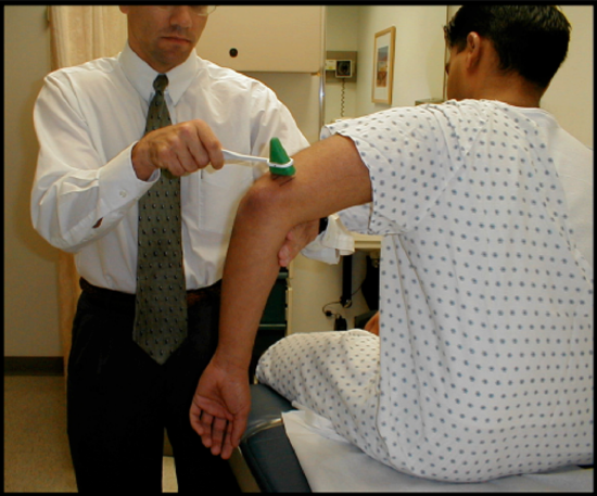
Procedure for Activity 15.3: Biceps Reflex Test (spinal reflex that tests the integrity of spinal nerves C5 and C6)

- Position The Subject:
- The elbow region should be visible and the proximal arm must be free of restrictive clothing (i.e., a tightly rolled shirt sleeve).
- Stand in front and to the side of the extremity to be examined.
- With the subject seated, allow the subject’s forearm to rest on their thigh with the elbow positioned in approximately 90 degrees of flexion and the forearm in about 40 degrees of supination.
- Locate the biceps tendon.
- Place your thumb firmly on the biceps brachii tendon.
- Allow the reflex hammer to swing loosely between your thumb and forefinger through a 45 to 60 degree arc.
- Strike your thumb (which is sitting firmly on the biceps brachii tendon) with the pointed end of the hammer.
- Palpate and visually observe the response to the provided stimulus (i.e., contraction of the biceps and slight elbow flexion). In the space below, document your observations (normal, hyporeactive, hyperreactive, anything interesting or odd).
- Repeat Steps 1—7 but have your subject execute Jendrassik’s maneuver. In the space below, document your observations (normal, hyporeactive, hyperreactive, anything interesting or odd).
Procedure for Activity 15.4: Triceps Reflex Test (spinal reflex that tests the integrity of spinal nerves C6-C7, primarily C7)
- Position The Subject:
- The elbow region should be visible and the proximal arm must be free of restrictive clothing (i.e., a tightly rolled up shirt sleeve).
- With the subject seated or standing, support his or her distal arm and allow the elbow to hang freely in approximately 90 degrees of elbow flexion.
- Stand in back and slightly to the side of the extremity being tested.
- Locate the triceps brachii tendon.
- Allow the reflex hammer to swing loosely between your thumb and forefinger through a 45 to 60 degree arc.
- Strike the triceps tendon (it is on the dorsal side of the distal arm about 2 cm above the olecranon process) with the blade of the reflex hammer positioned perpendicular to the tendon.
- Palpate and visually observe the response to the provided stimulus (i.e., elbow extension). In the space below, document your observations (normal, hyporeactive, hyperreactive, anything interesting or odd).
- Repeat Steps 1—5 but have your subject execute Jendrassik’s maneuver. In the space below, document your observations (normal, hyporeactive, hyperreactive, anything interesting or odd).
Procedure for Activity 15.5: Withdrawal Reflex Test (protective spinal reflex that tests the integrity of spinal nerves innervating the limbs tested)
The withdrawal reflex (nociceptive or flexor-withdrawal reflex) is a spinal reflex intended to protect the body from damaging stimuli. It is polysynaptic, and causes the stimulation of sensory, association, and motor neurons.
When a person touches a hot object and withdraws his hand from it without thinking about it, the heat stimulates temperature and nociceptors (pain receptors) in the skin, triggering a sensory impulse that travels to the central nervous system. The sensory neuron then synapses with interneurons that connect to motor neurons. Some of these send motor impulses to the flexor muscles to allow withdrawal.
Other motor neurons send inhibitory impulses to the extensor muscles so flexion is not inhibited—this is referred to as reciprocal innervation. Although this is a reflex, there are two interesting aspects to it:
-
The body can be trained to override that reflex. You may have seen this if you were splashed by hot water from a pot, but you did not let go of the pot to protect a child or pet.
-
An unconscious body (or even drunk or drugged bodies) will not exhibit the reflex.

- Position The Subject:
- Have the subject sit comfortably and rest the elbows on the lab bench with the palms facing forward
- Be prepared to observe the result when the noxious stimulus is applied in Step 3.
- Apply a noxious stimulus to the fingers or palm. Use can use any of the following for a noxious stimulus
- An electrical impulse from iWorx
- The point of a sanitized dissection probe
- The single point of a 2-point discrimination tool
- Visually observe the response to the provided stimulus (i.e., withdrawal of the hand to which the noxious stimulus was applied and extension of the contralateral elbow). In the space below, document your observations (normal, hyporeactive, hyperreactive, anything interesting or odd).
Procedure for Activity 15.6: Plantar Reflex Test (spinal reflex that tests the corticospinal tracts of the spinal cord)
The Plantar Reflex is often used to test the integrity of the corticospinal tracts of the spinal cord which carry motor impulses to skeletal muscles. A normal response is curling DOWN of the toes (Negative Babinski sign). An abnormal response is dorsiflexion of the big toe and flaring of the other four toes (Positive Babinski sign). The presence of the Babinski sign is always abnormal in adults, possibly indicating upper motor neuron lesions such as corticospinal tract damage, multiple sclerosis or amyotrophic lateral sclerosis (Lou Gehrig's disease). It is normal in infants under 1 year of age

Watch this video, then read the directions below: https://www.youtube.com/watch?v=2ySlW- FZMvw&feature=youtu.be
- Position The Subject:
- Have the subject remove shoes and socks. Have the subject sit on a table or chair and lift the foot so that you are holding the weight of the leg.
- Gently, but with firm pressure, use the handle side of the reflex hammer (the blunt side very near the metal tip of the reflex hammer) to begin.
- Start on the sole of the foot beginning with the heel, slowly moving up to under the pinky toe, then arcing medially from under the pinky toe to end under the big toe in one slow, continuous maneuver (it should take about 5-6 seconds to complete this procedure).
- Palpate and visually observe the response to the provided stimulus. In the space below, document your observations (normal, hyporeactive, hyperreactive, anything interesting or odd). This should include whether the subject showed a positive or negative Babinski’s sign.
Cranial Nerve Reflexes & Testing
As is true of somatic reflexes, cranial nerve testing is performed to assess the health of the cranial nerves themselves and the components (such as muscles and receptors) that they serve. In this part of the lab, you will perform several tests to learn about the nerves’ function and possible causes of abnormal results.
The glossopharyngeal nerve provides sensory supply to the palate. Testing of this nerve can be particularly important after a person has experienced a stroke or brain trauma. Lack of a gag reflex can indicate that changes in the person’s diet may be necessary to prevent choking or aspiration of food/fluid to the lungs where an infection could develop. However, sword swallowers will show a diminished gag reflex—can you explain why?
Shining a light into one eye activates sensory receptors in the retina of that eye. The signals are sent via the optic nerve to the midbrain both ipsilateral and contralateral. As a result, the neurons in the midbrain process the signal on both sides of the brain. Outflow from these neurons exits the brain within both oculomotor nerves but does not go to the relay centers in the thalamus that send signals to the cerebral cortex for visual processing. Components of these nerves are devoted to the pupillary light reflex synapse on parasympathetic neurons outside of the eye, activating the smooth muscle cells of the pupillary sphincter muscle. Pupils of both eyes constrict due to the bilateral processing in the midbrain. There is a direct pupillary light reflex in the eye that was originally stimulated and a consensual pupillary light reflex in the other eye. One cannot consciously suppress this protective reflex. Absence of the normal pupillary light reflex is generally an indication of severe trauma or deterioration of the vital brain stem tissue.
The corneal reflex, or blink reflex, is considered a superficial reflex. It tests the integrity of cranial nerve V (sensory) and cranial nerve VII (motor). The normal expected result is blinking of the eye to protect it from dust, debris, noxious chemicals, or insects. An absent or hyporeactive corneal reflex could be present in a person who is in a coma, has had a corneal transplant, or in someone who consistently wears contact lenses. Think about why a contact lens wearer might have a decreased corneal reflex. Both the photopupillary and corneal reflexes are vital because humans intake ~70% of our world through vision.
Since eyesight is so important to humans, cranial nerves III, IV, and VI (oculomotor, trochlear, and abducens, respectively) are also tested. These three nerves control the extrinsic, voluntary muscles of the eye; these muscles sit outside of the eye and help us to control movement of the eyes in activities such as tracking prey and predators. This is the test in which you are asked to follow an object with your eyes while keeping your head still. In part, you are testing the vestibulo-ocular reflex (VOR), which coordinates all of the components (Figure 16.10), both sensory and motor, that make it possible to maintain optimal visual acuity (focusing objects on the fovea centralis of the retina) with the eyes. Abnormalities in voluntary movement of the eyes can indicate brain trauma, stroke, brain cancers, local eye diseases affecting the muscles of ocular motion, or even myasthenia gravis. Fully testing the VOR would add another component: movement of the head and watching for symmetrical eye movement and focus. You will not perform this part today.

Procedure for Activity 15.7: Photopupillary Reflex Test (tests the integrity of Cranial Nerve II/ Optic Nerve)

Watch this video, then read the directions below: https://www.youtube.com/watch?v=NQxLsVM42HE
1) Position The Subject:
a) Have the subject sit or stand in an area where the lighting is dim.
b) Observe the size of the subject's pupils. You can measure the pupil size with a metric ruler. This is not necessary if your subject has light colored eyes.
Rt. Pupil = _____________ mm; Lt. Pupil = _____________ mm
c) Now, have the subject hold one hand vertically between the right and left eye along the midline of the nose. This is done to shield the left eye from direct light.
d) Stand to the left of the subject, then shine light into the subject's left eye. Observe the change in the left pupil and measure the size of the subject's left pupil. This is the direct photopupillary reflex.
Lt. Pupil = _____________ mm
Did the left pupil constrict or dilate? ____________________
e) Stay on the left of the subject, then shine light into the subject's left eye. Observe the change in the left pupil and measure the size of the subject's left pupil, if desired. This is the consensual photopupillary reflex.
Rt. Pupil = _____________ mm
Did the right pupil constrict or dilate? ____________________
2) Check for PERRLA (pupils equal, round, reactive to light and accommodation). So far, you have checked the P E R L, now you will check the A). To check for accommodation (that is proper pupil accommodation to near objects),
a) Hold your index finger about 12” in front of the subject
b) Slowly move your finger toward the subject’s nose and observe the pupils.
c) If the pupils constricts, the subject has normal accommodation
Does your subject exhibit PERRLA? _____________
3) Although we know that this test can tell us about the health of the optic nerve and midbrain, it is also essential for survival (especially to our ancient ancestors). Think about why the direct and consensual photopupillary reactions are essential to survival.
Procedure for Activity 15.8: Corneal Reflex Test (cranial reflex that tests the integrity of Cranial Nerve V / Trigeminal Nerve (sensory) and Cranial Nerve VII / Facial (motor))
Watch this video, then read the directions below: https://www.youtube.com/watch?v=XI4x0u1zc-U&feature=youtu.be
- Position The Subject:
- Have the subject sit in a quiet area and look straight ahead. Be mindful of those around, so that neither you nor the subject are bumped while performing this test.
- Don gloves and tease out a few wisps of a sterile cotton swab
- Gentle stroke the subject’s cornea (clear membrane over the white of the eye) or the eye lashes. In the space below, document your observations.
- Do you think a person who wears contact lenses will have a hyporeactive, normal, or hyperreactive response? Explain your answer.
Activity 15.9: Gag Reflex Test (cranial reflex that tests the integrity of Cranial Nerves IX / Glossopharyngeal Nerve)
- Position The Subject:
- Have the subject sit in a quiet area. Be mindful of those around, so that neither you nor the subject are bumped while performing this test.
- Use a sterile tongue depressor for this test. Unwrap the tongue depressor. Keep your fingers away from the end that will go in the subject’s mouth.
- Gentle stroke the subject’s uvula or the sides of the pharynx next to the uvula and observe the reaction. In the space below, document your observations.
- Discard the tongue depressor in the Sharps container.
Procedure for Activity 15.10: Motor Control of the Eyes Test (cranial reflex that tests the integrity of cranial nerves III/oculomotor nerve, IV/abducens nerve, and V/trigeminal nerve)
- Position The Subject:
- Have the subject sit comfortably in a chair with the face positioned straight ahead.
- Use your finger to slowly form a letter H in front of the subject. S/he should follow your motions with only the eyes (do not move the head)
- In the space below, document your observations.
Procedure for Activity 15.11: Testing a Learned Response (if time permits)
Materials: a metric ruler or meter stick, pen or pencil, timer, buckets with warm and ice water
Part 1: Baseline Responses
- The group recorder will record the subject’s performance on reaction time.
- The subject should stand with his/her arm straight out in front, parallel to the floor.
- A tester should hold a ruler vertically so that the bottom edge is even with and between the subject’s thumb and forefinger, which should be about 2 cm apart. The tester should give random and dissimilar verbal or non-verbal clues each time the ruler is about to be dropped.
- The subject should catch the ruler as quickly as possible after the tester drops it.
- Each time the subject catches the ruler, note the place where the fingers grasped the ruler. Use the information from Figure 1 on the next page to convert reaction time from centimeters (cm) to seconds (s). Repeat the procedure ten times (for ten trials); record the reaction times in Data Table 1 below.
Part 2: Learned Responses
- Again, the group recorder will record the subject’s performance on reaction time.
- The subject should stand with his/her arm straight out in front, parallel to the floor.
- A tester should hold a ruler vertically so that the bottom edge is even with and between the subject’s thumb and forefinger, which should be about 2 cm apart. For these trials, the tester should give the same clear verbal or non-verbal clue each time the ruler is about to be dropped. It is important to use the same cue (such as saying “Insta” each time.)
- The subject should catch the ruler as quickly as possible after the tester drops it.
- Each time the subject catches the ruler, note the place where the fingers grasped the ruler. Use the information from Figure 15.10 on the next page to convert reaction time from centimeters (cm) to seconds (s). Repeat the procedure ten times (for ten trials); record the reaction times in Data Table 1 below.
|
Baseline Reaction Times |
Learned Reaction Times |
||
|
Distance(cm) |
Reaction Time (s) |
Distance(cm) |
Reaction Time (s) |
|
1. __________ 2. __________ 3. __________ 4. __________ 5. __________ 6. __________ 7. __________ 8. __________ 9. __________ 10.__________
Avg: __________ |
1. __________ 2. __________ 3. __________ 4. __________ 5. __________ 6. __________ 7. __________ 8. __________ 9. __________ 10.__________
Avg: __________ |
1. __________ 2. __________ 3. __________ 4. __________ 5. __________ 6. __________ 7. __________ 8. __________ 9. __________ 10.__________
Avg: __________ |
1. __________ 2. __________ 3. __________ 4. __________ 5. __________ 6. __________ 7. __________ 8. __________ 9. __________ 10.__________
Avg: __________ |
Part :3 Data Analysis
1) How do these experiments demonstrate changes in learned reflexes, such as driving or being able to shoot a 3-pointer? Use your acquired data and observations to explain how the subject’s reaction speed changed from the baseline trials to the learned reaction trials.

Additional Learning Resources:
- Watch these videos to learn about reflexes before you perform them:
- Stretch Reflex https://www.youtube.com/watch?v=-xOFmaDd6nQ&feature=youtu.be
- Plantar Reflex (on normal 12 year old male) https://www.youtube.com/watch?v=2ySlW-FZMvw&feature=youtu.be
- Corneal Reflex https://www.youtube.com/watch?v=XI4x0u1zc-U&feature=youtu.be
- Pupillary Reflex (Look closely to see the pupillary change in response to light) https://www.youtube.com/watch?v=NQxLsVM42HE
- All Stretch Reflexes (look for the video demonstration in each module): http://healthcaresciencesocw.wayne.edu/reflex/start.htm