
3.4: Tips for Determining Left vs Right for Bones
- Page ID
- 72649
\( \newcommand{\vecs}[1]{\overset { \scriptstyle \rightharpoonup} {\mathbf{#1}} } \)
\( \newcommand{\vecd}[1]{\overset{-\!-\!\rightharpoonup}{\vphantom{a}\smash {#1}}} \)
\( \newcommand{\id}{\mathrm{id}}\) \( \newcommand{\Span}{\mathrm{span}}\)
( \newcommand{\kernel}{\mathrm{null}\,}\) \( \newcommand{\range}{\mathrm{range}\,}\)
\( \newcommand{\RealPart}{\mathrm{Re}}\) \( \newcommand{\ImaginaryPart}{\mathrm{Im}}\)
\( \newcommand{\Argument}{\mathrm{Arg}}\) \( \newcommand{\norm}[1]{\| #1 \|}\)
\( \newcommand{\inner}[2]{\langle #1, #2 \rangle}\)
\( \newcommand{\Span}{\mathrm{span}}\)
\( \newcommand{\id}{\mathrm{id}}\)
\( \newcommand{\Span}{\mathrm{span}}\)
\( \newcommand{\kernel}{\mathrm{null}\,}\)
\( \newcommand{\range}{\mathrm{range}\,}\)
\( \newcommand{\RealPart}{\mathrm{Re}}\)
\( \newcommand{\ImaginaryPart}{\mathrm{Im}}\)
\( \newcommand{\Argument}{\mathrm{Arg}}\)
\( \newcommand{\norm}[1]{\| #1 \|}\)
\( \newcommand{\inner}[2]{\langle #1, #2 \rangle}\)
\( \newcommand{\Span}{\mathrm{span}}\) \( \newcommand{\AA}{\unicode[.8,0]{x212B}}\)
\( \newcommand{\vectorA}[1]{\vec{#1}} % arrow\)
\( \newcommand{\vectorAt}[1]{\vec{\text{#1}}} % arrow\)
\( \newcommand{\vectorB}[1]{\overset { \scriptstyle \rightharpoonup} {\mathbf{#1}} } \)
\( \newcommand{\vectorC}[1]{\textbf{#1}} \)
\( \newcommand{\vectorD}[1]{\overrightarrow{#1}} \)
\( \newcommand{\vectorDt}[1]{\overrightarrow{\text{#1}}} \)
\( \newcommand{\vectE}[1]{\overset{-\!-\!\rightharpoonup}{\vphantom{a}\smash{\mathbf {#1}}}} \)
\( \newcommand{\vecs}[1]{\overset { \scriptstyle \rightharpoonup} {\mathbf{#1}} } \)
\( \newcommand{\vecd}[1]{\overset{-\!-\!\rightharpoonup}{\vphantom{a}\smash {#1}}} \)
Scapula: In order to determine if a scapula is right or left, orient it so the glenoid fossa (articulating surface) faces laterally (outward) and the spine is posterior (toward back) and superior (upper). The coracoid process and acromion process should be superior and anterior. Below is an example of a left clavicle

Clavicle: The acromial end should be lateral. The sternal end should be medial. The conoid tubercle should be positioned so that it is posterior and inferior. When you look down at the properly positioned clavicle, it will bow out close to the sternum, then angle curve posteriorly as you look at its lateral end. Below is an example of a left clavicle.

Humerus: The olecranon fossa should be on the posterior side and distal (farther away from the shoulder joint. This is where the olecranon process of the ulna fits in when the elbow is straightened. Make sure the head of the humerus is placed medially. Below is the posterior view of a right humerus.

Radius: First, put your arm in anatomical position. The head should be proximal (sitting at the elbow area). Now, place the radius so it’s on the lateral side of the forearm. Make sure the styloid process is lateral. Lastly, make sure the flat surface of the distal end is flat (flat side toward the front). The radius below is right.
Ulna: First, put your arm in anatomical position. Place the olecranon process proximal (sitting at the elbow area). Now, place the ulna so it’s on the medial side of the forearm. Make sure the styloid process is medial (you can feel this easily, just follow your pinky finger down to your wrist). Lastly, make sure that the radial notch (an indented area at the base of the olecranon) has to be medial where it will meet the head of the radius (this allows you to pronate and supinate the forearm). The ulna below is left.


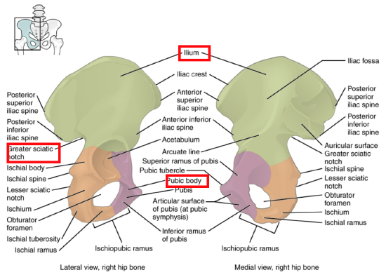
Ilium, ischium, pubis: Hold it with acetabulum facing laterally and the obturator foramen inferior and anterior. Now, make sure the pubic symphysis is anterior and the greater sciatic notches are posterior. Also make sure the ilium is superior. Below is a right bone
Femur: First, orient the bones so that the rounded head (femoral head) is superior and pointing medially. Then you will need to determine the anterior vs. posterior side. Look for the patellar surface, which should be distal and facing anteriorly. Also note how the articulating surfaces of the condyles extends far back on the posterior side. Lastly, make sure that the greater trochanter is lateral. Below is a left femur. You can view this femur rotating in 3D here: https://commons.wikimedia.org/wiki/F...r_-_close-up_- _animation.gif

Tibia: The larger flatter end (which contains the condyles and intercondylar eminence) should be superior. The anterior crest should, of course, be anterior—this is the part of your shin that you might hit on a drawer. Another way to determine the anterior side is to find the tibial tuberosity and make sure it faces anteriorly (it’s the little bump you feel just below the patella). The medial side can be determined by the medial malleolus (remember that the malleoli bracket the ankle and since the tibia is the medial bone of the lower limb, its malleolus must be medial). Below is a right tibia.

Fibula: The rounded surface of the lateral condyle should be distal and facing laterally (this is the outside bump of your ankle). The pointed end of the fibula’s head should point toward your head and be toward the posterior aspect of your body. Below is a right fibula.
