
3.6: Practice for Brain Models
- Page ID
- 72651
\( \newcommand{\vecs}[1]{\overset { \scriptstyle \rightharpoonup} {\mathbf{#1}} } \)
\( \newcommand{\vecd}[1]{\overset{-\!-\!\rightharpoonup}{\vphantom{a}\smash {#1}}} \)
\( \newcommand{\id}{\mathrm{id}}\) \( \newcommand{\Span}{\mathrm{span}}\)
( \newcommand{\kernel}{\mathrm{null}\,}\) \( \newcommand{\range}{\mathrm{range}\,}\)
\( \newcommand{\RealPart}{\mathrm{Re}}\) \( \newcommand{\ImaginaryPart}{\mathrm{Im}}\)
\( \newcommand{\Argument}{\mathrm{Arg}}\) \( \newcommand{\norm}[1]{\| #1 \|}\)
\( \newcommand{\inner}[2]{\langle #1, #2 \rangle}\)
\( \newcommand{\Span}{\mathrm{span}}\)
\( \newcommand{\id}{\mathrm{id}}\)
\( \newcommand{\Span}{\mathrm{span}}\)
\( \newcommand{\kernel}{\mathrm{null}\,}\)
\( \newcommand{\range}{\mathrm{range}\,}\)
\( \newcommand{\RealPart}{\mathrm{Re}}\)
\( \newcommand{\ImaginaryPart}{\mathrm{Im}}\)
\( \newcommand{\Argument}{\mathrm{Arg}}\)
\( \newcommand{\norm}[1]{\| #1 \|}\)
\( \newcommand{\inner}[2]{\langle #1, #2 \rangle}\)
\( \newcommand{\Span}{\mathrm{span}}\) \( \newcommand{\AA}{\unicode[.8,0]{x212B}}\)
\( \newcommand{\vectorA}[1]{\vec{#1}} % arrow\)
\( \newcommand{\vectorAt}[1]{\vec{\text{#1}}} % arrow\)
\( \newcommand{\vectorB}[1]{\overset { \scriptstyle \rightharpoonup} {\mathbf{#1}} } \)
\( \newcommand{\vectorC}[1]{\textbf{#1}} \)
\( \newcommand{\vectorD}[1]{\overrightarrow{#1}} \)
\( \newcommand{\vectorDt}[1]{\overrightarrow{\text{#1}}} \)
\( \newcommand{\vectE}[1]{\overset{-\!-\!\rightharpoonup}{\vphantom{a}\smash{\mathbf {#1}}}} \)
\( \newcommand{\vecs}[1]{\overset { \scriptstyle \rightharpoonup} {\mathbf{#1}} } \)
\( \newcommand{\vecd}[1]{\overset{-\!-\!\rightharpoonup}{\vphantom{a}\smash {#1}}} \)
The pictures below provide you with a low-tech way to practice finding and identifying anatomical parts of the brain and some examples of physiology questions you may encounter. The pictures are of RVCC’s lab models. Each picture will include labels and associated questions; the associated questions are similar to what you may expect to see on a practical or a lab assignment. If you have any questions or think you found an error in the answer key, please consult your instructor.
This practice work is optional, unless otherwise stated by your instructor. Answers for all figure and questions are at the end of the document.
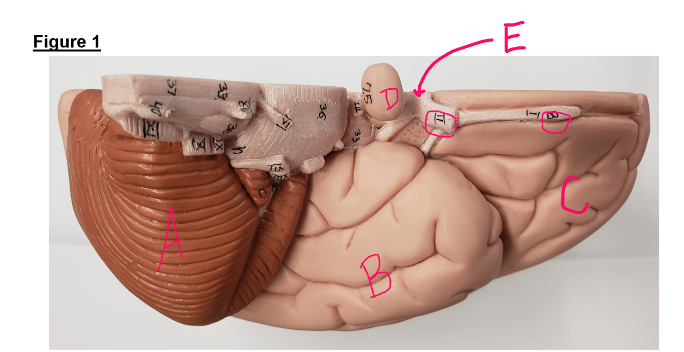
- What view (frontal, sagittal, bottom, top, internal external, transverse) are you looking at in this picture?
- Is this the left or the right side of the brain?
- Identify Structure A
- Identify Lobe B
- Identify Lobe C
- Identify Structure D
- Identify Structure E
- Identify Structure II
- Identify Structure 8
- Which structure helps plan movements; control learned, repetitious, or patterned motor skills; and, is involved with intellectual activities, personality, interpreting abstract ideas and making rational judgments?
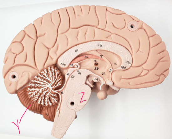
- What view (frontal, sagittal, bottom, top, internal external, transverse) are you looking at in this picture?
- Is this the left or the right side of the brain?
- Identify the structure labeled 10
- Identify Structure 26
- Identify Structure 11
- Identify Structure 23
- Identify Structure e
- Identify Structure 19 & 20
- Identify the structure labeled Z
- Identify the structure labeled Y
- Which structure (letter or number) plays a major role in balance and precise, coordinated skeletal muscle movement with visual, proprioceptive, auditory, and vestibular information?
- Which structure (letter or number) synthesizes cerebrospinal fluid?
- Which structure (letter or number) contains white matter tracts that convey action potentials between the left and right sides of the brain?
- Which structure (letter or number) synthesizes melatonin?
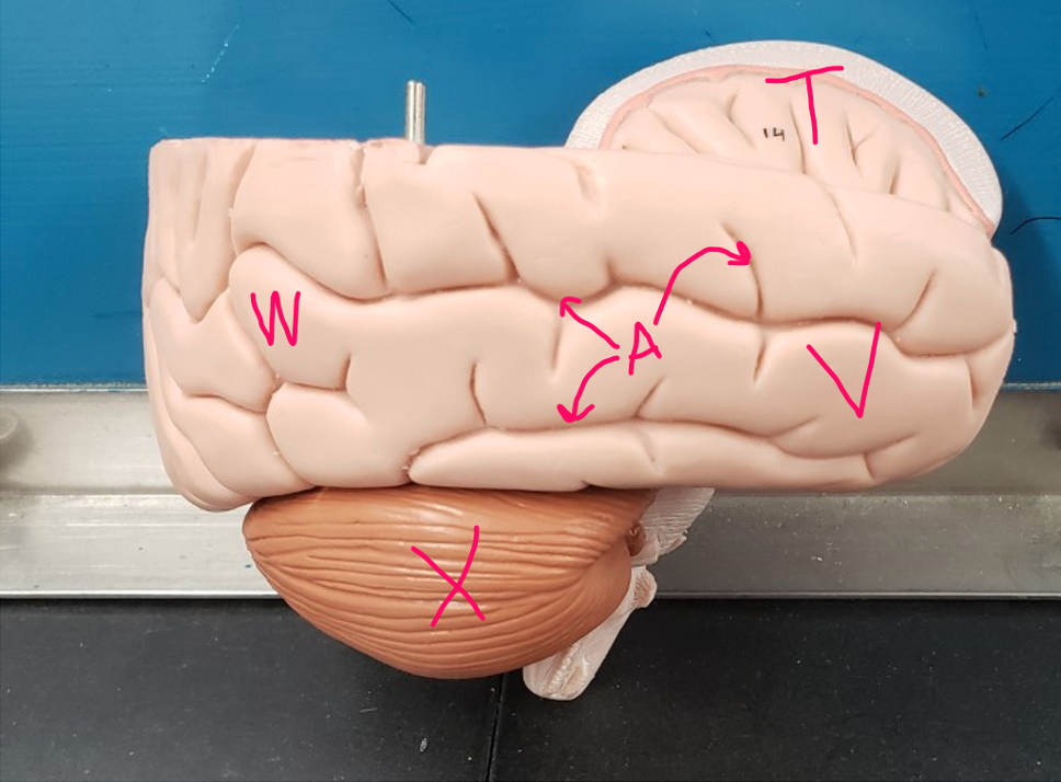
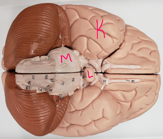
- What view (frontal, sagittal, bottom, top, internal external, transverse) are you looking at in this picture?
- Is this the left or the right side of the brain?
- Identify the lobe labeled W
- Identify the structure labeled X
- Identify the lobe labeled V
- Identify the lobe labeled T
- Does letter A point to gyri or sulci?
- Which structure contains Broca’s area?
- Which structure receives sensory signals about and interprets gustatory information and helps us understand visceral sensations (such as a stomach ache or nausea)?
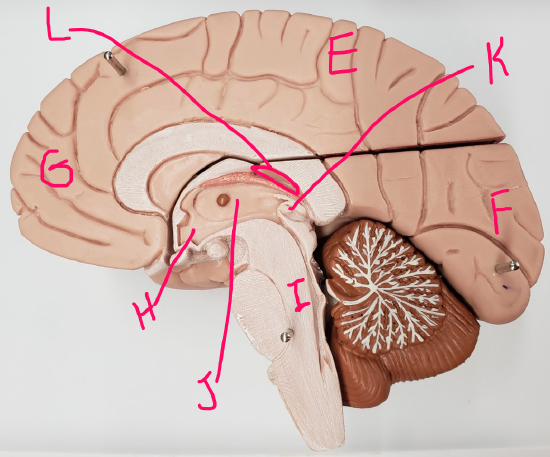
- What view (frontal, sagittal, bottom, top, internal external, transverse) are you looking at in this picture?
- Is this the left or the right side of the brain?
- Identify the structure labeled L
- Identify the lobe labeled E
- Identify the lobe labeled F
- Identify the lobe labeled G
- Identify the structure labeled H
- Identify the structure labeled I
- List one function of lobe F
- Which letter represents the structure with reflex centers for heart rate, blood pressure, and vomiting?
- Identify one function of structure H
- Which structure intakes somatosensory information and helps us to understand spatial discrimination?
- Identify a function of structure J
- Which structure (letter or number) regulates Circadian rhythms?
- What view (frontal, sagittal, bottom, top, internal external, transverse) are you looking at in this picture?
- Is this the left or the right side of the brain?
- Identify one function of structure L.
- Which structure helps to regulate breathing rate and depth with input from chemoreceptors?
- Identify one function of structure K
- Identify lobe K
- Identify structure L
- Identify structure M
- Identify one function of structure 8
- Which structure (letter or number) receives visual information from the rods and cones of the retina?
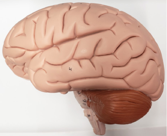
- What view (frontal, sagittal, bottom, top, internal external, transverse) are you looking at in this picture?
- Is this the left or the right side of the brain?
Figure 1 Answers
- This view shows the left side of the brain from the inferior view
- Left
- Cerebellum
- Temporal lobe (not just temporal)
- Frontal lobe (not just frontal)
- Pituitary gland
- Optic chiasma (not chiasm)
- Optic nerve
- Olfactory bulb
- C (frontal lobe)
Figure 2 Answers
- This is an internal sagittal section of the brain that has been cut in half
- Left
- Corpus collosum (you do not need to know specific areas of the corpus collosum)
- Choroid plexus
- Lateral ventricle
- Pineal gland
- 4th ventricle/ fourth ventricle
- Thalamus
- Pons
- Cerebellum
- Y
- Structure 26
- Structure 10
- Structure 23
Figure 3 Answers
- This is an external view of the right side of the brain; the section containing the superior frontal lobe, parietal lobe, and superior occipital lobe has been removed.
- Right
- Occipital lobe (not just occipital)
- Cerebellum
- Frontal lobe (not just frontal)
- Insula
- Sulci (which are the sunken areas formed by folding of the cerebral cortex during its early growth); gyri are the raised areas
- V
- T
Figure 4 Answers
- This is an internal sagittal section of the brain that has been cut in half
- Right
- 3rd ventricle / third ventricle
- Parietal lobe (not just parietal)
- Occipital lobe (not just occipital)
- Frontal lobe (not just frontal)
- Hypothalamus
- Medulla oblongota (regulated pulse, BP, and respiratory rate among other functions)
- This is the occipital lobe, whose functions include:
- Receives visual information from retinas
- Uses past visual experiences to interpret visual stimuli (color, form, or movement)
- Example: ability to recognize faces
- Structure I
- This is the hypothalamus, whose functions include:
- Controls autonomic nervous system
- blood pressure, rate and force of heartbeat, digestive tract motility, pupil size
- Initiates physical responses to emotions
- Regulates body temperature: sweating or shivering
- Regulates hunger and satiety in response to nutrient blood levels or hormones
- Regulates water balance and thirst
- Regulates sleep-wake cycles
- Controls endocrine system functions such as:
- Secretions of anterior pituitary gland (by use of releasing and inhibiting hormones to the anterior pituitary gland)
- Production of posterior pituitary hormones (ADH and oxytocin)
- Production of releasing and inhibiting hormones
- E (the parietal lobe)
- This is the thalamus, whose functions include:
- Relays, sorts, edits information coming into cortex
- Mediates sensation, motor activities, learning, and memory
- As part of Reticular Activating System, aids in maintaining cortical arousal (think of this as keeping the brain “awake” even when sleeping in case there’s an emergency—remember we used to live out in the wild with no walls or electronic security systems)
- K, (pineal gland), which regulates Circadian rhythms, which are also known as sleep-wake cycles
Figure 5 Answers
- This is an external view of the brain from its underside
- This view shows both the left (top part of picture) and right (bottom part of picture)
- This is the pituitary gland which secretes hormones
- M
- This is the temporal lobe, whose functions include:
- Receives and interprets auditory information (and sound memory)
- Contains part of Wernicke’s area (language, left)
- Conscious awareness of odors/olfaction
- Interpretation of smell as good/bad = frontal lobe
- Temporal lobe (not just temporal)
- Pituitary gland
- Pons
- This is the olfactory bulb (I is the olfactory nerve), its function is to receive olfactory/smell information from the olfactory receptors in the roof of the nose
- Structure II, which is the optic nerve
Figure 6 Answers
- This is an external view of the left side of the brain
- Left