
14.2E: Parasympathetic (Craniosacral) Division
- Page ID
- 7725

\( \newcommand{\vecs}[1]{\overset { \scriptstyle \rightharpoonup} {\mathbf{#1}} } \)
\( \newcommand{\vecd}[1]{\overset{-\!-\!\rightharpoonup}{\vphantom{a}\smash {#1}}} \)
\( \newcommand{\dsum}{\displaystyle\sum\limits} \)
\( \newcommand{\dint}{\displaystyle\int\limits} \)
\( \newcommand{\dlim}{\displaystyle\lim\limits} \)
\( \newcommand{\id}{\mathrm{id}}\) \( \newcommand{\Span}{\mathrm{span}}\)
( \newcommand{\kernel}{\mathrm{null}\,}\) \( \newcommand{\range}{\mathrm{range}\,}\)
\( \newcommand{\RealPart}{\mathrm{Re}}\) \( \newcommand{\ImaginaryPart}{\mathrm{Im}}\)
\( \newcommand{\Argument}{\mathrm{Arg}}\) \( \newcommand{\norm}[1]{\| #1 \|}\)
\( \newcommand{\inner}[2]{\langle #1, #2 \rangle}\)
\( \newcommand{\Span}{\mathrm{span}}\)
\( \newcommand{\id}{\mathrm{id}}\)
\( \newcommand{\Span}{\mathrm{span}}\)
\( \newcommand{\kernel}{\mathrm{null}\,}\)
\( \newcommand{\range}{\mathrm{range}\,}\)
\( \newcommand{\RealPart}{\mathrm{Re}}\)
\( \newcommand{\ImaginaryPart}{\mathrm{Im}}\)
\( \newcommand{\Argument}{\mathrm{Arg}}\)
\( \newcommand{\norm}[1]{\| #1 \|}\)
\( \newcommand{\inner}[2]{\langle #1, #2 \rangle}\)
\( \newcommand{\Span}{\mathrm{span}}\) \( \newcommand{\AA}{\unicode[.8,0]{x212B}}\)
\( \newcommand{\vectorA}[1]{\vec{#1}} % arrow\)
\( \newcommand{\vectorAt}[1]{\vec{\text{#1}}} % arrow\)
\( \newcommand{\vectorB}[1]{\overset { \scriptstyle \rightharpoonup} {\mathbf{#1}} } \)
\( \newcommand{\vectorC}[1]{\textbf{#1}} \)
\( \newcommand{\vectorD}[1]{\overrightarrow{#1}} \)
\( \newcommand{\vectorDt}[1]{\overrightarrow{\text{#1}}} \)
\( \newcommand{\vectE}[1]{\overset{-\!-\!\rightharpoonup}{\vphantom{a}\smash{\mathbf {#1}}}} \)
\( \newcommand{\vecs}[1]{\overset { \scriptstyle \rightharpoonup} {\mathbf{#1}} } \)
\(\newcommand{\longvect}{\overrightarrow}\)
\( \newcommand{\vecd}[1]{\overset{-\!-\!\rightharpoonup}{\vphantom{a}\smash {#1}}} \)
\(\newcommand{\avec}{\mathbf a}\) \(\newcommand{\bvec}{\mathbf b}\) \(\newcommand{\cvec}{\mathbf c}\) \(\newcommand{\dvec}{\mathbf d}\) \(\newcommand{\dtil}{\widetilde{\mathbf d}}\) \(\newcommand{\evec}{\mathbf e}\) \(\newcommand{\fvec}{\mathbf f}\) \(\newcommand{\nvec}{\mathbf n}\) \(\newcommand{\pvec}{\mathbf p}\) \(\newcommand{\qvec}{\mathbf q}\) \(\newcommand{\svec}{\mathbf s}\) \(\newcommand{\tvec}{\mathbf t}\) \(\newcommand{\uvec}{\mathbf u}\) \(\newcommand{\vvec}{\mathbf v}\) \(\newcommand{\wvec}{\mathbf w}\) \(\newcommand{\xvec}{\mathbf x}\) \(\newcommand{\yvec}{\mathbf y}\) \(\newcommand{\zvec}{\mathbf z}\) \(\newcommand{\rvec}{\mathbf r}\) \(\newcommand{\mvec}{\mathbf m}\) \(\newcommand{\zerovec}{\mathbf 0}\) \(\newcommand{\onevec}{\mathbf 1}\) \(\newcommand{\real}{\mathbb R}\) \(\newcommand{\twovec}[2]{\left[\begin{array}{r}#1 \\ #2 \end{array}\right]}\) \(\newcommand{\ctwovec}[2]{\left[\begin{array}{c}#1 \\ #2 \end{array}\right]}\) \(\newcommand{\threevec}[3]{\left[\begin{array}{r}#1 \\ #2 \\ #3 \end{array}\right]}\) \(\newcommand{\cthreevec}[3]{\left[\begin{array}{c}#1 \\ #2 \\ #3 \end{array}\right]}\) \(\newcommand{\fourvec}[4]{\left[\begin{array}{r}#1 \\ #2 \\ #3 \\ #4 \end{array}\right]}\) \(\newcommand{\cfourvec}[4]{\left[\begin{array}{c}#1 \\ #2 \\ #3 \\ #4 \end{array}\right]}\) \(\newcommand{\fivevec}[5]{\left[\begin{array}{r}#1 \\ #2 \\ #3 \\ #4 \\ #5 \\ \end{array}\right]}\) \(\newcommand{\cfivevec}[5]{\left[\begin{array}{c}#1 \\ #2 \\ #3 \\ #4 \\ #5 \\ \end{array}\right]}\) \(\newcommand{\mattwo}[4]{\left[\begin{array}{rr}#1 \amp #2 \\ #3 \amp #4 \\ \end{array}\right]}\) \(\newcommand{\laspan}[1]{\text{Span}\{#1\}}\) \(\newcommand{\bcal}{\cal B}\) \(\newcommand{\ccal}{\cal C}\) \(\newcommand{\scal}{\cal S}\) \(\newcommand{\wcal}{\cal W}\) \(\newcommand{\ecal}{\cal E}\) \(\newcommand{\coords}[2]{\left\{#1\right\}_{#2}}\) \(\newcommand{\gray}[1]{\color{gray}{#1}}\) \(\newcommand{\lgray}[1]{\color{lightgray}{#1}}\) \(\newcommand{\rank}{\operatorname{rank}}\) \(\newcommand{\row}{\text{Row}}\) \(\newcommand{\col}{\text{Col}}\) \(\renewcommand{\row}{\text{Row}}\) \(\newcommand{\nul}{\text{Nul}}\) \(\newcommand{\var}{\text{Var}}\) \(\newcommand{\corr}{\text{corr}}\) \(\newcommand{\len}[1]{\left|#1\right|}\) \(\newcommand{\bbar}{\overline{\bvec}}\) \(\newcommand{\bhat}{\widehat{\bvec}}\) \(\newcommand{\bperp}{\bvec^\perp}\) \(\newcommand{\xhat}{\widehat{\xvec}}\) \(\newcommand{\vhat}{\widehat{\vvec}}\) \(\newcommand{\uhat}{\widehat{\uvec}}\) \(\newcommand{\what}{\widehat{\wvec}}\) \(\newcommand{\Sighat}{\widehat{\Sigma}}\) \(\newcommand{\lt}{<}\) \(\newcommand{\gt}{>}\) \(\newcommand{\amp}{&}\) \(\definecolor{fillinmathshade}{gray}{0.9}\)Parasympathetic ganglia are the autonomic ganglia of the parasympathetic nervous system that lie near or within the organs they innervate.
- Describe features of the parasympathetic division of the autonomic nervous system
Key Points
- Each PSNS ganglion has three roots: a motor root, a sympathetic root, and a sensory root, as well as a number of exiting branches.
- Most are small terminal ganglia or intramural ganglia, so named because they lie near or within the organs they innervate.
- The parasympathetic system is referred to as having craniosacral outflow because of the location of PSNS fiber origins.
Key Terms
- lacrimal gland: One of a pair almond-shaped glands, one for each eye, that secrete the aqueous layer of the tear film.
- sympathetic root: This carries postsynaptic sympathetic fibers that traverse the ganglion without crossing a synapse.
- motor root: This carries presynaptic parasympathetic nerve fibers that terminate in the ganglion and create synapses for the postsynaptic fibers to travel to their target organs.
- sensory root: The proximal end of a dorsal afferent nerve that is attached to the spinal cord.
- parasympathetic: Relating to the part of the autonomic nervous system that inhibits or opposes the effects of the sympathetic nervous system.
EXAMPLES
Nerves that supply parasympathetic fibers to the parasympathetic ganglia of the head include the oculomotor nerve (ciliary ganglion); the facial nerve (pterygopalatine ganglion, submandibular ganglion); the glossopharyngeal nerve (otic ganglion); the vagus nerve (no named ganglion); and the pelvic splanchnic nerves (no named ganglion).
Parasympathetic ganglia are the autonomic ganglia of the parasympathetic nervous system, blue fibers). Most are small terminal ganglia or intramural ganglia, so named because they lie near or within (respectively) the organs they innervate.
The exceptions are the four paired parasympathetic ganglia of the head and neck. These paired ganglia supply all parasympathetic innervation to the head and neck: ciliary ganglion (spincter pupillae, ciliary muscle), pterygopalatine ganglion (lacrimal gland, glands of nasal cavity), submandibular ganglion (submandibular and sublingual glands), and otic ganglion (parotid gland).
Nerve innervation of the autonomic nervous system: The parasympathetic nervous system, shown in blue, is a division of the autonomic nervous system.
Each has three roots entering the ganglion (motor, sympathetic, and sensory roots) and a variable number of exiting branches.
- The motor root carries presynaptic parasympathetic nerve fibers (general visceral efferent fibers) that terminate in the ganglion by creating a synapse for the postsynaptic fibers traveling to target organs.
- The sympathetic root carries postsynaptic sympathetic fibers (general visceral efferent fibers) that traverse the ganglion without creating a synapse.
- The sensory root carries general sensory fibers (general somatic afferent fibers) that also do not create a synapse in the ganglion.
Some ganglia also carry special sensory fibers (special visceral afferent) for taste sensation.
The nerves that supply parasympathetic fibers to the parasympathetic ganglia of the head include the oculomotor nerve (ciliary ganglion), the facial nerve (pterygopalatine ganglion, submandibular ganglion), the glossopharyngeal nerve (otic ganglion), the vagus nerve, and the pelvic splanchnic nerves.
Because of its location, the parasympathetic system is commonly referred to as having craniosacral outflow, in contrast to the sympathetic nervous system, which is said to have thoracolumbar outflow.
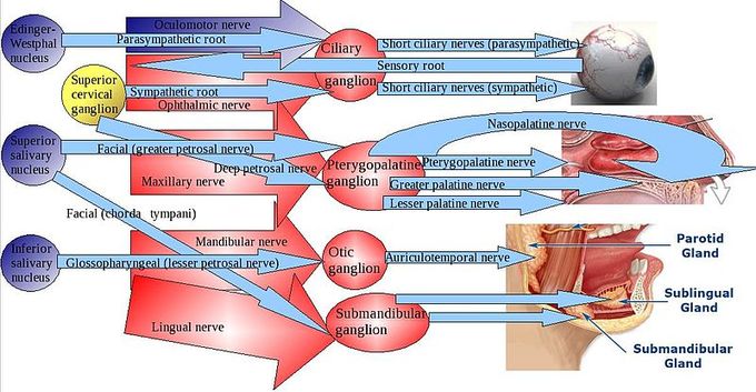
Parasympathetic ganglia of the head: The parasympathetic division has craniosacral outflow, meaning that the neurons begin at the cranial nerves (CN3, CN7, CN9, CN10) and the sacral spinal cord (S2–S4). Pre- and post-ganglionic fibers and targets are depicted.