
9.1: The Cardiovascular System
- Page ID
- 108121

\( \newcommand{\vecs}[1]{\overset { \scriptstyle \rightharpoonup} {\mathbf{#1}} } \)
\( \newcommand{\vecd}[1]{\overset{-\!-\!\rightharpoonup}{\vphantom{a}\smash {#1}}} \)
\( \newcommand{\dsum}{\displaystyle\sum\limits} \)
\( \newcommand{\dint}{\displaystyle\int\limits} \)
\( \newcommand{\dlim}{\displaystyle\lim\limits} \)
\( \newcommand{\id}{\mathrm{id}}\) \( \newcommand{\Span}{\mathrm{span}}\)
( \newcommand{\kernel}{\mathrm{null}\,}\) \( \newcommand{\range}{\mathrm{range}\,}\)
\( \newcommand{\RealPart}{\mathrm{Re}}\) \( \newcommand{\ImaginaryPart}{\mathrm{Im}}\)
\( \newcommand{\Argument}{\mathrm{Arg}}\) \( \newcommand{\norm}[1]{\| #1 \|}\)
\( \newcommand{\inner}[2]{\langle #1, #2 \rangle}\)
\( \newcommand{\Span}{\mathrm{span}}\)
\( \newcommand{\id}{\mathrm{id}}\)
\( \newcommand{\Span}{\mathrm{span}}\)
\( \newcommand{\kernel}{\mathrm{null}\,}\)
\( \newcommand{\range}{\mathrm{range}\,}\)
\( \newcommand{\RealPart}{\mathrm{Re}}\)
\( \newcommand{\ImaginaryPart}{\mathrm{Im}}\)
\( \newcommand{\Argument}{\mathrm{Arg}}\)
\( \newcommand{\norm}[1]{\| #1 \|}\)
\( \newcommand{\inner}[2]{\langle #1, #2 \rangle}\)
\( \newcommand{\Span}{\mathrm{span}}\) \( \newcommand{\AA}{\unicode[.8,0]{x212B}}\)
\( \newcommand{\vectorA}[1]{\vec{#1}} % arrow\)
\( \newcommand{\vectorAt}[1]{\vec{\text{#1}}} % arrow\)
\( \newcommand{\vectorB}[1]{\overset { \scriptstyle \rightharpoonup} {\mathbf{#1}} } \)
\( \newcommand{\vectorC}[1]{\textbf{#1}} \)
\( \newcommand{\vectorD}[1]{\overrightarrow{#1}} \)
\( \newcommand{\vectorDt}[1]{\overrightarrow{\text{#1}}} \)
\( \newcommand{\vectE}[1]{\overset{-\!-\!\rightharpoonup}{\vphantom{a}\smash{\mathbf {#1}}}} \)
\( \newcommand{\vecs}[1]{\overset { \scriptstyle \rightharpoonup} {\mathbf{#1}} } \)
\(\newcommand{\longvect}{\overrightarrow}\)
\( \newcommand{\vecd}[1]{\overset{-\!-\!\rightharpoonup}{\vphantom{a}\smash {#1}}} \)
\(\newcommand{\avec}{\mathbf a}\) \(\newcommand{\bvec}{\mathbf b}\) \(\newcommand{\cvec}{\mathbf c}\) \(\newcommand{\dvec}{\mathbf d}\) \(\newcommand{\dtil}{\widetilde{\mathbf d}}\) \(\newcommand{\evec}{\mathbf e}\) \(\newcommand{\fvec}{\mathbf f}\) \(\newcommand{\nvec}{\mathbf n}\) \(\newcommand{\pvec}{\mathbf p}\) \(\newcommand{\qvec}{\mathbf q}\) \(\newcommand{\svec}{\mathbf s}\) \(\newcommand{\tvec}{\mathbf t}\) \(\newcommand{\uvec}{\mathbf u}\) \(\newcommand{\vvec}{\mathbf v}\) \(\newcommand{\wvec}{\mathbf w}\) \(\newcommand{\xvec}{\mathbf x}\) \(\newcommand{\yvec}{\mathbf y}\) \(\newcommand{\zvec}{\mathbf z}\) \(\newcommand{\rvec}{\mathbf r}\) \(\newcommand{\mvec}{\mathbf m}\) \(\newcommand{\zerovec}{\mathbf 0}\) \(\newcommand{\onevec}{\mathbf 1}\) \(\newcommand{\real}{\mathbb R}\) \(\newcommand{\twovec}[2]{\left[\begin{array}{r}#1 \\ #2 \end{array}\right]}\) \(\newcommand{\ctwovec}[2]{\left[\begin{array}{c}#1 \\ #2 \end{array}\right]}\) \(\newcommand{\threevec}[3]{\left[\begin{array}{r}#1 \\ #2 \\ #3 \end{array}\right]}\) \(\newcommand{\cthreevec}[3]{\left[\begin{array}{c}#1 \\ #2 \\ #3 \end{array}\right]}\) \(\newcommand{\fourvec}[4]{\left[\begin{array}{r}#1 \\ #2 \\ #3 \\ #4 \end{array}\right]}\) \(\newcommand{\cfourvec}[4]{\left[\begin{array}{c}#1 \\ #2 \\ #3 \\ #4 \end{array}\right]}\) \(\newcommand{\fivevec}[5]{\left[\begin{array}{r}#1 \\ #2 \\ #3 \\ #4 \\ #5 \\ \end{array}\right]}\) \(\newcommand{\cfivevec}[5]{\left[\begin{array}{c}#1 \\ #2 \\ #3 \\ #4 \\ #5 \\ \end{array}\right]}\) \(\newcommand{\mattwo}[4]{\left[\begin{array}{rr}#1 \amp #2 \\ #3 \amp #4 \\ \end{array}\right]}\) \(\newcommand{\laspan}[1]{\text{Span}\{#1\}}\) \(\newcommand{\bcal}{\cal B}\) \(\newcommand{\ccal}{\cal C}\) \(\newcommand{\scal}{\cal S}\) \(\newcommand{\wcal}{\cal W}\) \(\newcommand{\ecal}{\cal E}\) \(\newcommand{\coords}[2]{\left\{#1\right\}_{#2}}\) \(\newcommand{\gray}[1]{\color{gray}{#1}}\) \(\newcommand{\lgray}[1]{\color{lightgray}{#1}}\) \(\newcommand{\rank}{\operatorname{rank}}\) \(\newcommand{\row}{\text{Row}}\) \(\newcommand{\col}{\text{Col}}\) \(\renewcommand{\row}{\text{Row}}\) \(\newcommand{\nul}{\text{Nul}}\) \(\newcommand{\var}{\text{Var}}\) \(\newcommand{\corr}{\text{corr}}\) \(\newcommand{\len}[1]{\left|#1\right|}\) \(\newcommand{\bbar}{\overline{\bvec}}\) \(\newcommand{\bhat}{\widehat{\bvec}}\) \(\newcommand{\bperp}{\bvec^\perp}\) \(\newcommand{\xhat}{\widehat{\xvec}}\) \(\newcommand{\vhat}{\widehat{\vvec}}\) \(\newcommand{\uhat}{\widehat{\uvec}}\) \(\newcommand{\what}{\widehat{\wvec}}\) \(\newcommand{\Sighat}{\widehat{\Sigma}}\) \(\newcommand{\lt}{<}\) \(\newcommand{\gt}{>}\) \(\newcommand{\amp}{&}\) \(\definecolor{fillinmathshade}{gray}{0.9}\)The Cardiovascular System
If your heart beats 75 times per minute, it would contract approximately 108,000 times in one day, more than 39 million times in one year, and nearly 3 billion times during a 75-year lifespan. Your heart is an amazing organ that works all day, every day, to ensure all of the cells, tissues, and organs in your body get the oxygen and nutrients needed to survive, while also removing carbon dioxide and waste by pumping about 2,000 gallons of blood around your body. This is a lot of work for an organ about the size of your fist that weighs less than a pound.
The heart is the power, or the pumping force, behind the circulation of blood through the body. Every time your heart beats it is contracting in order to push blood, much like you would squeeze a plastic ketchup bottle and push out ketchup. The heart is just one part of the cardiovascular system, the other part is the vessels through which blood travels through the body. The heart and blood vessels are a closed-loop system, where blood moves in one direction to and from your heart.
The heart has two halves, each with atria and ventricles known as the right atrium, right ventricle, left atrium, and left ventricle. As a closed-loop system, blood moves through the heart in a precise pattern.
- Deoxygenated blood enters the heart through the largest vein of the body called the superior vena cava
- The deoxygenated blood first enters the heart through the right atrium.
- The blood is then pushed down into the right ventricle.
- The deoxygenated blood now leaves the heart to go to the lungs and pick up oxygen.
- Oxygenated blood then returns to the heart through the left atrium.
- The blood is pushed into the left ventricle.
- Lastly, the oxygenated blood leaves the heart to go to the body through the largest artery of the body called the aorta.
Each side of the heart plays an important role in the circulation of blood. The right side of the heart is responsible for circulating the deoxygenated blood to the lungs to pick up oxygen, called pulmonary circulation, and the left side of the heart is responsible for circulating the blood to the rest of the body, called systemic circulation.

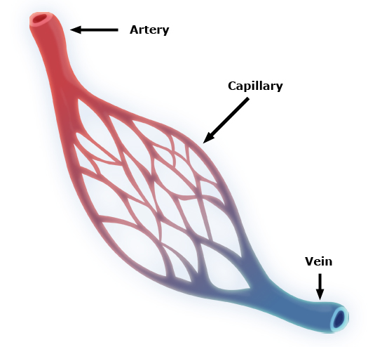
The three main types of blood vessels that transport blood throughout the body are the arteries, veins, and capillaries. In short, arteries are large blood vessels that carry blood away from the heart, veins are large blood vessels that carry blood to the heart, and capillaries are the small blood vessels that delivery oxygenated blood to organs and tissues and pick up deoxygenated blood from organs and tissues for the veins to take back to the heart. You might think of arteries and veins as big freeways or highways and the capillaries as the neighborhood streets, both working to provide a pathway to deliver food and take away waste.
In pictures of the circulatory system, arteries are typically represented by red blood vessels and veins are typically represented as blue blood vessels. Red is used for arteries because most arteries carry oxygenated blood, or red blood. Veins typically carry deoxygenated blood, or blue blood. The faster the heart beats, the more oxygen and nutrients are transported throughout the body.

Each beat of the heart helps to keep the blood moving within the closed system, however the heart has an additional feature that helps to keep the blood moving in one direction and these are called heart valves. Heart valves ensure unidirectional blood flow through the heart. Valves operate link French doors that open when the heart contracts and close when the heart is at rest. The four valves in the heart are the tricuspid valve, pulmonary valve, bicuspid valve, and aortic valve.

You do not tell your heart to beat, rather your heart beats automatically. For example, when your body needs more oxygen, such as when exercising, your body's autonomic nervous system tells your heart to speed up to pump more blood throughout your body. The Autonomic nervous system specifically sends messages to a specialized group of cells located high in the right atrium, called the Sinoatrial Node, or SA node. The SA node is commonly referred to as the pacemaker of the heart and is responsible for normal cardiac rhythm by sending an electrical signal that makes the heart contract.

The contraction of the heart is called systole and the relaxation of the heart is called diastole. Imagine clenching and relaxing your fist, when it is clenched that represents systole, and when it is relaxed that is diastole. When the heart contracts (systole) and pushes blood from the heart into the blood vessel (artery) it puts pressure on the blood vessel walls. When the heart relaxes (diastole) the pressure on the blood vessel walls lowers. By measuring the pressure in the blood vessel we can better understand the health of the heart and blood vessels. Measuring the pressure created in the blood vessel is known as your blood pressure. When you get your blood pressure tested they give you two numbers, the top number is the pressure created during systole and the bottom number is the pressure created during diastole. Thus, blood pressure readings are read as systolic over diastolic pressure. Average blood pressure is considered to be less than 120/80 (systolic/diastolic). If the pressure increases to 140/90 you are considered to have high blood pressure.
Blood pressure is a key indicator of heart health. When a doctor takes your blood pressure, they use a cuff to temporarily stop blood flow in an artery in your arm. As the cuff inflates, it squeezes the artery shut, stopping blood flow completely, which is why you don’t hear anything at first.
When the doctor slowly releases the pressure in the cuff, blood starts to flow again in spurts as the pressure in the cuff becomes slightly lower than the pressure in your artery. This turbulent, uneven blood flow creates sounds called Korotkoff sounds, which the doctor listens for with a stethoscope.
-
Systolic Pressure (the top number): The first Korotkoff sound is heard when blood starts to push through the artery with each heartbeat. This indicates the maximum pressure in your arteries when your heart contracts and pumps blood.
-
Diastolic Pressure (the bottom number): As the cuff continues to deflate, the sounds gradually fade and then disappear. This happens when the blood flow becomes smooth and steady again because the cuff pressure is lower than the resting pressure in your artery. This reflects the minimum pressure in your arteries when your heart is resting between beats.
So, the sounds are caused by changes in blood flow through the compressed artery, and they disappear when the artery is no longer squeezed enough to disrupt the flow.
Blood Pressure Categories
According to the American Heart Association, blood pressure falls into these categories:
| Systolic (mm Hg) | Diastolic (mm Hg) | |
| Normal | Less than 120 | Less than 80 |
| Elevated | 120–129 | Less than 80 |
| High Blood Pressure (Hypertension Stage 1) | 130-139 | 80-89 |
| High Blood Pressure (Hypertension Stage 2) | 140 or higher | 90 or higher |
| Hypertensive Crisis (Emergency care needed) | Higher than 180 | Higher than 120 |
Monitoring blood pressure regularly can help you detect and manage hypertension, reducing the risk of heart disease and stroke.