
3.3: Heinz Bodies
- Page ID
- 38781

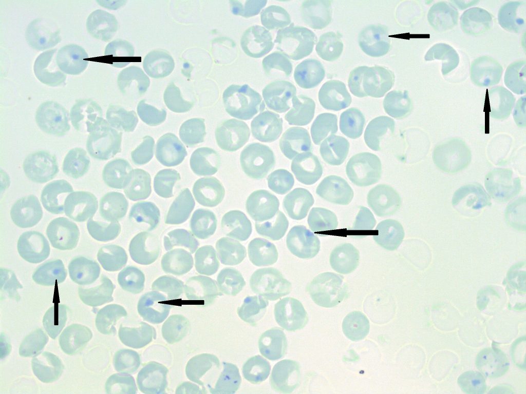
- A peripheral blood smear stained with a supravital stain demonstrating numerous heinz bodies (indicated by arrows). 100x oil immersion. From MLS Collection, University of Alberta, https://doi.org/10.7939/R35718396
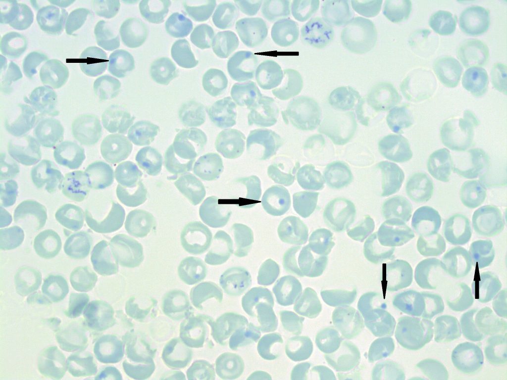
- A supravital stained peripheral blood smear showing numerous heinz bodies (indicated by arrows). 100x oil immersion. From MLS Collection, University of Alberta, https://doi.org/10.7939/R3901ZX4N
Appearance:
Inclusions are not visible on Wright or Romanowsky-stained blood smears. Inclusions can only be visualized with supravital stains. After staining, Heinz body inclusions appear dark blue-purple and are located at the periphery of the red blood cell at the membrane. The inclusions are round and look as if they are being ejected out of the cell.1,2
Note: Heinz bodies are usually not seen, as they are normally removed by splenic macrophages.3 Their presence indicates an increase in hemoglobin denaturation and precipitation, seen in numerous conditions that result in hemoglobin instability, oxidative damage, or excess globin chains.
Inclusion composition:1,2
Denatured or precipitated hemoglobin
Associated Disease/Clinical States:1,2
Glucose-6-phosphate dehydrogenase (G6PD) Deficiency
Hemoglobinopathies (may result in the formation of unstable hemoglobins)
Thalassemia
Post-splenectomy
Oxidizing drugs
Note: Appear in conditions where unstable hemoglobin can form.2
References:
1. Landis-Piwowar K, Landis J, Keila P. The complete blood count and peripheral blood smear evaluation. In: Clinical laboratory hematology. 3rd ed. New Jersey: Pearson; 2015. p. 154-77.
2. Rodak BF, Carr JH. Inclusions in erythrocytes. In: Clinical hematology atlas. 5th ed. St. Louis, Missouri: Elsevier Inc.; 2017. p. 107-14.
3. Bain BJ. Important supplementary tests. In: Blood cells: a practical guide [Internet]. 5th ed. Chichester, UK: John Wiley & Sons, Ltd; 2015 [cited 2018 Jul 10]: 277-94. Available from: http://doi.wiley.com/10.1002/9781118817322